

Книги по МРТ КТ на английском языке / Magnetic Resonance Imaging in Ischemic Stroke - K Sartor R 252 diger von Kummer Tobias Back
.pdfHemodynamic Infarcts and Occlusive Carotid Disease |
231 |
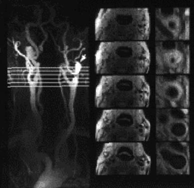
Fig. 15.6. Computerized registration and overlay of DWI lesions on PI images, to determine the exact location of the diffusion lesion with respect to the perfusion deficit. The alignment of the time-to-peak (TTP) and DWI images demonstrates that the lesions are indeed located in the borderzone area where the extent of the perfusion deficit is most pronounced
Fig. 15.7. The theoretical concepts of stroke in hemodynamic risk zones
– “dot-like” microembolic lesions in the most distal arterial branches resulting from more proximal vessel pathology and impaired emboli washout (left) and the complete infarction of the compromised tissue in the borderzone territory (right). The bottom row gives DWI examples of these lesion patterns
15.4
Evaluation of Vessel Pathology in Occlusive Carotid Disease
MRA is an emerging technique in carotid artery disease taking advantage of the differences of spin signals of flowing blood and static tissue, with the stationary tissue signal effectively suppressed relative to the blood flow signal (see Chap. 5). The ability of non-invasively imaging blood flow and thereby visualizing extraand intracranial vasculature offers the clinician valuable information. A single measurement is performed within a few minutes. Two techniques are most widely used to assess narrowing of the extracranial vessels and to visualize the intracranial vasculature: contrast-enhanced MRA (CE-MRA) and
time-of-flight (TOF) techniques. MRA source images are frequently postprocessed to provide a 3D visualization with an algorithm also termed maximum intensity projection (Mattle and Edelman 1992; Röther et al. 1993). This allows demonstration of the 3D vessel topography and offers the possibility to review the vessels in every desirable plane even after the investigation is concluded.
15.4.1
Obstruction of the Internal Carotid Artery
CE-MRA is a promising technique particularly for the evaluation of the proximal segments from the aortic arch covering the carotid bifurcation (Cloft

232 |
K. Szabo and M. G. Hennerici |
et al. 1996; Gass et al. 1997; Patel et al. 1995). Small intravenous doses of paramagnetic contrast agent strongly enhance the MR signal due to T1 shortening. While CE-MRA appears excellent to visualize the site and length of narrowing, it also shows a tendency to overemphasize arterial narrowing. Turbulent flow in case of a high-grade stenosis might therefore not be detected. Hence, complete signal loss over a short vessel segment can indicate highgrade vessel narrowing, but must not indicate vessel occlusion. From a practical standpoint CE-MRA is a very fast technique and therefore less susceptible to patient motion occuring with long acquisition times. Exact timing of the contrast injection is important to avoid obscuring the arterial vessels from hyperintensity due to early venous return. Although it has only recently become available CE-MRA has already contributed considerably to patient management as many vascular surgeons no longer require preoperative conventional contrast angiography, but may use the combination of duplex ultrasound studies and CE-MRA for visualization of the vascular pathology. Figure 15.8 gives an example for a high-grade stenosis identified with Duplex ultrasound and a signal loss over the proximal part of the affected vessel segment on CE-MRA (Gass et al. 2001).
High-resolution MRI has recently emerged as one of the most promising techniques for the noninvasive study of atherosclerosis of the ICA. This method can be used to characterize plaque composition and to monitor progression and is mainly performed using cross-sectional imaging of the vessel (Fig. 15.9). MR
techniques are not dependent on the angle of the imaging plane and are less dependent on the skill of the operator than is ultrasound. For the purposes of vascular imaging, MR is unique in that the modality can provide excellent contrast between the vessel wall and adjacent lumen by using flow-sensitive pulse sequences. Thus, MR can be used to image the vessel lumen (flowing blood) and, at the same time, produce tissue information that describes the vessel wall. By using T1-weighted and chemicalselective techniques (Corti et al. 2001; Yuan et al. 2001) the detection of lipid signals (cholesterol and cholesteryl esters) is possible. With the inclusion of sequences that are T2-weighted and T1or inter- mediate-weighted with a very short echo time, the specificity of MR imaging techniques has improved and allowed differentiation of necrotic cores from fibrous regions in ex vivo plaque specimens (Gold et al. 1993). Direct MRI of a thrombus has even been shown to be capable of detecting methemoglobin within intraplaque hemorrhage (Moody et al. 2003).
15.4.2
Role of the Anatomy of the Circle of Willis
MRA has been shown to be well suited to investigate the circle of Willis (CW) since abnormalities can be detected by MRI blood flow techniques (Anzola et al. 1995). 3D TOF sequences, although more time consuming than CE-MRA, are still the
Fig. 15.8. Contrast-enhanced MRA reveals high-grade proximal ICA stenosis with signal void at the level of the right carotid bifurcation (left). Color Doppler flow imaging demonstrates high-grade ICA stenosis with turbulent flow (light blue, top right) and power Doppler shows excellent delineation of the stenosis and increased blood flow velocity (yellow and red, bottom right)
Hemodynamic Infarcts and Occlusive Carotid Disease |
233 |
|
A |
|
B |
A |
|
B |
C |
C |
|
D |
|
E |
|
|
D |
|
E |
Fig. 15.9. Combined MRA (left) and plaque imaging (right) in a hypertensive patient with symptomatic left ICA stenosis. Cross-sectional black-blood imaging demonstrates the severely narrowed lumen (A and B) as well as the extension of the plaque to the carotid bifurcation (E), appearing normal on angiography
main method of visualizing the CW. To achieve a high contrast image, the stationary spins of background tissue are usually saturated with magnetization transfer pulses resulting in a low background signal, whereas unsaturated moving spins of blood produce a bright signal. TOF may be used either in a 2D or 3D sequence. In a 2D TOF, a set of thin slices is acquired sequentially, one slice after the other. Data in 3D TOF are acquired as volume (slab) or a set of volumes. The sensitivity of 3D TOF to slowly flowing blood is poor, but it can provide high spatial resolution and is therefore especially useful for the arterial angiography of small vessels with high-flow velocity. TOF sequences are mainly used to image the circle of Willis also covering the distal parts of the extracranial internal carotid and the vertebral arteries.
In patients with obstruction of the internal carotid artery, numerous collateral pathways that redistribute blood to the deprived site can maintain adequate cerebral blood flow. The CW is considered an important primary collateral pathway with its potential believed to be dependent on the presence and size of the component vessels, which vary among normal individuals (Alpers and
Berry 1963; Baumgartner et al. 1997; KrabbeHartkamp et al. 1998; Macchi et al. 1996; Patrux et al. 1994; Schomer et al. 1994). Using special 2D
phase-contrast techniques, hemodynamic information concerning blood flow direction is also achievable. In addition to imaging the extracranial arteries, the CW should also be assessed to detect patent collateral pathways.
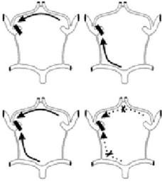
Hendrikse and coworkers (2001) investigated whether the presence of borderzone infarcts is related to the collateral ability of the CW in symptomatic and asymptomatic patients with unilateral occlusion of the ICA. They found that in patients with unilateral ICA occlusion, the presence of collateral flow via the posterior communicating artery in the circle of Willis is associated with a low prevalence of borderzone infarcts and that asymptomatic patients with an ICA occlusion do not have an increased collateral function of the CW. Figure 15.10 shows the four patterns of collateral flow via the CW to the hemisphere ipsilateral to the ICA occlusion.
Another study investigating the CW collateral flow by MRA showed that patients with ICA obstruction with minor neurological deficits demonstrated a higher prevalence of complete CW configurations (Hartkamp et al. 1999). It remains unclear whether these patients represent a special population leading to non-severe strokes or whether favorable CW configurations are acquired through secondary hemodynamic adaptation. There is also data pointing to the possibility that acute stroke patients suffering
234 |
K. Szabo and M. G. Hennerici |
A1 segment only |
PcomA only |
delay of the contrast bolus arrival to the entire MCA territory, while such asymmetry of the entire MCA territory is usually not seen in patients with small vessel disease or lacunar stroke. Cardiac embolism usually produces a pattern of multiple acute lesions in both hemispheres and both the anterior and posterior circulation, while multiple small lesions in the borderzones of a MCA territory may indicate highgrade carotid stenosis. Conversely in patients with known atrial fibrillation and co-existing high-grade ICA stenosis, PI and DWI may identify acute lesions in hemodynamic risk zones rather more suggestive of ischemia due to high-grade stenosis.
Both A1 segment and PcomA No collateral flow
Fig. 15.10. The four patterns of collateral flow via the circle of Willis to the hemisphere ipsilateral to the ICA occlusion. A1 segment indicates A1 segment of ipsilateral ACA, PcomA indicates posterior communicating artery
major stroke in underlying ICA disease may represent a subgroup with a high prevalence of hypoor aplastic CW, and may, therefore, be unable to adapt in an adequate manner or are destined by genetic factors to suffer stroke in the case of ICA disease (Szabo et al. 2001).
15.5
Use of MRI to Identify Other Causes of Stroke in Patients with Occlusive Carotid Disease
It is well known that in the age groups at risk for highgrade carotid stenosis concomitant myocardial and small vessel disease is often present. A recent analysis of the NASCET data indicated that approximately 20% of strokes in the territory of high-grade symptomatic carotid artery are of cardioembolic and lacunar origin (Barnett et al. 1998). Therefore, it appears important to determine the cause of acute deficits in patients with high-grade carotid stenosis and MRI; particularly DWI and PI seem useful to differentiate different mechanisms of acute ischemia providing a means to differentiate small vessel disease, lacunar stroke, and cardioembolic lesions. All these etiologies may produce multiple discrete lesions with different spatial distributions that may favor one or another stroke mechanism. Even in asymptomatic patients with ICA stenosis PI usually identifies a temporal
15.6
Treatment of Patients with Occlusive Carotid Disease
Surgical carotid endarterectomy is currently the accepted best standard of treatment for revascularization of extracranial carotid occlusive disease and has been validated by multiple randomized, controlled trials (Barnett et al. 1998; European Carotid Surgery Trialists’ Collaborative Group 1991). In the past years, however, carotid artery stenting has been started by different disciplines in an attempt to provide a potential therapeutic alternative to carotid endarterectomy for the treatment of atherosclerotic carotid artery disease. Reliable evaluation of the outcomes of carotid stenting is limited at this time because the outcomes have been reported from case series with few randomized trials and because longterm results are not yet established – randomized clinical studies are in progress (Brown and Hacke
2004; Featherstone et al. 2004; Higashida et al. 2004). A well-known problem of carotid stenting and of carotid surgery is procedure related embolism to the brain and several post-procedure DWI studies have repeatedly shown mainly asymptomatic DWI lesions (van Heesewijk et al. 2002). Different protection devices to prevent embolism are currently under investigation and no single best proceeding has been established to date.
Regardless of the treatment chosen, a dedicated MRI protocol including DWI as well as PI sequences should be performed beforehand in all patients with suspected symptomatic ICA disease. The combination of both methods offers the possibility of identifying patients with potential to benefit from surgery or stenting. In patients with high-grade stenosis, large perfusion deficits and a small acute ischemic
Hemodynamic Infarcts and Occlusive Carotid Disease |
235 |
lesion (DWI/PI “mismatch”), recanalization of the ICA should be achieved by all means. Results from a recent study suggested that the recanalizing procedure should ideally be performed within 2 weeks of the patient’s last symptoms (Rothwell et al. 2004). Possibly even more importantly, a dedicated MRI protocol may be able to demonstrate, that despite high-grade ICA stenosis intracranial collateralization well compensates for the proximal obstruction, questioning the use and necessity of endarterectomy in these situations.
15.7
Clinical Case Studies
After analyzing initial and final DWI and PI lesion size in stroke patients with > 70% internal carotid
artery stenosis, Neumann-Haefelin (1999) found, that most of the mismatch area identified on PI was not at high risk of irreversible tissue damage. In contrast to these findings, patients with initially small DWI lesions and an underlying carotid stenosis can deteriorate and develop considerably larger lesions. Figure 15.11 demonstrates the progressive course of hemodynamic stroke lesions in a 62-year-old patient with symptomatic left ICA occlusion who initially presented with only transient right hemiparesis, but 3 days later showed a progressive hemiparesis of the right side. Fluctuating blood pressure with intermittent systolic values < 140 mmHg were believed to have played a role in further infarct demarcation.
The following two examples show different configurations of the circle of Willis in two patients with occlusive ICA disease. While the 54-year-old patient in Fig. 15.12 suffering from left ICA occlusion shows good collateralization via the posterior communi-
a |
b |
c |
d |
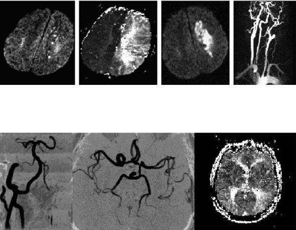
Fig. 15.11a–d. A 62-year-old patient with symptomatic left internal carotid artery occlusion and progressive stroke. Initial DWI shows only punctuate small lesions in the deep borderzone of the left hemisphere (a), while the hypoperfused area on the time-to-peak maps affects the complete left middle cerebral artery territory and is most pronounced in the deep borderzone area (b). On day 3, the acute lesion has grown considerably larger, paralleling progression of symptoms (c). Contrast-enhanced MRA shows proximal occlusion of the left internal carotid artery (d)
Fig. 15.12. An asymptomatic 54-year-old patient suffering from left internal carotid artery occlusion (left) shows excellent collateralization via the posterior communicating artery and a sufficient filling of the ipsilateral middle cerebral artery (middle). As a consequence, perfusion MRI (time-to-peak) detects only a slight asymmetry and delay of contrast agent arrival in the parietal parts of the middle cerebral artery territory (right)
236
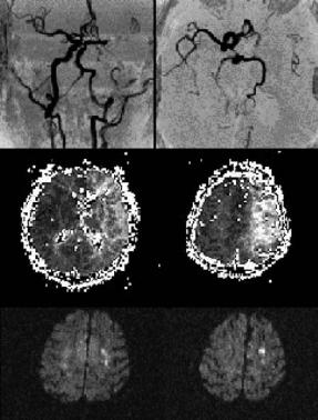
Fig. 15.13. A 76-year-old woman with a subtotal stenosis of the left internal carotid artery shows no sufficient collateral flow and only faint flow signal in the left middle cerebral artery (upper row), severe hypoperfusion (time-to-peak maps) of the left middle cerebral artery territory (middle row) and small acute hemodynamic stroke lesions on DWI (bottom row). The patient was later successfully treated with carotid endarterectomy
cating artery and a sufficient filling of the ipsilateral MCA on MRA, PI shows only a slight delay of contrast agent arrival in the posterior parts of the MCA territory. This patient was clinically asymptomatic. The 76-year-old woman with similar vessel pathology in Fig. 15.13, however, shows no sufficient collateral flow and only faint flow signal in the MCA, severe hypoperfusion of the left MCA territory and small acute hemodynamic stroke lesions on DWI. Duplex ultrasound demonstrated subtotal stenosis of the left ICA and the patient was later successfully treated with endarterectomy.
References
Adams JH, Brierley JB, Connor RCR et al (1966) The effects of systemic hypotension upon the human brain: clinical and neuropathological observations in 11 cases. Brain 89:235– 268
K. Szabo and M. G. Hennerici
Alpers BJ, Berry RG (1963) Circle of Willis in cerebral vascular disorders. Arch Neurol 8:398–402
Anzola GP, Gasparotti R, Magoni M et al (1995) Transcranial Doppler sonography and magnetic resonance angiography in the assessment of collateral hemispheric flow in patients with carotid artery disease. Stroke 26:214–217
Baird AE, Lövblad KO, Schlaug G et al (2000) Multiple acute stroke syndrome: marker of embolic disease? Neurology 54:674–678
Baumgartner RW, Baumgartner I, Mattle HP et al (1997) Transcranial color-coded duplex sonography in the evaluation of collateral flow through the circle of Willis. Am J Neuroradiol 18:127–133
Barnett HJ, Taylor DW, Eliasziw M et al (1998) Benefit of carotid endarterectomy in patients with symptomatic moderate or severe stenosis. North American Symptomatic Carotid Endarterectomy Trial Collaborators. N Engl J Med 339:1415–1425
Bäzner H, Hennerici M (2004) Georg Friedrich Händel’s strokes. Cerebrovasc Dis 17:326–331
Bogousslavsky J, Regli F (1986a) Borderzone infarctions distal to internal carotid artery occlusion: prognostic implications. Ann Neurol 20:346–350
Bogousslavsky J, Regli F (1986b) Unilateral watershed cerebral infarcts. Neurology 36:373–377
Brierley JB, Excell BJ (1966) The effects of profound systemic hypotension upon the brain of M. rhesus: physiological and pathological observations. Brain 89:235-268
Brown MM, Hacke W (2004) Carotid artery stenting: the need for randomised trials. Cerebrovasc Dis 18:57–61
Caplan LR, Hennerici M (1998) Impaired clearance of emboli (washout) is an important link between hypoperfusion, embolism, and ischemic stroke. Arch Neurol 55:1475–1482 Chambers BR, Norris JW (1986) Outcome in patients with
asymptomatic neck bruits. N Engl J Med 315:860–865 Chaves CJ, Silver B, Schlaug G et al (2000) Diffusionand
perfusion-weighted MRI patterns in borderzone infarcts. Stroke 31:1090–1096
Cloft HJ, Murphy KJ, Prince MR et al (1996) 3D gadoliniumenhanced MR angiography of the carotid arteries. Magn Reson Imaging 14:593–600
Corti R, Fuster V, Badimon JJ et al (2001) New understanding of atherosclerosis (clinically and experimentally) with evolving MRI technology in vivo. Ann NY Acad Sci 947:181–195
Del Sette M, Eliasziw M, Streifler JY et al (2000) Internal borderzone infarction: a marker for severe stenosis in patients with symptomatic internal carotid artery disease. Stroke 31:631–636
Edelman RR, Wielopolski P, Schmitt F (1994) Echo-planar MR imaging. Radiology 192:600–612
European Carotid Surgery Trialists’ Collaborative Group (1991) MRC European Carotid Surgery Trial: interim results for symptomatic patients with severe (70–99%) or with mild (0–29%) carotid stenosis. Lancet 337:1235–1243
Featherstone RL, Brown MM, Coward LJ (2004) International carotid stenting study: protocol for a randomised clinical trial comparing carotid stenting with endarterectomy in symptomatic carotid artery stenosis. Cerebrovasc Dis 18:69–74
Gass A, Gaa J, Schwartz A (1997) Cerebral infarction due to internal carotid artery dissection. J Neurol Neurosurg Psychiatry 63:420
Hemodynamic Infarcts and Occlusive Carotid Disease
Gass A, Gaa J, Sommer A et al (1999) Echo-planar diffusionweighted MRI in the diagnosis of acute ischemic stroke: characterisation of tissue abnormalities and limitations in the interpretation of imaging findings. Radiologe 39:695–702
Gass A, Meairs S, Neff W et al (2001) Towards visualization of symptomatic carotid stenosis. Arch Neurol 58:658–659
Gold GE, Pauly JM, Glover GH et al (1993) Characterization of atherosclerosis with a 1.5-T imaging system. J Magn Reson Imaging 3:399–407
Guckel FJ, Brix G, Schmiedek P et al (1996) Cerebrovascular reserve capacity in patients with occlusive cerebrovascular disease: assessment with dynamic susceptibility contrastenhanced MR imaging and the acetazolamide stimulation test. Radiology 201:405–412
Hartkamp MJ, van der GJ, van Everdingen KJ et al (1999) Circle of Willis collateral flow investigated by magnetic resonance angiography. Stroke 30:2671–2678
Heinsius T, Bogousslavsky J, van Melle G (1998) Large infarcts in the middle cerebral artery territory. Etiology and outcome patterns. Neurology 50:341–350
Hendrikse J, Hartkamp MJ, Hillen B et al (2001) Collateral ability of the circle of Willis in patients with unilateral internal carotid artery occlusion: borderzone infarcts and clinical symptoms. Stroke 32:2768–2773
Hennerici M, Hülsbömer HB Hefter H et al (1987) Natural history of asymptomatic extracranial arterial disease. Results of a long-term prospective study. Brain 110:777–791
Hennerici M, Daffertshofer M, Jakobs L (1998) Failure to identify cerebral infarct mechanisms from topography of vascular territory lesions. Am J Neuroradiol 19:1067–1074
Higashida RT, Meyers PM, Phatouros CC et al (2004) Reporting standards for carotid artery angioplasty and stent placement. Technology Assessment Committees of the American Society of Interventional; Therapeutic Neuroradiology and the Society of Interventional Radiology. Stroke 35:e112–e134
Hupperts RM, Warlow CP, Slattery J et al (1997) Severe stenosis of the internal carotid artery is not associated with borderzone infarcts in patients randomized in the European Carotid Surgery Trial. J Neurol 244:45–50
Kang DW, Chu K, Ko SB et al (2002) Lesion patterns and mechanism of ischemia in internal carotid artery disease: a diffu- sion-weighted imaging study. Arch Neurol 59:1577–1582
Kastrup A, Schulz JB, Mader I et al (2002) Diffusion-weighted MRI in patients with symptomatic internal carotid artery disease. J Neurol 249:1168–1174
Krabbe-Hartkamp MJ, van der GJ, de Leeuw FE et al (1998) Circle of Willis: morphologic variation on three-dimensional time-of-flight MR angiograms. Radiology 207:103–111
Le Bihan D (1991) Molecular diffusion nuclear magnetic resonance imaging. Magn Reson Q 7:1–30
Lee PH, Bang OY, Oh SH et al (2003) Subcortical white matter infarcts. Comparison of superficial perforating artery and internal borderzone infarcts using diffusion-weighted magnetic resonance imaging. Stroke 34:2630–2635
Macchi C, Catini C, Federico C et al (1996) Magnetic resonance angiographic evaluation of circulus arteriosus cerebri (circle of Willis): a morphologic study in 100 human healthy subjects. Ital J Anat Embryol 101:115–123
Maeda M, Yuh WT, Ueda T et al (1999) Severe occlusive carotid artery disease: hemodynamic assessment by MR perfusion imaging in symptomatic patients. Am J Neuroradiol 20:43–51
237
Mainwaring J (1760) Memoirs of the life of the late Georg Friedrich Händel. Dodsley, London
Mattle HP, Edelman RR (1992) Cerebral magnetic resonance angiography. Neurol Res 14:118–121
Moody AR, Murphy RE, Morgan PS et al (2003) Characterization of complicated carotid plaque with magnetic resonance direct thrombus imaging in patients with cerebral ischemia. Circulation 107:3047–3052
Mull M, Schwarz M, Thron A (1997) Cerebral hemispheric low-flow infarcts in arterial occlusive disease. Lesion patterns and angiomorphological conditions. Stroke 28:118– 123
Nakano S, Yokogami K, Ohta H et al (1995) CT-defined large subcortical infarcts: correlation of location with site of cerebrovascular occlusive disease. AJNR 16:1581–1585
Neumann-Haefelin T, Wittsack HJ, Fink GR et al (2000) Diffusionand perfusion-weighted MRI. Influence of severe carotid artery stenosis on the DWI/PI mismatch in acute stroke. Stroke 31:1311–1317
Ostergaard L, Sorensen AG, Kwong KK et al (1996) High resolution measurement of cerebral blood flow using intravascular tracer bolus passages, part II. Experimental comparison and preliminary results. Magn Reson Med 36:726–736
Patel MR, Kuntz KM, Klufas RA et al (1995) Preoperative assessment of the carotid bifurcation. Can magnetic resonance angiography and duplex ultrasonography replace contrast arteriography? Stroke 26:1753–1758
Patrux B, Laissy JP, Jouini S et al (1994) Magnetic resonance angiography (MRA) of the circle of Willis: a prospective comparison with conventional angiography in 54 subjects. Neuroradiology 36:193–197
Pessin MS, Hinton RC, Davis KR et al (1979) Mechanisms of acute carotid stroke. Ann Neurol 6:245–252
Pollanen MS, Deck JH (1989) Directed embolization is an alternate cause of cererbal watershed infarction. Arch Pathol Lab Med 113:1139–1141
Reith W, Heiland S, Erb G et al (1997) Dynamic contrastenhanced T2*-weighted MRI in patients with cerebrovascular disease. Neuroradiology 39:250–257
Ries S, Schminke U, Daffertshofer M, Hennerici M (1996) High intensity transient signals (HITS) in patients with carotid artery disease. Eur J Med Res 1:328–330
Rodda RA, Path FRC (1986) The arterial patterns associated with internal carotid infarcts. Stroke 17:69–75
Roh JK, Kang DW, Lee SH et al (2000) Significance of acute multiple brain infarction on diffusion-weighted imaging. Stroke 31:688–694
Rosen BR, Belliveau JW,Vevea JM et al (1990) Perfusion imaging with NMR contrast agents. Magn Reson Med 14:249–265 Röther J, Wentz KU, Rautenberg W et al (1993) Magnetic res-
onance angiography in vertebrobasilar ischemia. Stroke 24:1310–1315
Röther J, Guckel F, Neff W (1996) Assessment of regional cerebral blood volume in acute human stroke by use of singleslice dynamic susceptibility contrast-enhanced magnetic resonance imaging. Stroke 27:1088–1093
Rothwell PM, Warlow CP (1999) Prediction of benefit from carotid endarterectomy in individual patients: a risk-mod- elling study. European Carotid Surgery Trialists’ Collaborative Group. Lancet 353:2105–2110
Rothwell PM, Eliasziw M, Gutnikov SA et al (2004) Endarterectomy for symptomatic carotid stenosis in relation to clini-
238
cal subgroups and timing of surgery. Carotid Endarterectomy Trialists Collaboration. Lancet 363:915–924
Sacco RL.(2001) Clinical practice. Extracranial carotid stenosis. N Engl J Med 345:1113–1118
Sakuma H, Nomura Y, Takeda K et al (1991) Adult and neonatal human brain: diffusional anisotropy and myelination with diffusion-weighted MR imaging. Radiology 180:229–233
Schomer DF, Marks MP, Steinberg GK et al (1994) The anatomy of the posterior communicating artery as a risk factor for ischemic cerebral infarction. N Engl J Med 330:1565–1570 Szabo K, Kern R, Gass A et al (2001) Acute stroke patterns in patients with internal carotid artery disease. Stroke
32:1323–1329
Tanner JE (1983) Intracellular diffusion of water. Arch Biochem Biophys 224:416–428
Torvik A (1984) The pathogenesis of watershed infarcts in the brain. Stroke 15:221–223
Yuan C, Mitsumori LM, Beach KW et al (2001) Carotid ath-
K. Szabo and M. G. Hennerici
erosclerotic plaque: noninvasive MR characterization and identification of vulnerable lesions. Radiology 221:285–299 Van Heesewijk HP, Vos JA, Louwerse ES et al (2002) Carotid PTA and Stenting Collaborative Research Group. New brain lesions at MR imaging after carotid angioplasty and stent
placement. Radiology 224:361–365
Van der Zwan A, Hillen B (1991) Review of the variability of the territories of the major cerebral arteries. Stroke 22:1078–1084
Waterston JA, Brown MM, Butler P et al (1990) Small deep cerebral infarcts associated with occlusive internal carotid artery disease. A hemodynamic phenomenon? Arch Neurol 47:953–957
Weiller C, Ringelstein EB, Reiche W et al (1990) The large striatocapsular infarct. A clinical and pathophysiological entity. Arch Neurol 47:1085–1091
Zülch KJ (1963) New concepts concerning the pathogenesis of cerebral ischemia. Ann Radiol 6:7–14

Hypoxic–Ischemic Lesions |
239 |
16 Hypoxic–Ischemic Lesions
Karl Olof Lövblad
CONTENTS
16.1Introduction 239
16.2 |
Global Cerebral Ischemia and Hypoxia 239 |
16.2.1Pathophysiology 239
16.2.2Causes 239
16.2.3Neuropathology 240
16.3Neuroimaging 240
16.3.1Computed Tomography 240
16.3.2 |
Magnetic Resonance Imaging 241 |
16.3.2.1 |
Conventional MRI (T1-, T2-Weighted MRI) 242 |
16.3.2.2 |
Diffusion-Weighted MRI 243 |
16.4Conclusions 248 References 249
tomography (CT) can exclude hemorrhage and demonstrate diffuse ischemic edema; however, neither method may be sufficiently accurate to exactly delineate the extent of the damage incurred to the CNS. Since magnetic resonance imaging (MRI) appears superior in providing accurate information regarding brain anatomy and pathology, it should be used to assess its value in the diagnosis and management of patients with global cerebral ischemia and hypoxia.
16.1 Introduction
As medical technology progresses, more advanced life-support techniques are available. While this is an important benefit to the vast majority of patients, we are also confronted with a small number of patients sustaining lesions to the brain due to hypoxia and ischemia. Since the implications regarding outcome are very serious, it is of utmost importance to recognize these lesions early on, so that the appropriate measures can be taken. The lesions may be localized or diffuse (bilateral, infraand supra tentorial) depending on duration and acuity of ischemia. Significant acuity may suggest brain death clinically or radiologically (Lövblad and Bassetti 2000). Since these patients often receive various drugs and may be polymorbid (with both cardiac and cerebral pathologies), the physical examination can be difficult and frustrating, even for more experienced clinicians. Electroencephalography can assess central nervous system (CNS) dysfunction, and computed
16.2
Global Cerebral Ischemia and Hypoxia
16.2.1 Pathophysiology
At rest, the brain consumes 20% of the total oxygen that the whole body consumes. Global brain ischemia occurs when the arterial blood pressure cannot maintain a sufficient cerebral perfusion pressure. This happens with cardiac dysfunction, shock, and critical increase in intracranial pressure. Cerebral hypoxia is the deprivation of oxygen with a maintained cerebral blood flow. Pure hypoxia will occur in rare instances such as reduced atmospheric oxygen, which is an extremely rare cause of brain hypoxia, or as a result of drowning. Most cases occur in a combination of hypoxia and ischemia since pure hypoxia very often causes cardiac arrest and, thus, interruption of cerebral blood flow. The combination of both hypoxia and ischemia leads to more serious neuronal damage than hypoxia alone.
16.2.2 Causes
K. O. Lövblad, MD
Neuroradiology Unit, Department of Radiology and Medical Informatics, University Hospitals Geneva, 24 rue Micheli-du- Crest, 1211 Geneva 14, Switzerland
The causes of diffuse cerebral ischemia are: circulatory arrest (e.g., cardiac arrest), impaired circulation (strangulation, profound hypotension, extracranial
240 |
K. O. Lövblad |
vascular disease), impaired energy substrate supply (hypoxic hypoxia, hypoglycemia, carbon monoxide poisoning), diffuse small vessel disease (hypertension, eclampsia, types of small vessel arteritis) and micro-embolic disease (fat embolism, disseminated intravascular coagulation, cardiopulmonary bypass procedures) (Toole and Burrow 1990). Strangulation and hanging will lead to hypoxia due to oligemia, airway obstruction, and respiratory arrest in the case of brain stem compression by fractured vertebrae.
Laminar cortical necrosis is the end-stage of cortical ischemia and has been described in many conditions of cellular energy depletion, such as hypoxia and hypoglycemia. It may also be associated with a variety of other diseases affecting the nervous system (Valanne et al. 1996). Laminar cortical necrosis has been studied with nuclear medicine methods (Hawes and Mishkin 1972), but has recently been more extensively studied with CT and MRI.
16.2.3 Neuropathology
Acute cerebral ischemia affects neurons first and the more resistant glia and blood vessels in later stages. Deep cortical layers such as layer III of the cerebral cortex are especially vulnerable, mainly in the parietal and occipital regions and less in the frontal and temporal areas. The more vulnerable neurons are those of the caudate and putamen, the pyramidal cells of Sommer’s area and the Purkinje cells in the cerebellum. The thalamus and brainstem are more resistant to hypoxia and ischemia. White matter is generally considered to be more resistant than grey matter.
The watershed areas (see Chap. 15) are particularly sensitive to ischemia in cardiac arrest, especially in the parietal lobe area in the borderzone between anterior, posterior and middle cerebral artery territories.
16.3 Neuroimaging
CT and MRI have been used extensively to show acute cerebral ischemia (stroke) and its sequelae. While CT is mainly used to exclude hemorrhage, it is very specific in detecting changes in the water content of ischemic cerebral tissues.
16.3.1
Computed Tomography
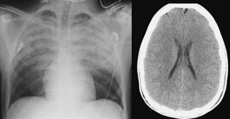
In the early stages of focal brain ischemia, CT can demonstrate areas of hypoattenuation in the brain tissue that correspond to acute severe ischemia (von Kummer et al. 2001). It may be more difficult to detect diffuse pathology (Fig. 16.1). While early changes such as hypoattenuation and sulcal effacement may be detected by experts, the inexperienced interpreter may miss them. The sensitivity of CT
Fig. 16.1. A 20-year-old army recruit, found comatose in the toilets. An AP chest radiograph shows signs of bilateral aspiration pneumonia (left). The unenhanced computed tomography image shows no signs of ischemia (right)