

Книги по МРТ КТ на английском языке / Magnetic Resonance Imaging in Ischemic Stroke - K Sartor R 252 diger von Kummer Tobias Back
.pdfStroke Syndromes |
3 |
1Stroke Syndromes
Georg Gahn
CONTENTS
1.1Introduction 3
1.2 |
Mechanisms of Ischemia 3 |
1.2.1Territorial Infarcts 4
1.2.1.1Large Vessel Occlusive Disease of the Anterior Circulation 4
1.2.1.2Large Vessel Occlusive Disease of the Posterior Circulation 6
1.2.2Lacunar Infarction 8
1.2.3Borderzone Infarction 9
1.3 |
Particular Etiological Stroke Syndromes 9 |
1.3.1Cardioembolic Stroke 9
1.3.2Dissection 10
1.3.3 Cerebral Venous Thrombosis 11
1.3.4Migraine 11
1.3.5Coma 12
1.3.6 Eye Movement Abnormalities 13
1.4Summary 13 References 13
1.1 Introduction
The major focus of this book is the evolving new technologies that help understand the underlying pathophysiological mechanisms of cerebral ischemia. Nevertheless, the anatomically based classification of stroke syndromes originally elaborated by C.M. Fisher still has a huge impact on the care of stroke patients. This chapter will give the reader a short overview of the most important stroke syndromes in the clinical setting. “Stroke” is defined as a sudden,
non-convulsive focal neurological deficit. The terms apoplexy originating from the Greek “αποπλεξ´ια”
and “insult” from the Latin “insultus” describe the same phenomenon and can be used synonymously. The term “neurovascular syndrome” may be a better term since a stroke is not unusually a slow progressing process rather than a “stroke” (Kennedy and Buchan 2004). The neurological deficit reflects both the loca-
G. Gahn, MD
Department of Neurology, University of Technology Dresden,
Fetscherstrasse 74, 01307 Dresden, Germany
tion and size of the ischemia or the hemorrhage, but may very well be due to an intracranial mass effect, a residuum after an epileptic seizure, a migraine attack, or an encephalitis. Combinations of neurological deficits are numerous, both in the hemispheres and in the brainstem (see as well chapter 14).
1.2
Mechanisms of Ischemia
Focal cerebral ischemia differs from global ischemia. In global ischemia irreversible neuronal damage occurs after 4–8 min at normal body temperature (Hochachka et al. 1996). In focal cerebral ischemia collateral vessels almost always provide some degree of residual blood flow, which may be insufficient to preserve neuronal survival (Coyle and Heistad 1991). Location of arterial occlusion affects the impairment of cerebral function: Obstruction below the circle of Willis often permits collateral flow through the anterior or the posterior communicating arteries. Vertebral artery obstruction can be bypassed through small deep cervical arteries which are residuals from the embryonic rete mirabilis in the posterior circulation. In obstructions of the cervical internal carotid artery, which derives from a branchial arch and not from a rete mirabilis, limited collateral flow can be provided through external carotid artery branches such as the periorbital or the ethmoidal arteries. Collateral flow mainly derives from the arteries of the circle of Willis.
Additional factors influence the extent of the final infarction. The speed of obstruction may allow collateral arteries to develop, if it occurs gradually (Busch et al. 2003), whereas complete sudden blockade of a major artery by an embolus leaves only some minutes to activate sufficient collateral flow. Hypoxia, hyperglycemia, acidosis, fever, hypotonia, and normal or abnormal variants in vascular anatomy my contribute to the resulting infarction (Hossmann 1999). Basically, the loss of oxygen and
4 |
G. Gahn |
glucose supply results in the collapse of cellular energy production with subsequent changes in cellular metabolism, degradation of cell membranes, and finally necrosis.
The margins of the infarction are usually hyperemic due to activated meningeal collaterals. The ischemic tissue swells rapidly because of increased intracellular and intercellular water content. During ischemia, the arteries first dilate to increase blood supply to the oligemic tissue, but will subsequently constrict due to ischemic damage. Reperfusion may then lead to hyperemia due to impaired autoregulative capacity of the damaged arteries. In prolonged ischemia sludging and endothelial damage will prevent reperfusion (Markus 2004).
1.2.1
Territorial Infarcts
1.2.1.1
Large Vessel Occlusive Disease of the Anterior Circulation
In 1951 C. Miller Fisher described the clinical findings associated with occlusion of the internal carotid artery (ICA) (Fisher 1951). In his report he first called attention to warning episodes preceding cerebral ischemia and called them “transient ischemic attack” (TIA). The major cause for occlusive disease of the ICA is atherosclerotic narrowing at the bifurcation of the common carotid artery (CCA) extending into the external and internal carotid arteries. Often atherosclerotic disease of the ICA is accompanied by atherosclerotic disease of the coronary and peripheral arteries (de Groot et al. 2004). Atherosclerotic plaques gradually narrow the vascular lumen. Ulceration of the plaque and hemorrhage into the plaque cause clotting of thrombocytes to the vessel wall and finally embolization to more distal arteries in the brain (Hennerici 2004). Progressive narrowing of the arterial lumen may also cause hemodynamic impairment of the cerebral territory supplied by the diseased artery if collateral flow through the arteries of the circle of Willis is not sufficient.
An important warning sign for occlusive ICA disease is an episode of transient monocular visual disturbance also called “amaurosis fugax” or “ocular TIA” (Wray 1993). Patients often describe this phenomenon as a dimming, darkening, obscuration or a curtain from above or from the side, which resolves after seconds or a few minutes. These attacks are
caused by a decrease in blood flow through the ophthalmic artery, distally to a hemodynamically relevant ICA obstruction. Alternatively small emboli, the so-called Hollenhorst plaques, may occlude retinal branches (Hollenhorst 1958).
Hemispheric TIAs display a variety of transient focal neurological deficits (Ferro 2004). Stereotyped TIAs point either to a hemodynamic rather than an embolic mechanism of cerebral ischemia or to an imminent lacunar infarction, the so-called capsular warning syndrome (see Sect. 1.2.2). Permanent ischemia with infarction within the MCA territory usually leads to weakness of the contralateral limbs more pronounced in the face and the arm than in the leg (Ferro 2004). Concomitant or isolated sensory symptoms are usually loss of position and pinprick sense and stereoagnosis. Neglect of the contralateral space and an attentional hemianopia are often prominent in larger hemispheric strokes which are then also accompanied by conjugate eye deviation towards the side of the brain lesion. Weakness mainly affecting the leg often occurs in anterior cerebral artery (ACA) territory stroke because of the representative area of the “homunculus” at the vertex (Bogousslavsky and Regli 1990). Left sided lesions are often accompanied by an aphasia (Kertesz 1993). Depending on the lesion location a more motor (frontal) or sensory (posterior) type of aphasia will occur (Pedersen et al. 2004).
1.2.1.1.1
Middle Cerebral Artery Occlusion
In Caucasians, the vast majority of MCA occlusions are of embolic origin with emboli arising from a carotid stenosis, the aortic arch or the heart (Heinsius et al. 1998) or from the venous side in case of a patent foramen ovale. In black or Asian patients a higher prevalence of intracranial occlusive disease is found with subsequent thrombotic arterial occlusion or stenosis (Feldmann et al. 1990).
Acute occlusion of the upper MCA trunk may lead to infarction in the frontal and superior parietal lobes. Hemiplegia, more pronounced in the face and arm, hemisensory loss, conjugate eye deviation and neglect to the contralateral side of space, especially to visual stimuli, will be the symptoms (Ferro 2004). Right sided lesions usually cause more severe visual neglect than left sided lesions (Karnath et al. 2002). Left sided lesions will also cause a motor aphasia in right handed patients (Kertesz 1993). Right sided lesions will also cause anosognosia more often than left sided lesions (Beis et al. 2004).
Stroke Syndromes |
5 |
In inferior MCA trunk occlusion infarctions of the lateral surface of the temporal lobe and the inferior parietal lobe may occur (Olsen 1991; Ringelstein et al. 1992). Motor or sensory deficits may not be severe, but visual field deficit and sensory aphasia in left sided infarctions and constructional apraxia in right sided lesions occur (Geschwind 1975; Spinazzola et al. 2003). Right temporal lesions cause agitation and confusion resembling an organic psychosis (Ferro 2001).
Deep infarctions in the MCA territory result from proximal MCA occlusion with blockage or hypoperfusion of the lenticulostriate arteries causing striatocapsular infarcts (Russmann et al. 2003). The basal ganglia have poor collateral supply, but leptomeningeal collaterals may prevent extension of infarction to the cortex (Ringelstein et al. 1992). Striatocapsular infarcts cause dense hemiplegia and less pronounced sensory deficits if the posterior capsule is spared (Donnan et al. 1991). Left sided infarcts may cause a temporary mutism or dysarthria (Urban et al. 2001). Right sided lesions cause neglect to the contralateral side (Karnath et al. 2002).
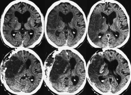
Complete occlusion of the MCA trunk with infarction of the entire MCA territory is a potentially devastating situation with severe paralysis, hemisensory loss, attentional hemianopia, conjugate eye deviation, and global aphasia in left sided lesions (Hacke et al. 1996). Because of its high mortality this type of
ischemic stroke has been called “malignant” MCA infarction (Fig. 1.1). The diagnosis of complete MCA territory infarction based on clinical judgment is unspecific and often requires neuroimaging studies (Berrouschot et al. 1998).
1.2.1.1.2
Anterior Cerebral Artery
The ACA supplies the head of the caudate nucleus, the anterior part of the internal capsule, the anterior perforated substance and the paramedian frontal lobe above the corpus callosum through the recurrent artery of Heubner (Ghika et al. 1990). Infarction of the paramedian frontal lobe causes weakness of foot, leg and shoulder and represents a typical pattern of neurological symptoms (Bogousslavsky and Regli 1990). Also apraxia of the left arm (“anterior disconnection syndrome”) may be a typical sign (Geschwind 1965). Transcortical motor and sensory aphasia, urinary incontinence in bilateral lesions occur. Abulia is seen in both unilateral and bilateral frontal lobe infarctions, sometimes of fluctuating intensity. The “alien hand sign” is also found in frontal lobe infarctions and may be another form of disconnection syndrome (Geschwind et al. 1995). Occasionally both ACA territories are supplied by only one ACA in a unilateral hypoplastic or aplastic A1 segment. Infarction of both ACA territories will
Fig. 1.1. Upper row, computed tomography of a patient with malignant right middle cerebral artery infarction on day one after onset of symptoms. Lower row, on day two massive edema with midline shift in spite of hemicraniectomy
6 |
G. Gahn |
cause a sudden onset of abulia, paraparesis, apathy, and incontinence, which may be misinterpreted as sudden onset of dementia (Ferro 2001). Infarction of the head of the caudate and the anterior internal capsule by occlusion of the Heubner artery will cause slight motor weakness, dysarthria, behavioral changes such as abulia or restlessness and hyperactivity. Cognitive and behavioral changes in these patients resemble the clinical signs found in patients with medial thalamic lesions (Bogousslavsky 1994).
1.2.1.1.3
Anterior Choroidal Artery
Blockade of the anterior choroidal artery may cause infarction of the lateral geniculate body causing hemianopia with preserved central vision and a prominent sensory loss with hemiparesis (Bruno et al. 1989).
1.2.1.2
Large Vessel Occlusive Disease of the Posterior Circulation
1.2.1.2.1
Obstruction of the Subclavian Artery
In severe occlusive disease of the subclavian artery (SCA) blood supply of the arm is mainly provided by reversed flow through the vertebral artery (VA) arising behind the obstruction. The so-called sub- clavian-steal syndrome consists of ischemic symptoms in the arm, especially after exercise, such as pain or numbness or coolness (Reivich et al. 1961). Consequently a diminished or delayed pulse in the radial artery or decreased blood pressure on the side of SCA stenosis can be palpated. Rarely neurological symptoms such as spells of dizziness may be brought about by exercise of the arm. Even more rare are ischemic brainstem strokes in subclavian-steal syndrome (Bornstein and Norris 1986).
1.2.1.2.2
Obstruction of the Vertebral Arteries
The origin of the VA at the SCA is the most common location of atherosclerotic VA disease. Dizziness, accompanied by other brainstem symptoms, such as diplopia, dysarthria, motor or sensory symptoms, suggest embolism towards the brainstem. Also hemianopia may occur due to VA embolism
when the emboli travel through the basilar artery into the posterior cerebral arteries (PCA). The pathophysiology of proximal VA obstruction is not well understood, since the VA is rarely operated on and therefore specimens as in carotid endarterectomy are rare (Caplan 1993). The mechanical situation in VA is very different form the ICA since it leaves the SCA at a 90° angle, whereas the ICA takes off the CCA at an almost 180° angle (Brandt et al. 2000).
VA obstruction causes hemodynamic problems in approximately one third of patients with posterior circulation ischemia (Caplan et al. 2004). Asymmetrical caliber of the two VAs is normal. In the neck multiple nuchal and muscular branches provide a network for potential collateral pathways, that can be activated in VA obstruction.
Intracranial atherosclerotic VA obstruction is mainly located at the origin of the posterior inferior cerebellar artery (PICA) and less frequent at the site of dura penetration. Consequently the most frequent clinical syndrome in VA occlusive disease is the dorsolateral medullary syndrome (“Wallenberg’s” syndrome) consisting of dizziness, retroorbital pain, facial numbness, dissociated sensory deficit, weakness, hoarseness, dysphagia and vomiting, nystagmus, Horner’s syndrome and failure of autonomic respiration (Vuilleumier et al. 1995).
With involvement of the cerebellar hemisphere supplied by the PICA, subsequent edema may cause obstruction of the 4th ventricle, hydrocephalus or compression of the medulla oblongata. Clinically, involvement of the entire cerebellar hemisphere can not be distinguished from partial cerebellar infarction (Amarenco and Hauw 1990). Therefore patients with neurological symptoms suggesting infarction within the PICA territory require neuroimaging studies and close clinical monitoring.
In blockade of the anterior spinal artery, ischemia of the medial medulla may occur with contralateral hemiparesis, ipsilateral tongue weakness and contralateral loss of posterior column sensation (Ho and Meyer 1981).
Isolated cerebellar infarctionwithoutinvolvement of the medulla is often difficult to identify, since gait ataxia, vomiting and dizziness may not be accompanied by typical brainstem symptoms (Barth et al. 1994). Cerebellar edema may compress the medulla and the pons leading to conjugate eye deviation to the side opposite the lesion without contralateral hemiparesis. This sign is probably pathognomonic for severe cerebellar mass effect and requires immediate intervention.
Stroke Syndromes
1.2.1.2.3
Basilar Artery Obstruction
Basilar artery (BA) occlusive disease is a highly lifethreatening condition first described by Kubik and Adams (1946). Atherosclerotic changes are mostly located at the origin of the BA, sometimes extending from the VAs. Often these patients experience brainstem TIAs such as diplopia, dizziness, weakness of both legs, or occipital headaches (von Campe et al. 2003). Obstruction of the BA often interrupts blood supply of the basis of the pons by the superior cerebellar arteries (SCA). Pseudobulbar paralysis by interruption of descending tracts to the bulbar nuclei is often seen. The spinothalamic tract and the cerebellar hemispheres are often spared from infarction. Disturbance of eye movements occur because of infarction of the lateral gaze centers in the paramedian pontine tegmentum, e.g. the medial longitudinal fasciculus (internuclear ophthalmoplegia), the parapontine reticular formation (PPRF), which generates lateral gazes, or the combination of both, resulting in the so called “one and a halfsyndrome” (Mehler 1989; Voetsch et al. 2004).
Infarction of the medial pontine tegmentum will cause coma and is a poor prognostic sign (Fisher 1977; Kataoka et al. 1997).
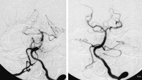
In most patients with BA thrombosis, obstruction is limited to the mid portion of the basilar artery (Fig. 1.2) (Voetsch et al. 2004). Embolic occlusion rather than thrombotic occlusion mainly blocks the distal part of the BA when it divides into the PCAs. The distal BA supplies the midbrain and
7
the diencephalon by small perforating arteries. Signs of dysfunction in the rostral brainstem are a variety of pupillary abnormalities (e.g. anisocoria, afferent pupillary deficit) (Martin et al. 1998; Mehler 1989). Vertical gaze palsy or skew deviation also point to the midbrain causing the “Top of the basilar” syndrome (Caplan 1980). Memory loss may occur in thalamic infarction as well as agitation, hallucinations mimicking frontal lobe disorders (Biller et al. 1985; Ghika-Schmid and Bogousslavsky 2000). The triad of hypersomnia, supranuclear vertical-gaze defect, and amnesia (the so-called “paramedian diencephalic syndrome”) is typically due to bilateral paramedian thalamic strokes in the territory of the anterior thalamic/subthalamic (or thalamoperforating) arteries (“en ailes de papillon”) (Meissner et al. 1987).
1.2.1.2.4
Posterior Cerebral Artery (PCA)
Historically, the French neurologist Charles Foix in 1923 first described the syndrome of infarction in the PCA territory as a thalamocapsular deficit (Foix and Masson 1923). The PCAs arise from the BA, but about 30% of patients have a hypoor aplastic P1 segment with the PCA nourished by the ICA through the posterior communicating artery (Margolis et al. 1971).
Headache in patients with PCA disease is often retro-orbital or above the eye reflecting innervation of the upper surface of the tentorium by the first division of the trigeminal nerve (Brandt et
Fig. 1.2. Digital subtraction angiography of a patient with basilar artery thrombosis before (left) and after (right) thrombolytic therapy. (Courtesy of Prof. von Kummer)
8 |
G. Gahn |
al. 2000; Ferro et al. 1995). Infarction in the PCA territory usually causes visual symptoms such as homonymous hemianopia or quadrantanopia and sensory deficits but seldom paralysis. Patients with hemianopia due to infarction of the striate cortex are fully aware of their deficit. In contrast patients with infarction in the parietal lobe within the MCA territory have visual neglect and are unaware of their deficit (Ferber and Karnath 2001). Proximal PCA-occlusion may simulate MCA-infarction because of thalamic involvement (Chambers et al. 1991). Optokinetic nystagmus is normal in patients with hemianopia but reduced towards the side of the visual defect in those with visual neglect (Morrow and Sharpe 1993).
Some neuropsychological syndromes can be present in PCA infarction (Ferro 2001):
Alexia without agraphia in left occipital lobe infarction and the splenium of the corpus callosum. Transfer of read words from the functional right visual cortex to the left sided language center is impossible due to interruption of the splenium. Transfer of primary language information for writing or speech is not impaired.
Transcortical sensory aphasia appears in patients with left PCA infarctions and displays difficulties in naming objects but no problems in repeating without understanding. Gerstmann’s syndrome with infarction of the angular gyrus consists of inability for right-left differentiation, finger anomia, constructional apraxia, agraphia and acalculia. In associative visual agnosia after left PCA infarctions visual but not tactile recognition of objects is impaired. Prosopagnosia is a problem with recognizing faces and occurs in right PCA infarctions. Cortical blindness occurs in bilateral PCA infarctions; however, pupillary reflexes are preserved (also see Chap. 14).
1.2.2
Lacunar Infarction
Cerebral microangiopathy accounts for 20%–30 % of all ischemic strokes and is mainly due to long lasting arterial hypertension. Narrowing of arterioles is caused by so called lipohyalinosis, a process collecting hyaline substances in the media of the small cerebral arteries (Fisher 1965c). These small arteries typically supply exclusively small territories of less than 1 or 2 cm in diameter. With progression of arterial narrowing blood flow towards the nourished territory diminishes until oligemia or ischemia occur. Eventually thrombotic mechanisms
may contribute to the blockade of the artery at this point. Finally a small infarction, called a “lacune”, will occur. Especially in the basilar artery atheromatous changes in the arterial wall may occlude the origins of the small penetrating arteries or be the origin of an expanding thrombosis adherent to the arterial wall (“microatheroma”) (Fisher and Caplan 1971). Usually this will cause blockade of several perforating arteries and subsequent small deep infarcts in contrast to lacunar infarcts which result from single perforating artery disease. An animal model to study cerebral microangiopathy in a standardized matter is presently not available (Caplan 1993). Conversely we rely on clinical and imaging data in humans to understand this disease.
Lacunar infarcts are typically located in the basal ganglia, the deep white matter and in the brainstem (Fisher 1965a, 1998). Depending on their location and their size circumscribed neurological symptoms will occur. C. Miller Fisher described the four classical lacunar syndromes:
•Pure motor stroke (Fisher and Curry 1964)
•Pure sensory stroke (Fisher 1965b)
•Ataxic hemiparesis (Fisher 1978)
•Dysarthria-clumsy-hand-syndrome (Fisher 1967; Fisher and Curry 1964)
All these syndromes are a consequence of small lacunes interrupting pathways in the white matter. Lacunar strokes almost never cause cortical symptoms such as aphasia or apraxia. Depending again on the location of the infarctions, patients may present with combined symptoms sometimes with preceding stereotyped TIAs. Lacunes within the internal capsule may cause dense hemiplegia, but never combined with impaired consciousness or conjugate eye deviation as in territorial infarcts with mass effect. At presentation, symptoms may be subtle or mild and may fluctuate or progress. This is probably related to the hemodynamic aspect in the pathogenesis of the disease. Many patients experience progression of the neurological deficits during the first 24 h after onset of ischemic symptoms, the so-called capsular warning syndrome (Donnan et al. 1993; Staaf et al. 2004). Often these patients wake up in the morning with neurological deficits which occurred sometimes during sleep and with comparatively low blood pressure (Chaturvedi et al. 1999). In contrast embolic strokes tend to occur after getting up, when activation of the cardiovascular system provokes plaque disruption or increased cardiac contractility. Notably, recent MRI studies
Stroke Syndromes |
9 |
showed no differences in stroke subtypes between waking strokes and strokes occurring during sleep (Donnan et al. 1993; Fink et al. 2002). Lacunar syndromes do not specifically help to localize the infarct to a certain territory in the brain and are not highly specific for the diagnosis of a “lacune” (Baumgartner et al. 2003; Gan et al. 1997).
1.2.3
Borderzone Infarction
Borderzone infarcts develop at the junction between different arterial territories (Adams et al. 1966). Pathophysiologically two different classes of borderzone infarcts can be identified: infarction between two arterial territories with a connecting arteriolar collateral network, so-called watershed infarcts, and infarcts between two arterial territories without arteriolar collaterals, so-called end-zone infarcts (Bogousslavsky and Moulin 1995). This classification is based on the anatomic distribution of the cerebral vascular supply consisting of two main systems: first, superficial arteries surround the brain parenchyma with an anastomotic network and send off perforating centripetal branches that do not anastomose (Moody et al. 1990). Second, deep branches originating from the major arterial branches penetrating the brain without anastomoses. Clinically relevant borderzones are the anterior borderzone between the MCA and the ACA and the posterior borderzone between the MCA and the PCA. They represent watershed areas and cause mainly cortical infarction. The subcortical borderzones between the deep and the superficial perforators represent endzone areas and cause subcortical infarctions (Read et al. 1998). In the posterior circulation both watershed and end-zone areas exist between the PICA and the SCA territories. The penetrating branches of the basilar artery are a potential source of end-zone infarctions comparable to the lenticulostriate arteries (Bogousslavsky and Moulin 1995).
The underlying mechanism of borderzone infarcts is the low flow situation (Ringelstein et al. 1983) in the most distal fields supplied by the cerebral circulation (“last meadows”) (also see Chap. 15). Hemodynamic impairment will consequently first cause ischemia in these areas. Typical clinical situations are a prolonged drop in systemic blood pressure, e.g. during cardiac surgery causing bilateral borderzone infarcts or severe occlusive disease of the internal carotid artery (Bladin and Chambers 1994). This may only lead to unilateral
borderzone infarction in the case of poor collateral supply through the circle of Willis (Powers 1991).
Clinically stereotyped TIAs or the opticocerebral symptom with simultaneous amaurosis and contralateral hemiparesis due to critically low blood supply through an occluded or almost occluded ICA herald a pending borderzone ischemia (Tsiskaridze et al. 2001). Rarely so-called limb shaking TIAs may point to a hemodynamic ischemia in ICA occlusive disease (Baquis et al. 1985).
1.3
Particular Etiological Stroke Syndromes
1.3.1
Cardioembolic Stroke
Thromboembolic stroke mainly derives from cardiac thrombus formation (Schneider et al. 2004). Less frequently the source is intra-arterial, from the distal end of a thrombus within the lumen of an obstructed carotid or vertebral artery or from an atheromatous plaque in the cervical arteries or in the aortic arch. The cardiac embolus usually arises in the anterior circulation through the internal carotid artery up into the middle cerebral artery. At a site of sudden lumen reduction either at the origin of the middle cerebral artery or more distally at the bifurcation into the middle cerebral artery branches, it gets stuck and blocks the lumen of the artery (Caplan 1993). Embolic infarction often turns into hemorrhagic transformation. Most often the middle cerebral artery, especially its inferior branch, is the site of embolic obstruction (Bogousslavsky et al. 1989). The embolic material may remain arrested and plug the artery solidly. It also may break into fragments and spread into smaller branches more peripherally. The phenomenon of a clot first lodging in the internal carotid artery, producing profound symptoms of hemispheric ischemia, and then migrating distally to a MCA pial branch has been called the “spectacular shrinking deficit” (Minematsu et al. 1992). The dramatic initial deficit diminishes to a minor deficit corresponding to the terminal branch artery.
At total of 75% of cardiac emboli reach the brain. Non-valvular atrial fibrillation with thrombus formation within the left atrial appendix or the left atrium is the most common reason for cardiac emboli (Ferro 2003). Cardio-embolic infarcts within the MCA territory carry a high risk for hemorrhagic transformation after reperfusion. Hemor-
10 |
G. Gahn |
rhagic transformation is a natural consequence of cerebral infarction, occurring in up to 65% of stroke patients and in up to 90% of patients with cardioembolic stroke within the first week after symptom onset (Molina et al. 2001). Hemorrhagic transformation does not impair neurological outcome after embolic stroke (Fiorelli et al. 1999). It may even suggest favorable outcome indicating early reperfusion of the blocked MCA (Molina et al. 2002).
Paradoxical embolism can occur in a patent foramen ovale with a right to left shunt. Embolic material arising from the pelvic or leg veins or elsewhere in the venous system may bypass the pulmonary system and reach the cerebral arteries (Braun et al. 2004).
1.3.2 Dissection
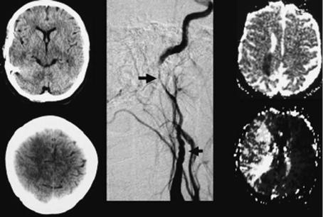
Spontaneous dissection of the internal carotid or the vertebral artery is an important cause of ischemic stroke in young adults (Fig. 1.3). In the late 1970s Fisher et al. (1978) and Mokri et al. (1979) described dissections of carotid and vertebral arteries as detected by modern diagnostic techniques rather than by post-mortem examination. This may occur
after both major or trivial traumatic head or neck injury (Schievink 2001). Many patients have preceding warning symptoms: the typical patient with carotid artery dissection presents with pain on one side of the head, face, or neck accompanied by a partial Horner’s syndrome and followed hours or days later by cerebral or retinal ischemia (Schievink 2001). Final infarction may arise mostly due to embolic and seldom due to hemodynamic mechanism (Benninger et al. 2004). In carotid artery dissection, Horner’s syndrome develops in less than half of patients as well as vagal, hypoglossal or accessorial nerve palsy. The underlying mechanism could be nerve compression, stretching or occlusion of small nourishing branches within an arterial wall by the intramural hematoma (Mokri et al. 1996). The pathogenesis of dissection remains obscure except in patients with obvious collagen tissue disease such as fibromuscular dysplasia. Other types of connective tissue alterations may also be associated with cervical artery dissections (Hausser et al. 2004). The site of dissection in adults is mainly the internal carotid artery at its distal extracranial course above the carotid bulb. In children the site of dissection is predominately intracranially (Fullerton et al. 2001). The risk for recurrent dissections is very low (Touze et al. 2003).
Fig. 1.3. Computed tomography (left), digital subtraction angiography (DSA, middle) and MRI (right) of a patient with internal carotid artery dissection. Note diffuse swelling of the frontal-parietal cortex on CT on day three after onset of symptoms. At this time point the patient suffered from a severe left hemiparesis. DSA shows classical “string sign” of cervical artery dissection with continuous narrowing of arterial lumen. MRI: ADC map (upper images) on day three after onset of symptoms shows small area of ischemia (dark). Time-to-peak parameter image (lower images) shows delayed contrast inflow to the entire right MCA territory. The patient underwent stent protected dilatation of the arterial stenosis 6 days after symptom onset and recovered almost completely from the severe hemiparesis. (Courtesy of Prof. von Kummer)
Stroke Syndromes |
11 |
1.3.3
Cerebral Venous Thrombosis
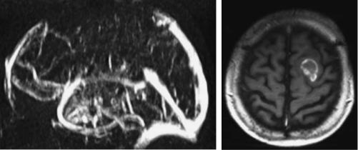
Thrombosis of the cerebral veins or sinuses may develop secondary to infections of the ear or the paranasal sinuses, to coagulation disorders or spontaneously (Bousser et al. 1985) (also see Chap. 18). Occlusion of the cerebral venous system may cause venous infarction stroke. The clinical signs are often unspecific and may be mainly caused by an obscure increase in intracranial pressure (Higgins et al. 2004). Fluctuating or permanent focal neurological deficits combined with headache and confusion may lead to the correct diagnosis. Chemosis and proptosis with cranial nerve III, IV and VI, and the ophthalmic division of 5th cranial nerve palsy are characteristic signs for thrombosis of the anterior cavernous sinus. Seizures and hemiparesis, predominantly of the leg, are suggestive of the sagittal sinus (Fig. 1.4). Involvement of the caudal cranial nerves indicate thrombosis of the posterior part of the cavernous sinus or the inferior petrous sinus. Bilateral thalamic infarction should raise the question of straight sinus thrombosis (Herrmann et al. 2004).
1.3.4 Migraine
Classical migraine with typical visual symptoms preceding unilateral headache are seldom a differential diagnosis with ischemic stroke. Increased awareness of patients of ischemic symptoms and rapid presentation to emergency rooms with immediate initiation of thrombolytic therapy may chal-
lenge the physician to identify an ongoing migraine attack with spreading depression mimicking cerebral ischemia. Accompanying vegetative symptoms are unspecific. Familiar hemiplegic migraine is a rare and even more challenging disorder due to its dramatic course in young patients (Ducros et al. 2001). The association between migraine and stroke is a dilemma for neurologists. Migraine is associated with an increased stroke risk and it is considered an independent risk factor for ischemic stroke in a particular subgroup of patients. The pathogenesis is not known, but several studies report some common biochemical mechanisms between the two diseases. A classification of migraine-related stroke that encompasses the full spectrum of the possible relationship between migraine and stroke has been proposed. It includes three main entities: coexisting stroke and migraine, stroke with clinical features of migraine, and migraine-induced stroke. The concept of migraine-induced stroke is well represented by migrainous infarction; it is described in the revised classification of the International Headache Society (IHS), and it represents the strongest demonstration of the relationship between ischemic stroke and migraine (Fig. 1.5). An interesting common condition in stroke and migraine is a patent foramen ovale which could play a pathogenetic role in both disorders. The association between migraine and cervical artery dissection is reported in recent studies. Migraine is more frequent in patients with cervical artery dissection (Tzourio et al. 2002). This supports the hypothesis that an underlying arterial wall disease could be a predisposing condition for migraine.
Basilar artery or vertebrobasilar migraine is not an uncommon type of migraine. Often young woman
Fig. 1.4. Patient with cerebral venous thrombosis. Venous MRA demonstrates occlusion of the sagittal sinus. MRI shows an intracranial hemorrhage, the typical complication of cerebral venous thrombosis. (Courtesy of Prof. von Kummer)
12 |
G. Gahn |
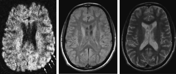
Fig. 1.5. Patient with hemiplegic migraine. Left, diffusion weighted MRI during migraine attack with severe right hemiparesis. Note the slight diffusion changes in the left temporal cortex suggesting ischemia. Middle and right, follow-up MRI (protondensityand T2-weighted) 1 year after migraine attack show no structural changes in the left temporal cortex. Clinically the patient recovered completely. (Courtesy of Prof. von Kummer)
experience visual disturbance similar to those in typical migraine but involving both visual fields. These symptoms may be accompanied by vertigo, ataxia, dysarthria, and sensory disturbances in both arms or legs bilaterally (Evans and Linder 2002).
1.3.5 Coma
In animals, destruction of the ascending reticular activating system (ARAS) induces a state of coma (Moruzzi and Magoun 1949). In men the ARAS is located in the paramedian tegmentum of the dorsal pons and the midbrain extending as a complex polysynaptic system from the upper half of the pons through the midbrain to the dorsal part of the hypothalamus and to the thalamic reticular formation (Vincent 2000).
In close vicinity of the ARAS the medial longitudinal fasciculus (MLF) and the oculomotor and trochlear nuclei are situated. Combined coma and oculomotor disturbances points to a brainstem lesion (Parvizi and Damasio 2003). Other clinical symptoms like respiratory pattern, pupillary reflex, and position or movement patterns of the limbs may help localize the site of the lesion.
Abnormal respiratory breathing patterns are of limited practical value, since a comatose patient due to cerebrovascular disease often requires immediate airway protection and mechanical ventilation. Urgent diagnostic imaging will provide the appro-
priate diagnostic information (Brazis et al. 1990). “Cheyne-Stokes respiration” consists of brief periods of hyperventilation regularly combined with short episodes of apnea. During hyperventilation periods the patients may become more alert. The cause of Cheyne-Stokes respiration can be a large bilateral cortical lesion, bilateral thalamic lesions, as well as metabolic disturbances in uremia, anoxia, heart failure (Cherniack and Longobardo 1973). “Hyperventilation” may occur in midbrain or pontine lesions and is often accompanied by severe respiratory distress. It can also be found in brainstem tumors leading to local pH lowering because of their high metabolism and thereby providing a breathing stimulus to the medullary respiratory center (Plum 1972). A lesion in the lateral tegmentum of the lower pons may cause a “apneustic breathing” with long inspiratory pauses (Plum and Alvord 1964). Low pontine and medullary lesions may cause “cluster breathing” with irregular breathing sequences. “Ataxic breathing” displays completely irregular breathing patterns, often seen in terminally ill patients with impairment of the dorsomedial respiratory centers (Brazis et al. 1990).
The pupillary light reflex may help differentiating metabolic cause from structural brainstem lesion in comatose patients (Tokuda et al. 2003). The light reflex is very resistant to metabolic dysfunction. An abnormal light reflex, especially when unilateral, points to a midbrain lesion. Bilateral diencephalic lesions or metabolic coma may cause bilateral small pupils well reacting to light (“diencephalic pupils”).