

Книги по МРТ КТ на английском языке / Magnetic Resonance Imaging in Ischemic Stroke - K Sartor R 252 diger von Kummer Tobias Back
.pdf24 |
|
M. W. Parsons and S. M. Davis |
|
Table 3.1. Stroke trials underway using MRI |
|
|
|
|
|
|
|
Trial name |
Validation of |
Use of MRI as |
Use of MRI as a |
|
perfusion-diffusion |
Selection tool |
Surrogate Outcome/ |
|
mismatch |
|
Proof of concept |
|
|
|
|
Thrombolytic trials |
|
|
|
Echoplanar Imaging Thrombolysis Evaluation Trial (EPITHET) |
Yes |
No |
Yes |
DWI evolution for understanding stroke aetiology |
Yes |
No |
No |
(DEFUSE) |
|
|
|
Stroke evaluation for late endovascular cerebral thrombolysis |
No |
Yes |
Yes |
with MR (SELECT MR) |
|
|
|
Desmoteplase in acute stroke (DIAS/DEDAS) |
No |
Yes |
Yes |
MR and recanalization of stroke clots using embolectomy |
Yes |
Yes |
Yes |
(MR RESCUE) |
|
|
|
Combination approach to lysis using eptifibatide and rt-PA |
No |
No |
Yes |
(CLEAR) |
|
|
|
Neuroprotective trials |
|
|
|
Glucose Reduction and Cerebral Events (GRACE) |
No |
No |
Yes |
AMPA receptor antagonist treatment in ischaemic stroke |
No |
No |
Yes |
(Artist-MRI) |
|
|
|
Magnetic Resonance Intravenous Magnesium Efficacy in Stroke |
No |
No |
Yes |
(MR IMAGES) |
|
|
|
|
|
|
|
stroke. Furthermore, the available data suggest that treatment is promising for a far greater range of patients than the current restricted approvals, particularly those 3–6 h after stroke onset (Donnan et al. 2003; Wardlaw 2001).
Current thrombolysis guidelines are based on a rigid time clock (< 3 h) without any imaging of the ischaemic penumbra. This time window is quite restrictive and very few stroke patients receive thrombolytic treatment worldwide. Extension of the therapeutic window beyond 3 h could substantially increase the number of patients able to receive thrombolysis. However, for this to occur with improved outcomes better selection criteria based on identification of the ischaemic penumbra using rapid and accessible neuroimaging techniques are required (Baron et al. 1995; Davis and Donnan 2004). Although there are many definitions of the ischaemic penumbra, the most practical for stroke clinicians is tissue that is at risk of infarction but still salvageable and that is the target of acute stroke therapy (Astrup et al. 1981; Ginsberg and Pulsinelli 1994; Hossmann 1994). Therefore, to select patients for acute stroke treatment, neuroimaging must distinguish penumbral tissue from the ischaemic core and benign oligaemic tissue.
The presence and extent of the ischaemic penumbra is time-dependent. However, stroke is a heterogeneous disorder, and survival of the penumbra can vary from less than 3 h to well beyond 48 h from patient to patient (Darby et al. 1999; Read et al. 2000). Penumbral survival is dependent upon many factors, such as location of vessel occlusion, state of collateral blood
supply, extent of grey versus white matter ischaemia, and physiologic variables such as blood glucose, body temperature and blood pressure (Davis and Donnan 2004). Approximately 90%–100% of patients with supratentorial large artery occlusion have penumbral tissue at 3 h after stroke onset (which likely explains the strongly positive sub-3 h tPA data) (Darby et al. 1999; Read et al. 2000). By 6 h, 75%–80% of patients still have penumbral tissue (Darby et al. 1999; Read et al. 2000). This is a substantial number, although a significant minority of patients in the 3- to 6-h window are unlikely to benefit from thrombolysis if the standard clinical and non-contrast CT criteria are applied (and may explain the less convincing 3- to 6-h tPA trial results to date) (Hacke et al. 1995, 1998). Indeed, it was recently demonstrated that the size of the infarct core varied dramatically between patients in the first 6 h after middle cerebral artery (MCA) occlusion, and was the strongest predictor of outcome (Jovin et al. 2003).
Thus, a ‘tissue clock’ where the extent of both irreversible (core) and reversible ischaemia (penumbra) is determined, rather than a rigid time window, would seem the ideal guide to patient selection for thrombolysis. A practical and accessible method of imaging the penumbra could also have a major impact on the institution of other new acute stroke therapies (Davis and Donnan 2002). Neuroprotective drugs have shown great promise in animal stroke models, but have failed many times to translate into positive human studies. These negative results may at least partly relate to the fact that no method of penumbral imaging has been used to select patients
Therapeutic Impact of MRI in Acute Stroke |
25 |
for treatment, despite the penumbra being the target for these drugs (Davis and Donnan 2002).
Combining diffusion-weighted and perfusion MRI with MR angiography (MRA) (multimodal echoplanar MRI) has revolutionised the evaluation of patients with acute stroke and appears as the best technique currently available to guide acute therapy. Diffusion-weighted MRI (DWI) delineates ischemic brain tissue within minutes, and perfusion MRI (PI) defines the area of cerebral hypoperfusion. PI and DWI have higher sensitivities for ischaemic change in the crucial first hours following symptom onset than conventional MRI or CT. This is the window when acute stroke therapies are likely to be of most benefit. Furthermore, the loci and patterns of PI and DWI lesions enable inferences to be made about stroke pathophysiology, and likely lesion evolution and outcome at times when conventional imaging is normal or any ischaemic changes are subtle. In particular, the difference in volume between a larger PI lesion compared to the DWI lesion may outline the extent of ischaemic tissue at risk of infarction. Such ‘per- fusion-diffusion mismatch’ is a simple and practical approach to define the ischaemic penumbra (Fig. 3.1). Other, more complicated (and perhaps less widely applicable) MRI models of the ischaemic penumbra have also been proposed (Kidwell et al. 2003).
3.1
MR Perfusion-Diffusion Mismatch: An Approach to Identifying the Ischaemic Penumbra
There is now a significant body of clinical data indicating that early DWI lesion volumes not only correlate well with final infarct size, but also with clinical outcome (Baird et al. 1997, 2000; Barber et al. 1998a; Lovblad et al. 1997; Saunders et al. 1995; Tong et al. 1998; Warach et al. 1996). Serial diffusion-weighted imaging has revealed progressive enlargement of the DWI lesion if the acute PI lesion is greater than the acute DWI lesion with a reduction of a perfusion-diffusion mismatch over time. Others have observed a rather constant extent of these lesions (Fiehler 2004). The eventual DWI lesion volume correlates closely with final T2weighted lesion volume and neurologic outcome (Baird et al. 1997; Barber et al. 1998a; Tong et al. 1998; Warach et al. 1996).
Perfusion imaging (PI) is complementary to DWI in acute stroke assessment. In animal models, PI lesions are visible immediately after vessel occlusion and resolve rapidly after successful thrombolysis or reperfusion (Muller et al. 1995; Yenari et al. 1997). In stroke patients, serial PI studies can document
a 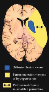
 b
b
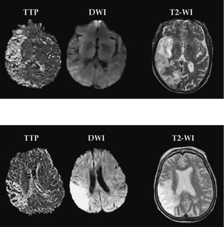
Fig. 3.1a,b. a The ischaemic penumbra. The perfusion (PI) lesion delineates the extent of hypoperfusion and the diffusion (DWI) lesion outlines the infarct core. The difference between the two lesions (perfusion-diffusion mismatch) represents the ischaemic penumbra, tissue at risk of progression to infarction. b Patient imaged at 3 h after onset of left hemiparesis and neglect with large PI (time to peak - TTP) lesion and smaller DWI lesion. At day 3 reperfusion has not occurred and the infarct core (diffusion lesion) has expanded greatly into the region of acute perfusion-diffusion mismatch. This is consistent with the perfusion-diffusion mismatch area representing the ischaemic penumbra
26 |
M. W. Parsons and S. M. Davis |
the evolution of perfusion lesions and clarify when reperfusion occurs. Earlier reperfusion correlates strongly with improved stroke outcome (Barber et al. 1998b). Most patients who do not receive thrombolytics have persisting perfusion deficits for > 24 h, whereas early reversal of PI lesions may be seen following thrombolysis (Kidwell et al. 2000; Marks et al. 1999).
Early PI lesions are typically larger than early DWI lesions (perfusion-diffusion mismatch) (Barber et al. 1998a; Warach et al. 1996). In acute stroke patients, the volume of the early PI lesion correlates more closely with the acute neurological deficit, suggesting that the acute PI lesion provides a more accurate estimate of the amount of brain tissue with impaired function (Baird et al. 1997; Barber et al. 1998a). However, if early reperfusion does not occur, the DWI lesion will expand to fill most of the volume of the acute PI lesion (Barber et al. 1998a). This expanded DWI lesion volume correlates closely
with the patient’s final neurological deficit and final infarct volume on T2-weighted imaging (Barber et al. 1998a). Thus, there is substantial evidence to indicate that perfusion-diffusion mismatch tissue is ‘at risk’ of progressing to infarction if rapid reperfusion does not occur (Baird et al. 1997; Barber et al. 1998a; Beaulieu et al. 1999; Schlaug et al. 1999; Warach et al. 1996). These observations provide compelling support for the mismatch model of the ischaemic penumbra as the natural history of early DWI lesions in untreated patients is to grow over time into the area of the initial PI lesion as the penumbra progressively fails and becomes recruited into the infarct (Fig. 3.2).
Some patients may undergo rapid irreversible injury to larger regions of brain, presumably due to poor collateral circulation. These patients may present with large acute DWI lesions that are of similar size to the acute PI lesion (non-mismatch pattern, see Fig. 3.3). It has been suggested that this group of
Fig. 3.2. Acute PI (TTP) and DWI scans demonstrate perfusion-diffusion mismatch. Follow-up T2-weighted imaging at 1 month documents major expansion of the infarct core into the original mismatch region
Fig. 3.3. There is no significant difference in size between the acute perfusion (TTP) and diffusion (DWI) lesion scans, nonmismatch. On follow-up T2-weighted imaging there has been no expansion of the infarct core

Therapeutic Impact of MRI in Acute Stroke |
27 |
patients are unlikely to benefit from thrombolysis as there is little or no tissue at risk (Baird et al. 1997; Darby et al. 1999; Fisher 1997). Another PI/DWI pattern seen in stroke is where the acute DWI lesion is substantially larger than the PI lesion (Baird et al. 1997; Barber et al. 1998a). These patients may have experienced partial or complete reperfusion, implying that thrombolytic therapy will not be of benefit (Caplan et al. 1997).
Further natural history studies supported the mismatch model of the penumbra by demonstrating that if early reperfusion of the PI lesion occurs, growth of the acute DWI lesion is inhibited. Thus, there is salvage of the mismatch region and these patients may experience substantial clinical improvement (Barber et al. 1998b).
Of great relevance to the potential use of multimodal MRI in extending the time window for thrombolysis have been the observations that per- fusion-diffusion mismatch was present for up to 24 h after symptom onset (Table 3.2). Whilst the presence and volume of mismatch decreases over time, at least 50% of patients still have significant tissue at risk 24 h after stroke onset (Fig. 3.4). This correlates well with positron emission tomography studies that show penumbral tissue persists up to 48 h after symptom onset, and that spontaneous survival of this tissue results in clinical improvement (Markus et al. 2003; Read et al. 2000). There is mounting evidence that the time window for salvage of the penumbra is well beyond 3 h and multimodal MRI can identify such patients.
The evolution of the mismatch model of the penumbra has led to a number of studies examining the response of acute DWI/PI patterns to thrombolytic therapy, particularly with respect to treatment beyond 3 h after stroke onset (Jansen et al. 1999; Kidwell et al. 2000; Parsons et al. 2002a; Schellinger et al. 2000). These studies supported the mismatch-penumbra hypothesis by demonstrating that thrombolysis rescues mismatch tissue
from infarction. Furthermore, some reports have provided evidence that the natural history of tissue at risk can be altered by thrombolysis, and, that this may translate into improved clinical outcome (Hacke 2004; Parsons et al. 2002a).
Our group compared patients with and without perfusion-diffusion mismatch treated with intravenous tPA within 6 h to a group of matched controls (Parsons et al. 2002a). Mismatch patients treated with tPA had greater vessel recanalisation, enhanced reperfusion, greater penumbral salvage, and less infarct expansion than controls (Fig. 3.5). The positive effects of thrombolysis on lesion growth were reflected in mismatch patients treated with tPA having improved measures of clinical outcome. Of note, the MR endpoints of enhanced reperfusion, greater penumbral salvage, and reduced infarct expansion all correlated with a greater chance of clinically meaningful improvement. A particularly provocative finding was that the improvement in clinical outcome seen in the mismatch patients treated with tPA compared to controls was completely negated when non-mismatch patients were included in the analysis (Parsons et al. 2002a). This implies that patients with perfusion-diffusion mismatch have the most to gain from thrombolytic therapy.
Fig. 3.4. Graph showing the proportion of patients with per- fusion-diffusion mismatch at various time points after stroke onset. Although the proportion of patients with mismatch declines with time, 50% of patients still have at-risk tissue 24 h after symptom onset (Darby et al. 1999)
Table 3.2. Studies demonstrating the persistence of perfusion-diffusion mismatch
Author |
Time |
Percentage with perfusion- |
Type of PI map used to assess hypoperfusion |
|
window |
diffusion mismatch |
|
Baird et al. (1997) |
24 h |
70% |
First moment time |
Darby et al. (1999) |
24 h |
80% at 6 hours |
First moment time |
|
|
65% at 12 hours |
|
|
|
50% at 24 hours |
|
Neumann-Haefelin et al. (1999) |
24 h |
80% |
Time to peak |
Beaulieu et al. (1999) |
07 h |
50% |
Time to peak |
Sorenson et al. (1999) |
12 h |
65% |
Mean transit time and cerebral blood flow |
Schellinger et al. (2000) |
06 h |
80% |
Time to peak |
Parsons et al. (2001) |
06 h |
75% |
Mean transit time and cerebral blood flow |

28 |
M. W. Parsons and S. M. Davis |
a |
b |
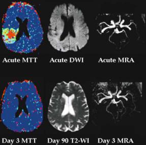
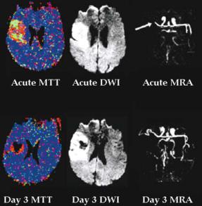
Fig. 3.5a,b. a Patient imaged at 3 h after stroke onset. There is a large perfusion lesion and extensive perfusion-diffusion mismatch. The PI map is a contrast mean transit time (MTT), green tissue is moderate contrast delay (4–6 s) and red is severe delay (> 6 s). The patient did not receive tPA, and at day 3 had persisting vessel occlusion on MRA (arrow) and large perfusion lesion. Consequently, there was major infarct expansion. b Patient treated with t-PA 5 h after symptom onset following MRI. Pre-therapy there is an occluded proximal M1 segment. Following tPA there is recanalization and reperfusion. In contrast to (a), there is salvage of a large amount of acute perfusion-diffusion mismatch tissue from infarction. Of interest, there is minor reduction in the DWI lesion after thrombolysis
A phase II study of a new thrombolytic agent, desmoteplase, has also recently been completed (Hacke 2004). A total of 104 patients were randomised to desmoteplase or placebo in a dose escalation study. Perfusion-diffusion mismatch was used as an inclusion criteria for the study. Patients who received desmoteplase had significantly higher reperfusion and reduced DWI lesion growth. A phase III study using perfusion-diffusion mismatch and perfusion CT to select patients is planned.
3.2
Refining the Mismatch Model of the Penumbra
In our view, the MRI patterns for identifying the ideal candidate for thrombolysis are straightforward. This particularly applies to treatment beyond the 3-h window. A small infarct core identified by DWI and a large perfusion deficit on PI indicate the potential for a major benefit from thrombolysis. This simple, practical mismatch model has, however, been challenged by partial normalisation of
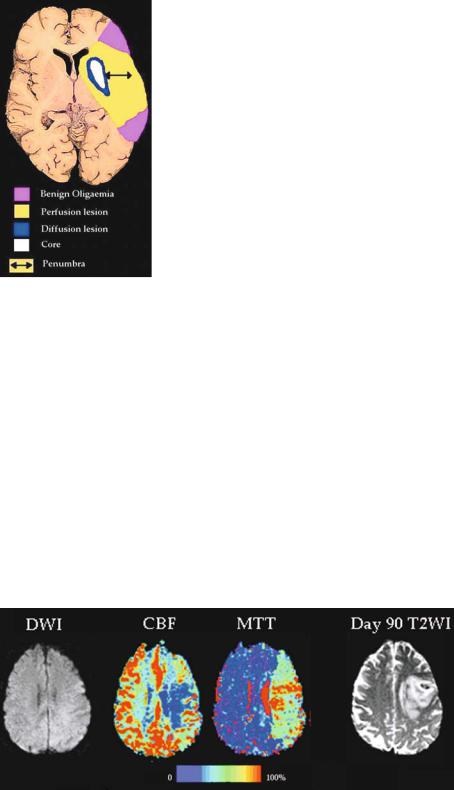
the DWI lesion, and because PI can exaggerate the area of truly ischaemic tissue (Fig. 3.6) (Kidwell et al. 2003; Parsons et al. 2001).
3.2.1
Perfusion Thresholds
PI may not necessarily differentiate between true penumbra and benign oligaemic tissue. The original experimental work by Astrup et al. (1981) identified three thresholds of hypoperfused tissue based on cerebral blood flow (CBF): core (CBF < 6–10 ml/100 g/ min), penumbra (CBF < 10–20 ml/100 g/min), and oligaemic tissue (CBF below normal range but not at risk of infarction). It is clear that the commonly used PI contrast transit maps, time to peak (TTP) and mean transit time (MTT) include some benign oligaemic tissue at the border of the PI lesion (Barber et al. 1998a; Neumann-Haefelin et al. 1999; Parsons et al. 2001). Our group, and others, have demonstrated that the natural history of acute DWI lesion is to grow into the mismatch region, and that this growth encompasses a large proportion of the visible acute MTT or TTP lesion (Barber et al. 1998a; Neumann-
Therapeutic Impact of MRI in Acute Stroke |
29 |
Fig. 3.6. The potential flaws in the mismatch hypothesis. The perfusion lesion probably overestimates truly hypoperfused tissue and includes some benign oligaemic tissue. The DWI lesion may overestimate the infarct core and be partially reversible
Haefelin et al. 1999; Parsons et al. 2001). However, these PI maps significantly overestimate final infarct size (Fig. 3.7) (Parsons et al. 2001).
How to best distinguish between penumbra and benign oligaemia at the border of the perfusion lesion has been the subject of intense research and debate. A range of perfusion (and apparent diffusion coefficient) thresholds have been identified that distinguish between penumbra and benign oligaemia with varying degrees of accuracy (Fig. 3.7) (Desmond et al. 2001; Grandin et al. 2002;
Neumann-Haefelin et al. 1999; Parsons et al. 2001; Thijs et al. 2001). However, there is a number
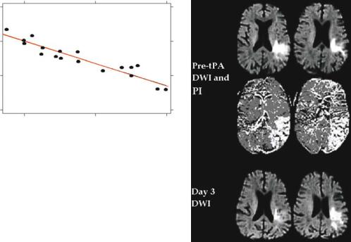
of difficulties with the predictive value of isolated ADC and perfusion thresholds. One major problem is that absolute quantification of cerebral perfusion with PI is not really possible, hence correlation with Astrup’s experimental thresholds are impossible (Calamante et al. 1999, 2002). Simplistically, this relates to the fact that there is a non-linear relationship between contrast concentration and signal intensity with gadolinium-based PI (Calamante et al. 1999, 2002). Another major issue with perfusion thresholds is that they change with time. This has been demonstrated both experimentally, and in human stroke with PI (Butcher et al. 2003; Heiss 1983; Hossmann 1994; Hossmann et al. 1977). In research performed by our group, penumbral tissue with a contrast transit delay of up to 15 s was salvaged from infarction at 2 h after stroke onset, but at 6 h the threshold for salvage was less than 6 s delay in contrast transit (Fig. 3.8) (Butcher et al. 2003).
3.2.2
DWI Reversal
Much debate exists over the accuracy of the acute DWI lesion identifying the ischaemic core, that is, tissue that is irreversibly damaged. There is no doubt that diffusion lesions may be partially reversed with early reperfusion. This has been demonstrated in both animal and human stroke (Chalela et al. 2003; Kidwell et al. 2000; Li et al. 1999, 2000). However, in humans these lesion reversals in most instances are only minor or partial, and are quite often not permanent. Indeed, in our thrombolytic series, less than 5% of patients had what would be considered any significant reduction in ischaemic lesion volume between the pre-treatment DWI and outcome T2-weighted
Fig. 3.7. Acute DWI and colour PI maps compared to outcome infarct size. Note that the acute CBF lesion is closest to final infarct size and the MTT lesion overestimates final infarct size, suggesting that the MTT map is demonstrating benign oligaemia as well as ‘true penumbra’
30
Mean transit time delay (seconds)
M. W. Parsons and S. M. Davis
15
10
5
0
2 |
4 |
6 |
Time to initial MRI (hours)
Fig. 3.8. Graph demonstrating that the perfusion threshold for mismatch tissue to be salvaged from infarction declines with time. These patients all had major reperfusion on follow-up imaging and tissue with severe hypoperfusion (mean transit time 15-s delay) was able to be salvaged from infarction within 3 h, but at 6 h, the threshold for salvage declined to only 3 s delay in mean transit time (Butcher et al. 2003)
scan (Parsons et al. 2002a). Most of these patients also had a large volume of acute perfusion-diffusion mismatch tissue salvaged from infarction. In fact, the reduction in volumes from the acute DWI lesion to outcome T2-weighted infarct were very small compared to the amount of mismatch tissue that was salvaged from infarction (Fig. 3.5b). Only one patient with non-mismatch was an exception to this rule (Fig. 3.9). We believe that acute perfusion-diffusion mismatch identifies the great majority of tissue at risk of infarction. The acute DWI lesion does not necessarily represent infarcted tissue. However, it is a very useful predictor of the minimum extent of tissue that eventually infarcts.
It has been proposed that the phenomenon of DWI lesion reversal means that patients without perfusion-diffusion mismatch may still benefit from thrombolytic therapy (Kidwell et al. 2000). However, others consider more research is needed before adopting such an approach into clinical practice (Schellinger et al. 2003). We have not found that non-mismatch patients benefited from thrombolysis compared to historical controls (Parsons et al. 2002a). The ongoing Australasian Echoplanar Imaging Thrombolysis Evaluation Trial (EPITHET) has been designed, in part, to answer the question of whether non-mismatch patients benefit from thrombolysis (Parsons et al. 2002a).
In an attempt to address the issues regarding PI overestimating at risk tissue and DWI overestimating the ischaemic core, multivariate models using different perfusion and ADC measures have been developed by several groups (Kidwell et al. 2003;
Fig. 3.9. Patient without acute perfusion-diffusion mismatch and significant DWI reversal following intravenous tPA
Warach 2001). Kidwell et al. (2003) have shown that such a model using ADC and TTP values predicted infarction and salvage with 81% accuracy compared to 53% with the simple mismatch model. Although combined tissue viability models may be more accurate at predicting tissue fate than the mismatch model, their generalisability is yet to be proven. It is uncertain whether the improved precision is of clinical significance for patient selection. For example, one visualises an extensive area of acute perfusion-diffusion mismatch using PI and DWI maps at the MR console, how often would the availability of a co-registered quantitative tissue outcome map (appropriately) change the decision to treat with thrombolysis? This question can only be answered with further research. It is also important to recognise that perfusion-diffusion mismatch provides a good, rapidly available estimation of the penumbra and a practical means of selecting candidates for acute stroke therapy.
3.2.3
What Does MRA Add to PI/DWI/?
A previous study concluded that PI/DWI tissue at risk was always associated with a proximal vessel
Therapeutic Impact of MRI in Acute Stroke |
31 |
occlusion on MRA (Schellinger et al. 2001). However, as our group has previously reported, about 25% of patients with perfusion-diffusion mismatch do not have an observable lesion on MRA (Barber et al. 1999b). These patients have PI lesions indicating probable branch MCA occlusion below the resolution of the MRA sequence. In our experience with acute stroke patients, the time invested in using longer MRA sequences (> 4 min) which may have greater resolution beyond the MCA division, is not productive. In our thrombolytic series, considerable at risk tissue was still present in the mismatch patients without visible MRA lesions (Parsons et al. 2002a). Indeed, this group of patients seems to have the most dramatic response to tPA, often with major reperfusion, complete penumbral salvage, and no infarct expansion (Fig. 3.10). Therefore, thrombolysis should still be considered for patients without a visible MRA lesion, provided that perfusion-diffu- sion mismatch is present.
Furthermore, a number of groups have now demonstrated that patients with distal vessel occlusion (including those without visible MRA lesions) have more complete reperfusion in response to tPA compared to those with proximal occlusion (Linfante et al. 2002; Neumann-Haefelin et al. 2004; Parsons
Fig. 3.10. Patient received intravenous tPA following MRI. PI map is a colour coded MTT map where mildly hypoperfused tissue is light blue, moderate hypoperfusion is green, and severe is red. Despite no visible vessel occlusion on acute MRA, there is still significant tissue at risk. Following thrombolysis, there is complete reperfusion and all mismatch tissue is salvaged from infarction on follow-up imaging
et al. 2002a). This is consistent with conventional angiographic data (Furlan et al. 1999). These results emphasise that thrombolysis should not be withheld from patients with distal arterial occlusion (if per- fusion-diffusion mismatch is present), but also that alternative treatment approaches might need to be considered for patients with ICA or proximal MCA occlusion. These might include intra-arterial (IA) thrombolysis and combined IV/IA thrombolysis. Such approaches might increase the risk of haemorrhagic transformation, however it is clear that the natural history of untreated distal ICA or proximal M1 MCA occlusion is very poor (Baird et al. 1997; Barber et al. 1999b; Darby et al. 1999; Linfante et al. 2002). Thus, MRA is complementary to DWI/PI in acute stroke. It is particularly useful in determining whether more aggressive reperfusion strategies are appropriate. However, it does not provide the valuable pathophysiologic information about cerebral perfusion in acute stroke that PI can.
3.2.4
Clinical Diffusion Mismatch?
The relationship of sub-6 h PI and DWI lesion volumes with acute and outcome National Institutes of Health Stroke Scale (NIHSS) has been a source of recent controversy (Schellinger et al. 2001). Most reports, however, show a strong correlation between acute PI lesions and baseline clinical scores (Barber et al. 1998a; Tong et al. 1998; Warach et al. 1996, 1999). This has led to the suggestion that the discrepancy between stroke severity, assessed with the NIHSS, and the volume of the DWI lesion (‘clinical-diffusion mismatch’) could be used as a surrogate for perfusion-dif- fusion mismatch. Our group has examined this hypothesis and have found that an NIHSS > 7 and DWI lesion volume < 25 cm3 predicts the presence of perfusion-diffusion mismatch with > 90% specificity, but low sensitivity. If PI had been omitted, more than 60% of patients with perfusion-diffu- sion mismatch would have been misdiagnosed as not having tissue at risk (Prosser et al. 2004). Indeed, other groups have demonstrated that up to 25% of patients with hyperacute stroke would have been incorrectly categorised as not having tissue at risk if PI was disregarded and DWI information alone was used (Sunshine et al. 2001). Therefore, combining PI with DWI dramatically increases the number of patients potentially eligible for acute stroke therapy.
32 |
M. W. Parsons and S. M. Davis |
3.2.5 False-Negative DWI
The other circumstance where PI is invaluable in acute stroke patients is where DWI is false-nega- tive (Sunshine et al. 2001). This is a more common occurrence in hyperacute stroke, where decision making for therapy is critical (Sunshine et al. 2001). Overall, the sensitivity and specificity of DWI for ischemic lesions in acute stroke (< 24 hours) is very high, although false-negative DWI is more common in the posterior fossa (Ay et al. 1999; Lovblad et al. 1998; Oppenheim et al. 2000). In hyperacute stroke, the false-negative DWI rate may be above 10%, even in the anterior circulation (Sunshine et al. 2001). In this time window, combining PI with DWI provides essential diagnostic and therapy-guiding information that DWI alone does not.
3.3
Vertebrobasilar Stroke
The great majority of hyperacute stroke MRI studies have been limited to patients with supratentorial ischaemia. While much less frequent, vertebrobasilar infarction has a high mortality, up to 70%–80%. Both IA and IV thrombolysis in small, uncontrolled series have dramatically improved outcomes (Brandt et al. 1996; Grond et al. 1998; Hacke et al. 1988). Sensitivity of DWI, and especially PI, at the base of the skull is probably lower due to susceptibility artefacts. Recently of note, substantial regions of perfusion-diffusion mismatch have been demonstrated in the posterior fossa, with salvage of these regions after thrombolysis (Ostrem et al. 2004). Many stroke centres throughout the world use multimodal stroke MRI to confirm vertebrobasilar occlusion and exclude major brainstem and/or thalamic and occipital infarction before proceeding to thrombolytic therapy (Schellinger et al. 2003).
3.3.1
Prediction of Haemorrhagic Transformation with MRI
It is controversial whether ischaemic changes on CT involving > 33% of the MCA territory increases the risk of haemorrhagic transformation (HT) associated with tPA (Patel et al. 2001; von Kummer et al. 1997). This may partially reflect the insensitivity
of CT for brain ischemia above 10 ml per 100 g/min. DWI more clearly identifies acute ischaemia below 30 ml per 100 g/min and thus may be a more reliable predictor of risk of thrombolysis-related HT (Barber et al. 1999a). A number of studies have now suggested that DWI findings can help to identify patients at increased risk for HT (Selim et al. 2002; Tong et al. 2001). In general, they have found that the number of voxels with an ADC < 550 × 10–6 mm2/s is the best predictor. However, this is essentially a marker of overall DWI lesion volume (Selim et al. 2002). The problem with these predictors is that there is no absolute cut-off DWI/ADC lesion volume, and the overlap between patients with and without HT is significant. In our experience, an acute DWI lesion volume > 100 cm3 (or qualitatively greater than 50% of the MCA territory) particularly if it is associated with no perfusion-diffusion mismatch (and regardless of vessel status on MRA), should probably be a contraindication to thrombolysis (Fig. 3.11). This requires further study, and EPITHET may provide more definitive answers to the question of HT prediction. The presence of prior microbleeds on T2*-weighted MRI may also be a risk factor for thrombolysis-related HT (Kidwell et al. 2002; Nighoghossian et al. 2002). This is a relatively infrequent finding (less than 10% of patients), and the association is not yet firmly established.
Fig. 3.11. Patient who received t-PA following MRI. PI, = quantitative MTT map. There is a distal M1 occlusion on MRA, but no obvious tissue at risk (no perfusion-diffusion mismatch). The baseline DWI lesion is quite large. Thrombolysis successfully achieves recanalization and reperfusion, but there is also haemorrhagic transformation apparent on day 3 imaging
Therapeutic Impact of MRI in Acute Stroke |
33 |
3.4
Detection of ICH with MRI
Emerging data suggests that MRI may be able to detect hyperacute intra-parenchymal haemorrhage at least as accurately as CT (Fiebach et al. 2004; Linfante et al. 1999). So-called susceptibil- ity-weighted T2* (gradient echo) sequences detect haemorrhage due to a profound signal loss caused by the paramagnetic effects of deoxyhaemoglobin, even when present in trace amounts (Linfante et al. 1999). In practice, the unprocessed gradient echo (T2*) images performed for PI, or the b = 0 images obtained from DWI echoplanar sequences can be used to sensitively detect ICH without adding any extra time to the stroke MRI protocol (Lin et al. 2001; Perl et al. 1999). Thus, stroke MRI probably obviates the need for a screening CT before thrombolysis.
3.4.1
Uncertain Stroke Onset
A not uncommon scenario is the acute stroke patient who has woken from sleep with a neurologic deficit, or is aphasic/obtunded and there is no corroborative history. Traditionally, such patients have been excluded from acute therapies, as the exact time of stroke onset is unclear. However, a significant proportion of these patients have perfusion-diffu- sion mismatch, suggesting recent stroke onset, and potential benefit from thrombolysis (Fink et al. 2002). As we are advocating that selection of patients for acute stroke therapy should be based on cerebral pathophysiology rather than a rigid time window it may well be that such patients will be treated in the future. However, at this point further evidence from randomised trials such as EPITHET and DIAS will be required to make such a major practice change.
3.4.2
Hyperacute Stroke MRI as a Practical Tool
Whilst the practicality of MRI as a neuroimaging tool for hyperacute stroke has been questioned in the past, there is now substantial data demonstrating its feasibility and practicality (Powers 2000; Powers and Zivin 1998). Current stroke MRI protocols take 15–20 min to perform (total ‘table’ time) and are part of the routine stroke workup in many academic centres (Kidwell et al. 2000; Parsons
et al. 2001; Schellinger et al. 2003; Sunshine et al. 1999; Warach 2001). Although protocols vary somewhat, a streamlined hyperacute stroke MRI protocol need only include a T1 sagittal localising sequence, DWI, PI, T2* gradient echo, and MRA (usually time of flight). This allows determination of the presence of perfusion-diffusion mismatch, site of vessel occlusion, and exclusion of ICH.
3.4.2.1
Echoplanar MRI Thrombolysis Evaluation Trial
Although the mismatch hypothesis is a compelling one, randomised, prospective blinded trials are required as definitive evidence. The Royal Melbourne Hospital MRI Stroke Study group has designed the echoplanar imaging thrombolysis evaluation trial (EPITHET). This is a multicentre double blind, randomized, placebo controlled trial of tPA versus placebo in 100 eligible patients with hemispheric infarction 3–6 h after stroke onset (Davis and Donnan 2004). The trial is an example of a ‘proof of concept’ study discussed below, using (now established) biologically meaningful MR surrogate outcomes. The major hypothesis underlying EPITHET is that in patients treated with tPA between 3 and 6 h after stroke onset, the presence and extent of perfusion-diffusion mismatch will predict a therapeutic response. This response will be defined by the major MR outcome: reduced expansion of the ischaemic core between the acute DWI and outcome T2-weighted studies. Extent of reperfusion and penumbral salvage will also be used as MR outcome measures. In addition, an important secondary hypothesis being tested is that larger acute infarct cores (as represented by the DWI lesion) will be associated with an increased risk of haemorrhagic transformation in tPA treated patients. Twelve centres are currently involved (ten in Australia and two in New Zealand), it is anticipated that recruitment will be completed by the end of 2005.
It is important to note that this trial is not using perfusion-diffusion mismatch to select patients, but as a surrogate outcome measure. The rationale for this is that the mismatch hypothesis, whilst strongly supported by much data, remains unproven. Similarly, it is not yet established that patients with nonmismatch do not respond to thrombolysis (as there is at least a theoretical possibility of partial DWI reversal). Should either the mismatch hypothesis and/or the hypothesis that a large acute DWI lesion predicts ICH be proven in this trial, a logical next