

Книги по МРТ КТ на английском языке / Magnetic Resonance Imaging in Ischemic Stroke - K Sartor R 252 diger von Kummer Tobias Back
.pdf34 |
M. W. Parsons and S. M. Davis |
step would be to use PI/DWI to select patients for tPA in the 3- to 6-h window in a phase III placebocontrolled RCT with primary clinical endpoints.
3.4.2.2
Future Stroke Patient Management: A Potential Thrombolysis Algorithm
Although ongoing trials may modify the following treatment algorithm, a possible role of combined PI and DWI as part of a multimodal MRI protocol in the near future for the selection of acute ischaemic stroke patient for thrombolysis is presented below. Indeed, many centres do use stroke MRI to select patients for thrombolysis beyond 3 h (Schellinger et al. 2003). At present, as the evidence is not conclusive, we prefer to randomise post-3-h patients to thrombolytic trials.
The thrombolysis algorithm is based on the assumption that the hypotheses discussed above are proven to be correct. That is, primarily: (1) the presence of significant perfusion-diffusion mismatch (qualitatively +/– quantitatively assessed) predicts treatment response, and (2) a ‘large’ DWI lesion (qualitatively > ½ MCA territory, and/or quantitatively > 100 cm3) predicts haemorrhagic transformation. Patients excluded from thrombolysis because of extensive DWI lesions with or without perfusion-dif- fusion mismatch may be considered for more aggressive measures such as hypothermia or decompressive hemicraniectomy (Schwab et al. 1998, 2001).
A further possible scenario (particularly when thrombolysis later than 3 h after symptom onset is being considered) is that PI/DWI and MRA might be used to decide on the dose of tPA. This postulate is based on the early dose-finding tPA studies that smaller doses of tPA appeared to have significant efficacy, and possibly also a lesser risk of haemorrhage (Brott et al. 1992; Haley et al. 1992). Furthermore, our work, and angiographic studies, demonstrate that more proximal sites of occlusion are less likely to recanalize with thrombolysis (Alexandrov et al. 2001; Furlan et al. 1999; Parsons et al. 2002a). Therefore, a smaller dose of tPA might be as efficacious for distal vessel occlusions, and for more proximal occlusions (ICA, MCA stem) a higher dose of IV tPA, or (where available) a combined IV/IA approach might be used (Ernst et al. 2000). The risk of a higher dose of IV tPA (1.1 mg/kg as used in ECASS I) could be justified on two counts, patients with large baseline DWI lesions would be excluded, and the natural history of untreated proximal vessel occlusion is very
poor (Hacke et al. 1995). New thrombolytic agents, or combined lower dose tPA and a platelet glycoprotein IIb/IIIa inhibitor (e.g., abciximab) might be alternative treatment arms (Abciximab in Ischemic Stroke Investigators 2000).
It is predicted that EPITHET will provide more information regarding the site of vessel occlusion and response to tPA. Should this trial confirm a lower rate of major reperfusion (and correspondingly greater infarct expansion and lower penumbral salvage) with intravenous tPA for proximal occlusions, then trials of more aggressive reperfusion strategies would be indicated. A safety proof of concept study using PI/DWI (and MRA) both to select patients and for surrogate measures of outcome, would be ideal for this purpose. In such a study, patients without large pre-treatment DWI lesions and perfusion-diffusion mismatch might, for example, be randomised to 0.9 mg/kg or 1.1 mg/kg IV tPA in a blinded manner. Rates of recanalization, major reperfusion, and ICH would be important outcome measures.
Pending safety confirmation of the higher tPA dose, the following thrombolysis algorithm could be also be tested in a randomised controlled trial, again using MRI surrogate outcomes, in the 3- to 6-h window (see below). The control group would probably receive standard dose tPA. It is envisaged that the following treatment algorithm might equally apply to ischaemic stroke patients presenting within 3 h of symptom onset. However, some might argue that
|
|
Yes |
Large DWI lesion (>1/2 MCA territory) |
|
No tPA |
||||
|
|
|||
|
|
|
No |
|
|
|
|
||
|
|
|
|
|
|
|
No |
Perfusion-Diffusion mismatch |
|
No tPA |
||||
|
|
|||
|
|
|
Yes |
|
|
|
|
Site of MRA vessel occlusion
ICA/proximal M1 |
Distal M1/M2 |
No visible occlusion |
1.1 mg/kg IV |
|
0.9 mg/kg IV |
|
0.6 mg/kg IV |
or 0.6 mg/kg |
|
|
|
|
followed by IA |
|
|
|
|
|
|
|
|
|
Potential future thrombolysis treatment algorithm using PI/ DWI and MRA
Therapeutic Impact of MRI in Acute Stroke |
35 |
clinically eligible patients presenting within 3 h do not need MRI to make a decision regarding thrombolysis. At the other end of the spectrum, if patients presenting after 6 h still have significant perfusion-diffusion mismatch without a large DWI lesion, it might also be reasonable to treat these patients as below. After all, the rationale for PI/DWI in acute stroke is to image ischaemic pathophysiology. Thus, treatment should be based on a ‘tissue clock’ rather than a time clock (Kidwell et al. 2000) (see Fig. 3.3).
Potential future thrombolysis treatment algorithm using PI/DWI and MRA for patients with unknown time of stroke onset or being admitted after 3 h.
3.4.2.3
Improving Acute Stroke Trial Design Using MRI
3.4.2.3.1
Patient Selection Using MRI
In the last few years, many unsuccessful acute stroke trials have been reported. Trials of intravenous tPA initiated up to 6 h from symptom onset have failed to demonstrate efficacy in patients without imaging confirmation of the diagnosis or pathophysiologic features most amenable to treatment (Albers and Clark 1999; Hacke et al. 1995, 1998). To demonstrate efficacy in stroke trials with a treatment time window greater than 3 h (including neuroprotective agents), trial design needs to be improved (Davis and Donnan 2002).
A number of factors may predict tissue response and clinical efficacy in stroke trials. Clearly, time is an important factor (Marler et al. 2000). However, the amount of ischaemic tissue at risk of infarction that is still potentially salvageable (the ischaemic penumbra) is another factor that is highly likely to predict clinical response. Much of the data presented above: (1) demonstrates that PI/DWI can identify such tissue at-risk, and (2) establishes that the ischaemic penumbra is a prime locus of ischaemic injury and a key target for acute stroke therapies. For example, we have shown that patients with significant perfusion-diffusion mismatch treated with thrombolysis have a significant degree of reperfusion and tissue salvage compared to similar controls (Parsons et al. 2002a). This also translated into a statistically significant clinical response (Parsons et al. 2002a). The sample size of this study (albeit not a randomised, placebo-controlled trial) was markedly smaller than that needed to show clinical benefit in the NINDS trial, which did not
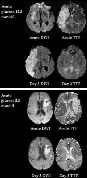
Fig. 3.12. Patient (top) with acute perfusion-diffusion mismatch and an acute blood glucose level of 12.3 mmol/l. Despite major reperfusion, much of the at-risk mismatch tissue progressed to infarction. In contrast, the bottom patient with an acute blood glucose of 5.3 mmol/l and a large area of mismatch, had major penumbral (mismatch) salvage on follow-up imaging
select an homogenous sample of patients based on imaging of pathology (The National Institute of Neurological Disorders and Stroke rt-PA Stroke Study Group 1995).
It is important to note that MRI selection of patients does not just apply to thrombolytic trials. In fact, selecting patients with an ischaemic penumbra may be critically important for trials of neuroprotective drugs, and other therapies such as manip-
36 |
M. W. Parsons and S. M. Davis |
ulation of physiologic variables (blood glucose, hypothermia) and decompressive hemicraniectomy (Fisher and Brott 2003). For example, our group has found that the adverse effect of acute hyperglycaemia on stroke outcome was seen predominantly in patients with an MR-defined ischaemic penumbra (Fig. 3.12). Acute hyperglycaemia in patients with perfusion-diffusion mismatch increased the progression of this at-risk tissue to infarction and was associated with worse clinical outcome, compared to normoglycaemic patients with mismatch (Parsons et al. 2002b). These results suggest that a trial of acute, aggressive glycaemic control in stroke might be optimised by using PI/DWI to select patients with salvageable at-risk tissue (Baird et al. 2003; Parsons et al. 2002b).
In a similar vein, decompressive hemicraniectomy has been shown to reduce mortality after malignant MCA infarction (Schwab et al. 1998). It appears the earlier the surgery is performed, the better the outcome. MRI is promising in predicting the likelihood of this complication within 6 h of stroke onset (Thomalla et al. 2003). An extensive DWI lesion predicts malignant MCA infarct very accurately, allowing ultra-early decompressive surgery. As suggested in the above thrombolysis treatment algorithm, patients with extensive acute DWI lesions would be excluded from thrombolytic treatment. However, such patients may still have perfusion-diffusion mismatch and other aggressive experimental treatments such as early decompression or hypothermia may salvage remaining at risk tissue (Schwab et al. 1998, 2001). This area deserves further study.
There seems little doubt that trials of neuroprotective drugs, of which there have been many negative results to date, might be more likely to succeed if patients with an ischaemic penumbra were selected using perfusion-diffusion mismatch (Lees et al. 2000; Muir and Lees 1995; Yamaguchi et al. 1998). This has a rational basis, as neuroprotective drugs are targeted at ‘preserving’ the penumbra (Davis and Donnan 2002; Fisher and Brott 2003).
3.4.2.3.2
PI/DWI as Surrogate Markers of Treatment Response
– ‘Proof of Concept’
As well as using PI/DWI to select an ‘ideal’ patient population to optimise the chances of efficacy of a therapeutic agent, PI/DWI may also have a role in the monitoring of ischaemic lesion evolution and therapeutic response in acute stroke trials. Reducing
the functional disability associated with cerebral infarction is the clinical goal of acute stroke therapy. Therefore, reduction of infarct volume is the biological objective. A relative reduction in infarct volume in animal models is required to advance a drug into clinical trials. Replicating these observations in a clinical sample is the next logical step. However, this has often been bypassed in favour of very expensive, large clinical endpoint trials that have failed for treatments given beyond the 3-h window. Pilot ‘proof-of-concept’ studies using a biologically meaningful outcome variable, similar to EPITHET, may provide an important bridge between animal studies and the large phase III trials (Barber et al. 2004; Davis and Donnan 2002).
Although it seems intuitive, the surrogate outcome measures used in such proof-of-concept trials must have a tight correlation with clinical outcome. The first randomised-controlled trial of PI/DWI for outcome assessment did not definitively demonstrate efficacy of the experimental agent, but it did show a clear association between infarct expansion and poorer clinical outcome (Warach et al. 1999). The results of our case-control tPA series strengthen the case for using PI/DWI as biologically meaningful outcome measures (Parsons et al. 2002a). In the comparison between tPA treated patients and untreated controls; baseline to outcome infarct expansion, reperfusion, and penumbral salvage all had strong correlations with clinically meaningful improvement. These correlations were independent of treatment group, although a higher proportion of tPA treated patients had better MRI and clinical outcomes.
Other studies have also demonstrated the potential value of using PI/DWI in proof concept studies. Our group has shown, based on statistical modelling from a natural history stroke cohort with serial PI/DWI, that a therapy postulated to reduce infarct expansion by 50% would need 100 patients in each group to show a significant difference between treatment and placebo groups (Barber et al. 2004). Similarly, a therapy postulated to increase the chances of major reperfusion from acute to subacute PI by 50% would only need 50 patients in each group to demonstrate a significant difference between active treatment and placebo (Barber et al. 2004). Others have shown that the presence of recanalization (on MRA) or reperfusion (on PI) after thrombolysis are powerful predictors of eventual clinical outcome (Chalela et al. 2003, 2004; Schellinger et al. 2000). In fact, extent of reperfusion from preto post thrombolysis PI was a more sensitive predictor
Therapeutic Impact of MRI in Acute Stroke
of excellent outcome than MRA recanalization, and appears to be the ideal surrogate outcome for testing new reperfusion therapies (Chalela et al. 2004).
3.5 Conclusions
MRI is being increasingly used as a selection tool and an outcome measure in stroke trials, reflecting the growing evidence that direct pathophysiologic imaging may provide a more rational approach to acute stroke therapy than clinical diagnosis (and non-contrast CT scanning) alone. Stroke MRI is practical and feasible. Perfusion-diffusion mismatch provides a reliable estimation of the ischaemic penumbra. The future holds great promise. It is only a matter of time before MRI routinely assists the stroke clinician in making individual therapeutic decisions, and guides stroke researchers in identifying effective therapies that can be delivered well beyond the current 3-h window.
References
Abciximab in Ischemic Stroke Investigators (2000) Abciximab in acute ischemic stroke: a randomized, double-blind, pla- cebo-controlled, dose-escalation study. Stroke 31:601–609 Albers GW, Clark WM (1999) For the ATLANTIS Study Investigators. The ATLANTIS rt-PA (Alteplase) acute stroke trial:
final results. Cerebrovasc Dis 9:126
Alexandrov AV, Burgin WS, Demchuk AM, El-Mitwalli A, Grotta JC (2001) Speed of intracranial clot lysis with intravenous tissue plasminogen activator therapy: sonographic classification and short-term improvement. Circulation 103:2897–2902
Astrup J, Symon L, Siesjo BK (1981) Thresholds in cerebral ischemia - the ischemic penumbra. Stroke 12:723–725
Ay H, Buonanno FS, Rordorf G, Schaefer PW, Schwamm LH, Wu O, Gonzalez RG, Yamada K, Sorensen AG, Koroshetz WJ (1999) Normal diffusion-weighted MRI during stroke like deficits. Neurology 52:1784–1792
Baird AE, Benfield A, Schlaug G, Siewert B, Lovblad K, Edelman R, Warach S (1997) Enlargement of human cerebral ischemic lesion volumes measured by diffusion-weighted magnetic resonance imaging. Ann Neurol 41:581–589
Baird AE,Lovblad KO,Dashe JF,Connor A,Burzynski C,Schlaug G, Straroselskaya I, Edelman RR, Warach S (2000) Clinical correlations of diffusion and perfusion lesion volumes in acute ischemic stroke. Cerebrovasc Dis 10:441–448
Baird TA, Parsons MW, Phanh T, Butcher KS, Desmond PM, Tress BM, Colman PG, Chambers BR, Davis SM (2003) Persistent poststroke hyperglycemia is independently associated with infarct expansion and worse clinical outcome. Stroke 34:2208–2214
37
Barber PA, Darby DG, Desmond PM, Yang Q, Gerraty RP, Jolley D, Donnan GA, Tress BM, Davis SM (1998a) Prediction of stroke outcome with echoplanar perfusionand diffu- sion-weighted magnetic resonance imaging. Neurology 51:418–426
Barber PA, Davis SM, Infeld B, Baird AE, Donnan GA, Jolley D, Lichtenstein M (1998b) Spontaneous reperfusion following ischemic stroke is associated with improved outcome. Stroke 29:2522–2528
Barber PA, Darby DG, Desmond PM, Gerraty RP, Yang Q, Li T, Jolley D, Donnan GA, Tress BM, Davis SM (1999a) Identification of major ischemic change. Diffusion-weighted imaging versus computed tomography. Stroke 30:2059–2065
Barber PA, Darby DG, Desmond PM, Yang Q, Gerraty RP, Jolley D, Donnan GA, Tress BM, Davis SM (1999b) Absent middle cerebral artery flow predicts the presence and evolution of the ischemic penumbra. Neurology 52:1125-1132
Barber PA, Parsons MW, Desmond PM, Bennet DA, Donnan GA, Tress BM, Davis SM (2004) The use of PWI and DWI measures in the design of “proof of concept” stroke trials. J Neuroimaging 14:123-132
Baron JC, von Kummer R, del Zoppo GJ (1995) Treatment of acute ischemic stroke: challenging the concept of a rigid and universal time window. Stroke 26:2219–2221
Beaulieu C, de Crespigny A, Tong DC, Moseley ME, Albers GW, Marks MP (1999) Longitudinal magnetic resonance imaging study of perfusion and diffusion in stroke: evolution of lesion volume and correlation with clinical outcome (see comments). Ann Neurol 46:568–578
Brandt T, Grau AJ, Hacke W (1996) Severe stroke. Baillieres Clin Neurol 5:515–541
Brott TG, Haley EC Jr, Levy DE, Barsan W, Broderick J, Sheppard GL, Spilker J, Kongable GL, Massey S, Reed R et al (1992) Urgent therapy for stroke. Part I. Pilot study of tissue plasminogen activator administered within 90 minutes. Stroke 23:632–640
Butcher K, Parsons M, Baird T, Barber A, Donnan G, Desmond P, Tress B, Davis S (2003) Perfusion thresholds in acute stroke thrombolysis. Stroke 34:2159–2164
Calamante F, Thomas DL, Pell GS, Wiersma J, Turner R (1999) Measuring cerebral blood flow using magnetic resonance imaging techniques. J Cereb Blood Flow Metab 19:701–735 Calamante F, Gadian DG, Connelly A (2002) Quantification of perfusion using bolus tracking magnetic resonance imaging in stroke: assumptions, limitations, and potential impli-
cations for clinical use. Stroke 33:1146–1151
Caplan LR, Mohr JP, Kistler JP, Koroshetz W (1997) Should thrombolytic therapy be the first-line treatment for acute ischemic stroke? Thrombolysis-not a panacea for ischemic stroke. N Engl J Med 337:1309–1310; discussion 1313
Chalela JA, Ezzeddine M, Latour L, Warach S (2003) Reversal of perfusion and diffusion abnormalities after intravenous thrombolysis for a lacunar infarction. J Neuroimaging 13:152–154
Chalela JA, Kang DW, Luby M, Ezzeddine M, Latour LL, Todd JW, Dunn B, Warach S (2004) Early magnetic resonance imaging findings in patients receiving tissue plasminogen activator predict outcome: insights into the pathophysiology of acute stroke in the thrombolysis era. Ann Neurol 55:105–112
Darby DG, Barber PA, Gerraty RP, Desmond PM, Yang Q, Parsons M, Li T, Tress BM, Davis SM (1999) Pathophysiological topography of acute ischemia by combined diffu- sion-weighted and perfusion MRI. Stroke 30:2043–2052
38
Davis SM, Donnan GA (2002) Neuroprotection: establishing proof of concept in human stroke. Stroke 33:309–310
Davis SM, Donnan GA (2004) Advances in penumbra imaging with MR. Cerebrovasc Dis 17 [Suppl 3]:23–27
Desmond PM, Lovell AC, Rawlinson AA, Parsons MW, Barber PA, Yang Q, Li T, Darby DG, Gerraty RP, Davis SM, Tress BM (2001) The value of apparent diffusion coefficient maps in early cerebral ischemia. AJNR Am J Neuroradiol 22:1260–1267
Donnan GA, Howells DW, Markus R, Toni D, Davis SM (2003) Can the time window for administration of thrombolytics in stroke be increased? CNS Drugs 17:995–1011
Ernst R, Pancioli A, Tomsick T, Kissela B, Woo D, Kanter D, Jauch E, Carrozzella J, Spilker J, Broderick J (2000) Combined intravenous and intra-arterial recombinant tissue plasminogen activator in acute ischemic stroke. Stroke 31:2552–2557
Fiebach JB, Schellinger PD, Gass A, Kucinski T, Siebler M, Villringer A, Olkers P, Hirsch JG, Heiland S, Wilde P, Jansen O, Rother J, Hacke W, Sartor K (2004) Stroke magnetic resonance imaging is accurate in hyperacute intracerebral hemorrhage: a multicenter study on the validity of stroke imaging. Stroke 35:502–506
Fink JN, Kumar S, Horkan C, Linfante I, Selim MH, Caplan LR, Schlaug G (2002) The stroke patient who woke up: clinical and radiological features, including diffusion and perfusion MRI. Stroke 33:988–993
Fisher M (1997) Characterizing the target of acute stroke therapy. Stroke 28:866–872
Fisher M, Brott TG (2003) Emerging therapies for acute ischemic stroke: new therapies on trial. Stroke 34:359–361 Furlan A, Higashida R, Wechsler L, Gent M, Rowley H, Kase C, Pessin M, Ahuja A, Callahan F, Clark WM, Silver F, Rivera F (1999) Intra-arterial prourokinase for acute ischemic stroke. The PROACT II study: a randomized controlled trial. Prolyse in acute cerebral thromboembolism. JAMA
282:2003–2011
Ginsberg MD, Pulsinelli WA (1994) The ischemic penumbra, injury thresholds, and the therapeutic window for acute stroke. Ann Neurol 36:553–554
Grandin CB, Duprez TP, Smith AM, Oppenheim C, Peeters A, Robert AR, Cosnard G (2002) Which MR-derived perfusion parameters are the best predictors of infarct growth in hyperacute stroke? Comparative study between relative and quantitative measurements. Radiology 223:361–370
Grond M, Rudolf J, Schmulling S, Stenzel C, Neveling M, Heiss WD (1998) Early intravenous thrombolysis with recombinant tissue-type plasminogen activator in vertebrobasilar ischemic stroke. Arch Neurol 55:466–469
Hacke W (2004) Desmoteplase In Acute Stroke Study (DIAS) - results of phase II trial. International Stroke Conference, San Diego
Hacke W, Warach S (2000) Diffusion-weighted MRI as an evolving standard of care in acute stroke. Neurology 54:1548–1549
Hacke W, Zeumer H, Ferbert A, Bruckmann H, del Zoppo GJ (1988) Intra-arterial thrombolytic therapy improves outcome in patients with acute vertebrobasilar occlusive disease. Stroke 19:1216–1222
Hacke W, Kaste M, Fieschi C, Toni D, Lesaffre E, von Kummer R, Boysen G, Bluhmki E, Hoxter G, Mahagne MH, Henerici M (1995) Intravenous thrombolysis with recombinant tissue plasminogen activator for acute hemispheric stroke. The
M. W. Parsons and S. M. Davis
European Cooperative Acute Stroke Study (ECASS). JAMA 274:1017–1025
Hacke W, Kaste M, Fieschi C (1998) Randomised double-blind placebo controlled trial of thrombolytic therapy with intravenous alteplase in acute ischaemic stroke (ECASS II). Lancet 352:1245–1251
Haley EC Jr, Levy DE, Brott TG, Sheppard GL, Wong MC, Kongable GL, Torner JC, Marler JR (1992) Urgent therapy for stroke. Part II. Pilot study of tissue plasminogen activator administered 91–180 minutes from onset. Stroke 23:641–645
Heiss WD (1983) Flow thresholds for functional and morphological damage of brain tissue. Stroke 14:329–331
Hossmann K-A (1994) Viability thresholds and the penumbra of focal ischemia. Ann Neurol 36:557–565
Hossmann K-A, Sakaki S, Zimmermann V (1977) Cation activities in reversible ischemia of the cat brain. Stroke 8:77–81
Jansen O, Schellinger P, Fiebach J, Hacke W, Sartor K (1999) Early recanalisation in acute ischaemic stroke saves tissue at risk defined by MRI. Lancet 353:2036–2037
Jovin TG, Yonas H, Gebel JM, Kanal E, Chang YF, Grahovac SZ, Goldstein S, Wechsler LR (2003) The cortical ischemic core and not the consistently present penumbra is a determinant of clinical outcome in acute middle cerebral artery occlusion. Stroke 34:2426–2433
Kidwell CS, Saver JL, Mattiello J, Starkman S, Vinuela F, Duckwiler G, Gobin YP, Jahan R, Vespa P, Kalafut M, Alger JR (2000) Thrombolytic reversal of acute human cerebral ischemic injury shown by diffusion/perfusion magnetic resonance imaging. Ann Neurol 47:462–469
Kidwell CS, Saver JL, Villablanca JP, Duckwiler G, Fredieu A, Gough K, Leary MC, Starkman S, Gobin YP, Jahan R, Vespa P, Liebeskind DS, Alger JR, Vinuela F (2002) Magnetic resonance imaging detection of microbleeds before thrombolysis: an emerging application. Stroke 33:95–98
Kidwell CS, Alger JR, Saver JL (2003) Beyond mismatch: evolving paradigms in imaging the ischemic penumbra with multimodal magnetic resonance imaging. Stroke 34:2729– 2735
Lees KR, Asplund K, Carolei A, Davis SM, Diener HC, Kaste M, Orgogozo JM, Whitehead J (2000) Glycine antagonist (gavestinel) in neuroprotection (GAIN International) in patients with acute stroke: a randomised controlled trial. GAIN International Investigators. Lancet 355:1949–1954
Li F, Han SS, Tatlisumak T, Liu K-F, Garcia JH, Sotak C, Fisher M (1999) Reversal of acute apparent diffusion coefficient abnormalities and delayed neuronal death following transient focal cerebral ischemia in rats. Ann Neurol 46:333– 342
Li F, Silva MD, Liu KF, Helmer KG, Omae T, Fenstermacher JD, Sotak CH, Fisher M (2000) Secondary decline in apparent diffusion coefficient and neurological outcomes after a short period of focal brain ischemia in rats. Ann Neurol 48:236–244
Lin DD, Filippi CG, Steever AB, Zimmerman RD (2001) Detection of intracranial hemorrhage: comparison between gradient-echo images and b(0) images obtained from dif- fusion-weighted echo-planar sequences. AJNR Am J Neuroradiol 22:1275–1281
Linfante I, Llinas RH, Caplan LR, Warach S (1999) MRI features of intracerebral hemorrhage within 2 hours from symptom onset. Stroke 30:2263–2267
Linfante I, Llinas RH, Selim M, Chaves C, Kumar S, Parker RA,
Therapeutic Impact of MRI in Acute Stroke
Caplan LR, Schlaug G (2002) Clinical and vascular outcome in internal carotid artery versus middle cerebral artery occlusions after intravenous tissue plasminogen activator. Stroke 33:2066–2071
Lovblad K, Baird AE, Schlaug G, Benfield A, Siewert B, Voetsch B, Connor A, Burzynski C, Edelman R, Warach S (1997) Ischemic lesion volumes in acute stroke by diffu- sion-weighted magnetic resonance imaging correlate with clinical outcome. Ann Neurol 42:164–170
Lovblad KO, Laubach HJ, Baird AE, Curtin F, Schlaug G, Edelman RR, Warach S (1998) Clinical experience with dif- fusion-weighted MR in patients with acute stroke. AJNR Am J Neuroradiol 19:1061–1066
Marks MP, Tong DC, Beaulieu C, Albers GW, de Crespigny A, Moseley ME (1999) Evaluation of early reperfusion and i.v. tPA therapy using diffusionand perfusion-weighted MRI (see comments). Neurology 52:1792–1798
Markus R, Reutens DC, Kazui S, Read S, Wright P, Chambers BR, Sachinidis JI, Tochon-Danguy HJ, Donnan GA (2003) Topography and temporal evolution of hypoxic viable tissue identified by 18F-fluoromisonidazole positron emission tomography in humans after ischemic stroke. Stroke 34:2646–2652
Marler JR, Tilley BC, Lu M, Brott TG, Lyden PC, Grotta JC, Broderick JP, Levine SR, Frankel MP, Horowitz SH, Haley EC Jr, Lewandowski CA, Kwiatkowski TP (2000) Early stroke treatment associated with better outcome: the NINDS rtPA stroke study. Neurology 55:1649–1655
Muir KW, Lees KR (1995) A randomized, double-blind, pla- cebo-controlled pilot trial of intravenous magnesium sulfate in acute stroke. Ann NY Acad Sci 765:315–316
Muller TB, Haraldseth O, Jones RA, Sebastiani G, Godtliebsen F, Lindboe CF, Unsgard G (1995) Combined perfusion and diffusion-weighted magnetic resonance imaging in a rat model of reversible middle cerebral artery occlusion. Stroke 26:451–457; discussion 457–458
National Institute of Neurological Disorders and Stroke rt-PA Stroke Study Group (1995) Tissue plasminogen activator for acute ischemic stroke. N Engl J Med 333:1581–1587
Neumann-Haefelin T, Wittsack HJ, Wenserski F, Siebler M, Seitz RJ, Modder U, Freund HJ (1999) Diffusionand per- fusion-weighted MRI. The DWI/PWI mismatch region in acute stroke. Stroke 30:1591–1597
Neumann-Haefelin T, du Mesnil de Rochemont R, Fiebach JB, Gass A, Nolte C, Kucinski T, Rother J, Siebler M, Singer OC, Szabo K, Villringer A, Schellinger PD (2004) Effect of incomplete (spontaneous and postthrombolytic) recanalization after middle cerebral artery occlusion: a magnetic resonance imaging study. Stroke 35:109–114
Nighoghossian N, Hermier M, Adeleine P, Blanc-Lasserre K, Derex L, Honnorat J, Philippeau F, Dugor JF, Froment JC, Trouillas P (2002) Old microbleeds are a potential risk factor for cerebral bleeding after ischemic stroke: a gradient-echo T2*-weighted brain MRI study. Stroke 33:735–742
Oppenheim C, Logak M, Dormont D, Lehericy S, Manai R, Samson Y, Marsault C, Rancurel G (2000) Diagnosis of acute ischaemic stroke with fluid-attenuated inversion recovery and diffusion-weighted sequences. Neuroradiology 42:602–607
Ostrem JL, Saver JL, Alger JR, Starkman S, Leary MC, Duckwiler G, Jahan R, Vespa P, Villablanca JP, Gobin YP, Vinuela F, Kidwell CS (2004) Acute basilar artery occlusion: diffusion-perfusion MRI characterization of tissue salvage
39
in patients receiving intra-arterial stroke therapies. Stroke 35:e30–e34
Parsons MW, Yang Q, Barber PA, Darby DG, Desmond PM, Gerraty RP, Tress BM, Davis SM (2001) Perfusion magnetic resonance imaging maps in hyperacute stroke: relative cerebral blood flow most accurately identifies tissue destined to infarct. Stroke 32:1581–1587
Parsons MW, Barber PA, Chalk J, Darby DG, Rose S, Desmond PM, Gerraty RP, Tress BM, Wright PM, Donnan GA, Davis SM (2002a) Diffusionand perfusion-weighted MRI response to thrombolysis in stroke. Ann Neurol 51:28–37
Parsons MW, Barber PA, Desmond PM, Baird TA, Darby DG, Byrnes G, Tress BM, Davis SM (2002b) Acute hyperglycemia adversely affects stroke outcome: a magnetic resonance imaging and spectroscopy study. Ann Neurol 52:20–28
Patel SC, Levine SR, Tilley BC, Grotta JC, Lu M, Frankel M, Haley EC, Jr., Brott TG, Broderick JP, Horowitz S, Lyden PD, Lewandowski CA, Marler JR, Welch KM (2001) Lack of clinical significance of early ischemic changes on computed tomography in acute stroke. JAMA 286:2830–2838
Perl J 2nd, Tkach JA, Porras-Jimenez M, Lieber M, Obuchowski N, Ross JS, Ding XP, Ruggieri PM, Shearer DM, Khajavi K, Masaryk TJ (1999) Hemorrhage detected using MR imaging in the setting of acute stroke: an in vivo model. AJNR Am J Neuroradiol 20:1863–1870
Powers WJ (2000) Testing a test. A report card for DWI in acute stroke. Neurology 54:1549–1551
Powers WJ, Zivin JA (1998) Magnetic resonance imaging in acute stroke. Not ready for prime time. Neurology 50:842– 843
Prosser J, Butcher K, Allport L, Parsons MW, Baird TA, MacGregor L, Tress BM, Davis SM (2004) Clinical-diffusion mismatch predicts the penumbra with high specificity. World stroke congress, Vancouver
Read SJ, Hirano T, Abbott DF, Markus R, Sachinidis JI, TochonDanguy HJ, Chan JG, Egan GF, Scott AM, Bladin CF, McKay WJ, Donnan GA (2000) The fate of hypoxic tissue on 18Ffluoromisonidazole positron emission tomography after ischemic stroke. Ann Neurol 48:228–235
Saunders DE, Howe FA, van den Boogaart A, McLean MA, Griffiths JR, Brown MM (1995) Continuing ischemic damage after acute middle cerebral artery infarction in humans demonstrated by short-echo proton spectroscopy. Stroke 26:1007–1013
Schellinger PD, Jansen O, Fiebach JB, Heiland S, Steiner T, Schwab S, Pohlers O, Ryssel H, Sartor K, Hacke W (2000) Monitoring intravenous recombinant tissue plasminogen activator thrombolysis for acute ischemic stroke with diffusion and perfusion MRI. Stroke 31:1318–1328
Schellinger PD, Fiebach JB, Jansen O, Ringleb PA, Mohr A, Steiner T, Heiland S, Schwab S, Pohlers O, Ryssel H, Orakcioglu B, Sartor K, Hacke W (2001) Stroke magnetic resonance imaging within 6 hours after onset of hyperacute cerebral ischemia. Ann Neurol 49:460–469
Schellinger PD, Fiebach JB, Hacke W (2003) Imaging-based decision making in thrombolytic therapy for ischemic stroke: present status. Stroke 34:575–583
Schlaug G, Benfield A, Baird AE, Siewert B, Lovblad KO, Parker RA, Edelman RR, Warach S (1999) The ischemic penumbra: operationally defined by diffusion and perfusion MRI. Neurology 53:1528–1537
Schwab S, Steiner T, Aschoff A, Schwarz S, Steiner HH, Jansen O, Hacke W (1998) Early hemicraniectomy in patients
40
with complete middle cerebral artery infarction. Stroke 29:1888–1893
Schwab S, Georgiadis D, Berrouschot J, Schellinger PD, Graffagnino C, Mayer SA (2001) Feasibility and safety of moderate hypothermia after massive hemispheric infarction. Stroke 32:2033–2035
Selim M, Fink JN, Kumar S, Caplan LR, Horkan C, Chen Y, Linfante I, Schlaug G (2002) Predictors of hemorrhagic transformation after intravenous recombinant tissue plasminogen activator: prognostic value of the initial apparent diffusion coefficient and diffusion-weighted lesion volume. Stroke 33:2047–2052
Sorensen AG, Copen WA, Ostergaard L, Buonanno FS, Gonzalez RG, Rordorf G, Rosen BR, Schwamm LH, Weisskoff RM, Koroshetz WJ (1999) Hyperacute stroke: simultaneous measurement of relative cerebral blood volume, relative cerebral blood flow, and mean tissue transit time. Radiology 210:519–527
Sunshine JL, Tarr RW, Lanzieri CF, Landis DM, Selman WR, Lewin JS (1999) Hyperacute stroke: ultrafast MR imaging to triage patients prior to therapy. Radiology 212:325– 332
Sunshine JL, Bambakidis N, Tarr RW, Lanzieri CF, Zaidat OO, Suarez JI, Landis DM, Selman WR (2001) Benefits of perfusion MR imaging relative to diffusion MR imaging in the diagnosis and treatment of hyperacute stroke. AJNR Am J Neuroradiol 22:915–921
Thijs VN, Adami A, Neumann-Haefelin T, Moseley ME, Marks MP, Albers GW (2001) Relationship between severity of MR perfusion deficit and DWI lesion evolution. Neurology 57:1205–1211
Thomalla GJ, Kucinski T, Schoder V, Fiehler J, Knab R, Zeumer H, Weiller C, Rother J (2003) Prediction of malignant middle cerebral artery infarction by early perfusionand diffusion-weighted magnetic resonance imaging. Stroke 34:1892–1899
M. W. Parsons and S. M. Davis
Tong DC, Yenari MA, Albers GW, O’Brien M, Marks MP, Moseley ME (1998) Correlation of perfusionand diffusionweighted MRI with NIHSS score in acute (<6.5 hour) ischemic stroke (see comments). Neurology 50:864–870
Tong DC, Adami A, Moseley ME, Marks MP (2001) Prediction of hemorrhagic transformation following acute stroke: role of diffusionand perfusion-weighted magnetic resonance imaging. Arch Neurol 58:587–593
Von Kummer R, Allen KL, Holle R, Bozzao L, Bastianello S, Manelfe C, Bluhmki E, Ringleb P, Meier D, Hacke W (1997) Acute stroke: usefulness of early CT findings before thrombolytic therapy. Radiology 205:327–333
Warach S (2001) Tissue viability thresholds in acute stroke: the 4-factor model. Stroke 32:2460–2461
Warach S, Gaa J, Siewert B, Wielopolski P, Edelman R (1995) Acute human stroke studied by whole brain echoplanar dif- fusion-weighted magnetic resonance imaging. Ann Neurol 37:231–141
Warach S, Dashe JF, Edelman RR (1996) Clinical outcome in ischemic stroke predicted by early diffusion-weighted and perfusion magnetic resonance imaging: a preliminary analysis. J Cereb Blood Flow Metab 16:53–59
Warach S, Pettigrew LC, Dashe JF, Pullicino P, Sabounjian L (1999) The effect of citicholine on lesion volume in acute stroke: a multicenter double blind placebo controlled trial. Stroke 30:243
Wardlaw JM (2001) Overview of Cochrane thrombolysis metaanalysis. Neurology 57:S69–S76
Yamaguchi T, Sano K, Takakura K, Saito I, Shinohara Y, Asano T, Yasuhara H (1998) Ebselen in acute ischemic stroke: a placebo-controlled, double-blind clinical trial. Ebselen Study Group. Stroke 29:12–17
Yenari MA, de Crespigny A, Palmer JT, Roberts S, Schrier SL, Albers GW, Moseley ME, Steinberg GK (1997) Improved perfusion with rt-PA and hirulog in a rabbit model of embolic stroke. J Cereb Blood Flow Metab 17:401–411

Insights from Experimental Studies |
41 |
4Insights from Experimental Studies
Tobias Back
CONTENTS
4.1Basic Pathophysiology 41
4.1.1 Dimensions of Injury 41
4.1.2Infarct Maturation 43
4.1.3 Thresholds of Cerebral Blood Flow 44
4.1.4Cerebrovascular Reactivity and Functional Activation 46
4.1.5 |
Mechanisms of Injury 47 |
4.2 |
Evolution of Ischemic Lesions 49 |
4.2.1Global Ischemia 49
4.2.2Focal Ischemia 51
4.2.2.1 |
Comparison of Diffusion Imaging with Changes |
|
|
in Relaxation Times 51 |
|
4.2.2.2 |
Correlation Between Changes in Diffusion, |
|
|
Cerebral Metabolites and Tissue Injury |
53 |
4.2.2.3 |
Composition of Focal Ischemic Lesions |
55 |
4.2.2.4 |
Transient Occlusion: Reversibility of Changes 57 |
|
4.3 |
Treatment of Focal Cerebral Ischemia |
59 |
4.3.1 |
Recanalizing Strategies and Risk of Hemorrhage 59 |
|
4.3.2Neuroprotective Strategies 62
4.4 |
Delayed Effects After Cerebral Ischemia 63 |
4.4.1 |
Effects on Functional Activation 64 |
4.4.2 |
Brain Plasticity and Stem Cell Implantation 65 |
4.5 |
Outlook on Future Research 66 |
|
References 67 |
4.1
Basic Pathophysiology
Our insight into the development, evolution and the mechanisms of damage in cerebral ischemia is mainly based on animal studies. A large variety of experimental models have been developed that imitate conditions of stroke and cardiac arrest (Hossmann 1991). In the past, experiments had to be terminated at certain timepoints to obtain invasive measurements of lesion size, blood flow, metabolism or other markers of injury. Therefore, longitudinal observations required large animal numbers and the inter-individual differences complicated the analysis of results. The advent of MR techniques of imaging
T. Back, MD
Department of Neurology, University Hospital Mannheim,
Ruprecht-Karls University Heidelberg, Theodor-Kutzer-Ufer
1–3, 68167 Mannheim, Germany
(MRI) and spectroscopy (MRS) has enabled us to perform longitudinal studies that document intraindividual disease progression and provide excellent contrast between normal and injured tissue, even in small animals such as rodents (Hoehn-Berlage 1995; Le Bihan et al. 1986; Moseley et al. 1990).
Especially the development of diffusion-weighted MR imaging (DWI) has triggered many investigations of cerebrovascular disease because ischemic lesions could be clearly delineated already in the hyperacute phase of ischemia (Moseley et al. 1990) when conventional staining methods are unable to display clearcut tissue changes (Garcia et al. 1993). It is, therefore, conceivable that MRI has been widely applied to diagnose and study brain ischemia not only in animal experiments, but also in the clinical environment (Moseley et al. 1995; Warach et al. 1992, 1995) where it is now going to replace conventional CT scanning in many instances. The fact that ischemic brain lesions can be measured repeatedly by non-invasive MR techniques can be used not only to observe the natural course of stroke (Baird et al. 1997) or anoxic ischemia after cardiac arrest (Arbelaez et al. 1999), but also to detect treatment effects, e.g., by recanalizing interventions like thrombolytic therapy in stroke patients (Parsons et al. 2002). In the future, possibly only one complex MR investigation may provide information on all of the following: the morphology, perfusion state and metabolic condition of ischemic tissue as well as the functional status of perilesional brain. It may also enable us to distinguish old from new lesions, estimate the age of the latter and provide information on the prognosis of different tissue compartments in terms of viability. But this knowledge is largely, not exclusively, based on experimental studies that will be reviewed in this chapter.
4.1.1
Dimensions of Injury
It has been believed that ischemic lesions are basically static in nature unless reperfusion is rapidly initiated, and that it would be difficult to treat them
42 |
T. Back |
because the process of irreversible tissue damage evolves within several minutes. We know today that brain ischemia behaves differently in a very dynamic manner with regard to its spatial and temporal patterns of evolution. This notion is particularly true for territorial infarcts, i.e., ischemia occurring in the territory of a major cerebral artery that is obstructed by a thrombus or embolus. However, our current knowledge is limited for other types of focal ischemia such as lacunar infarcts or hemodynamic infarction in which other patterns of lesion evolution might be seen. Repeated DWI combined with metabolic and blood flow imaging could show that territorial infarcts expand early over the initial 6–8 h (Gyngell et al. 1995; Kohno et al. 1995b). Similar observations were made in a cat model of middle cerebral artery (MCA) occlusion (Heiss et al. 1994) and patients with hemispheric stroke (Heiss et al. 1992) by using sequential multitracer positron emission tomography (PET).
Based on those and other investigations it has been suggested that primary ischemic damage (occurring within 8-12 h after onset of ischemia) should be differentiated from secondary damage that appears at later stages of infarct maturation. The search for mechanisms of damage has revealed that early injury is probably in large parts due to excitotoxic
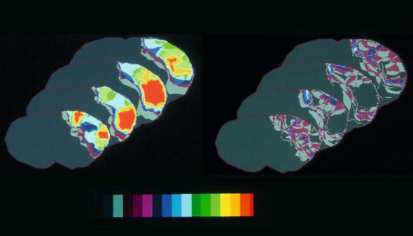
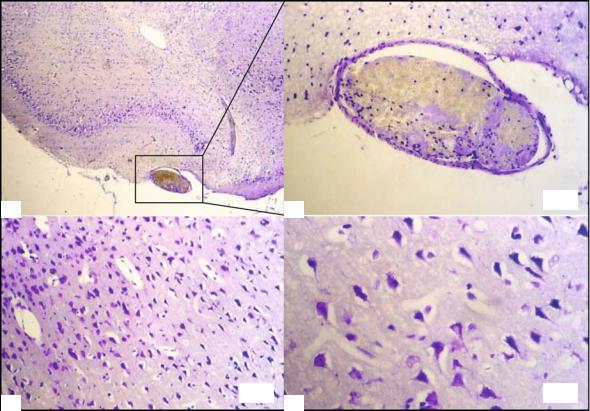
processes related to excessive glutamate release and calcium overload of cells whereas molecular and cellular responses like inflammation and the occurrence of pro-apoptotic gene products typically occur at later stages of the disease process. The obstruction of a major cerebral vessel causes a blood flow gradient that declines towards the center of the territory affected. The high vulnerability of neurons, that also shows a topical preference, leads to a similar gradient of neuronal loss: whereas in the periphery of ischemic regions neurons are selectively injured with normal appearing neuropil and astroglia (socalled scattered neuronal injury), the regions with dense ischemia suffer cell loss of all brain structures including neurons, glial cells and blood vessels also named pan-necrosis. Figure 4.1 shows frequency maps of complete and incomplete (scattered neuronal) infarction in a rat model of MCA occlusion to illustrate the spatial heterogeneity of tissue changes that may also change over time resulting in a small rim of selective neuronal damage that in the subacute phase may account for about 15% of the total lesion (Back et al. 1996). Figure 4.2 demonstrates histological sections that were obtained 6 h post embolic MCA occlusion in a rat: the clot obstruction of the MCA is visualized as well as the selective neuronal cell loss occurring in the infarct borderzone.
Frequency maps of ischemic damage
Complete infarction |
Scattered neuronal injury |
0.00 |
# of rats |
9.00 |
Fig. 4.1. Frequency maps of two types of ischemic damage: complete infarction (left) and scattered neuronal injury (right). Histology was obtained 24 h after left MCA occlusion in rats. Total number of animals was 9. Pseudocolor representation denotes the number of animals that showed the respective type of injury at this pixel. Note the widespread distribution of incomplete infarction over the affected left hemisphere. [Adapted from Alexis et al. (1996)]
Insights from Experimental Studies |
43 |
×16
a |
b |
×16 |
×40 |
c |
d |
Fig. 4.2a-d. Histological staining with cresyl violet 6 h after embolic stroke in rats. Coronal section of the right temporobasal region (affected side, a) shows a fibrin-rich thrombus in the middle cerebral artery, MCA (b). Within the infarct borderzone of the same animal, selective neuronal loss is seen (condensed dark neurons with surrounding vacuoles) in the presence of preserved neurons and intact neuropil (c, higher magnification in d). This histological picture is typical for the ischemic penumbra of brain infarcts
Especially in models of transient cerebral ischemia, apoptotic cell death has been observed after 3–7 days post insult in selected brain regions in which basal energy metabolism has been preserved (Chen et al. 1997; Du et al. 1996). In the meantime, molecular “switches” have been identified that gate different populations of neurons with regard to the type of cell death they eventually undergo (Nicotera 2003). However, there is little doubt that in animal stroke the vast majority of cells would die from necrosis or, alternatively, secondary energy failure even in the presence of a pro-apop- totic genetic balance. The concept of thresholds of cerebral blood flow (CBF) for various functions of brain parenchyma (see below) explains why the infarct core suffers from pan-necrosis whereas the periinfarct border in which function is suppressed, but structure initially preserved (the so-called ischemic penumbra), may show apoptotic cell death or a combination of both.
4.1.2
Infarct Maturation
In focal brain ischemia, the evolution of lesions has been investigated by using histopathological methods (Dereski et al. 1993; Garcia et al. 1993). These studies were able to demonstrate the phenomenon of infarct maturation, i.e., that there is time needed for the macroscopic and microscopic changes to appear on histological sections of the brain. At least 2-3 h following ischemia are necessary for cells to show distinct ischemic changes such as generalized swelling, shrinkage and scalloping of neurons and the formation of vacuoles in dendrites (Garcia et al. 1993). During the initial hours of ischemia, it is difficult to assess the lesion size by using histological means because the ischemic changes are sparse and in the more peripheral regions not well demarcated. Initial evidence of cell loss can be obtained as early as 1 h post occlusion (stria-