


CASE
5Surgery for temporal lobe epilepsy
Victoria Wykes and Anna Miserocchi
 Expert commentary Andrew McEvoy
Case history
A 30-year-old, left-handed woman was referred to the epilepsy service with frequent seizures occurring three or four times per month, which had proved refractory to medication. She described daily episodes of gradual onset of ‘butterflies in the stomach’ followed by ‘a bitter taste’ lasting several minutes. Witnesses stated that she would lose awareness. Sometimes she turned her head to the right prior to flexing both elbows to 90 degrees, with clenching of her left hand. The episodes lasted several minutes and occurred once per week. After the event, she would rub her nose with her right hand, had speech difficulty for up to 5 minutes and experienced left arm weakness, which fully recovered after several hours. Triggers included sleep deprivation and the menstrual cycle. Seizures have resulted in four recent episodes of skin burns. Two secondary generalized tonic clonic seizures (GTCS) had occurred 8 years ago, but no episodes of status epilepticus.
 Learning point
In 1981, the International League Against Epilepsy (ILAE) established a standardized classification and terminology based on seizure manifestations and electroencephalography (EEG) to aid diagnosis, and communication between health professionals and patients [1]. With ongoing advances, new proposals are being made to this classification scheme [2]. Both ILAE classifications are based on the widelyaccepted concept that seizures may be generalized or focal.
Generalized seizures arise from large areas of cortex, and consciousness is lost. However, focal seizures arise in specific small areas of the cortex in one hemisphere, can spread within the same hemisphere or cross to the contralateral side, and may progress into a generalized seizure. Focal seizures can be further divided into simple partial seizures (SPS), which occur without alteration in consciousness
and complex partial seizures (CPS) in which consciousness is impaired (see Tables 5.1 and 5.2 for characteristics). CPS arising in the temporal lobe typically demonstrates a tripartite seizure pattern with aura, loss of awareness and automatisms
Table 5.1 Simple partial seizure characteristics
Manifestations |
Ictal semiology |
Hemispheric localisation |
Motor |
Clonus, spasms, or posturing |
Contralateral to side of seizure |
|
Jacksonian march |
semiology |
|
Progression of seizure along primary motor |
|
|
cortex affecting corresponding muscles |
|
|
Todd’s paresis |
|
|
Post-ictal unilateral motor weakness fully |
|
|
reversible within 24 hours |
(continued) |
|
|
|
|
|
|
|
|
|

44 |
Challenging concepts in neurosurgery |
|
|
|
|
Table 5.1 Simple partial seizure characteristics (continued)
Manifestations |
Ictal semiology |
Hemispheric localisation |
Somatosensory |
Epigastric rising |
Mesial temporal lobe |
|
Tingling, numbness, electrical shock |
Contralateral central or parietal |
|
sensation, burning |
region |
|
Simple visual phenomena, flashing |
Calcarine cortex |
|
lights or colours |
|
Autonomic |
Change in pupil size, skin colour, blood |
|
pressure, heart rate, pilo-erection |
May be isolated to simple partial seizures, but more common in CPS and GTCS
Table 5.2 Complex partial seizure characteristics
Manifestation |
Ictal semiology |
Hemispheric localization |
|
|
|
Aura |
Short-lived event lasting seconds (may last days) |
|
|
occurring in isolation or proceed into CPS or |
|
|
GTCS |
|
Altered consciousness |
Absence with motor arrest |
|
Automatism, |
Oro-alimentry: chewing, lip smacking, |
Mesial temporal lobe |
involuntary motor |
swallowing drooling |
|
action. May be |
Gestural: fiddling hand movements, tapping/ |
Contralateral temporal |
purposeful, complex |
patting/ rubbing. Complex actions include |
and frontal lobe |
activity with interaction |
undressing and genital-directed actions |
|
with the local |
Verbal: meaningless sounds, whistling, word |
|
environment. |
repetition or formed sentences. |
|
|
Mimicry: excitement, laughter, anger, or fear |
|
|
Ambulatory: walking, running, circling |
|
|
Responsive: semi-purposeful behaviour, with |
|
|
interaction with environment |
|
|
Violence: consider as acutely confused patient. |
|
|
Never premeditated, remembered, skilful, and |
|
|
rarely goal direct (important features in forensics) |
|
|
|
|
Her past medical history revealed a normal birth and achievement of normal developmental milestones. There was no history of infection of the CNS or head injury. She had suffered a single febrile convulsion, at age 18 months
 Expert comment
Childhood febrile seizures (FS) are common, occurring in approximately 5% of children. Simple FS are unlikely to be associated with long-term sequelae. Complex FS are defined as prolonged (greater than 15 minutes), multiple within 24 hours, lateralized, or associated with post-ictal neurological deficits, in the absence of a defined cause. Overall, the risk of developing long-term epilepsy following atypical FS is reported as 6% (ten times higher than average). 40–60% of patients with temporal lobe epilepsy (TLE) will report an atypical FS. It is unclear as to whether this reflects a causal association or indicates pre-existing neurological disease, which is responsible for both the complex febrile seizure and later epilepsy [3].

Case 5 Surgery for temporal lobe epilepsy |
45 |
Seizures re-emerged when she was 18 years old and she was started on antiepileptic drugs (AED). When seen, she was taking three AED: carbamazepine 1600mg, levetiracetam 4000mg and pregabalin 300mg daily; however she had tried five other AED, which either did not improve her symptoms or caused intolerable side effects.
 Expert comment
There is debate regarding what constitutes an ‘adequate trial of therapy’ before a patient is diagnosed with medically refractory epilepsy. The current consensus is failure of two first-line AED at an appropriate dose, appropriate to the type of epilepsy, over 2 years [4]. Patients presenting for epilepsy surgery assessment have frequently had extensive and prolonged medication trials. There is a drive to identify medically refractory epileptic patients early, especially children, and offer them surgery.
Her additional past medical history was unremarkable. She felt that both her shortand long-term memory had deteriorated since commencing AED. There was no family history of seizures or neurological abnormality. General and neurological examinations were unremarkable. There were no psychiatric contraindications to surgery. Her neuropsychology assessment demonstrated that she functioned intellectually at an average level. Her episodic memory for both verbal and non-verbal material was good. Most noteworthy was the finding of relative weakness in verbal reasoning, working memory, and literacy skills. This profile suggested dominant hemisphere cerebral dysfunction.
 Expert comment
Favourable outcome from surgery relies on accurate pre-surgical evaluation of the ictal onset zone, through a variety of investigations including:
●Clinical semiology (localizing signs).
●Structural and functional radiological imaging.
●Electrical localization with inter-ictal and ictal EEG, performed most commonly via video-telemetry.
●Detailed neuropsychological profiling to ascertain any localizing functional deficit.
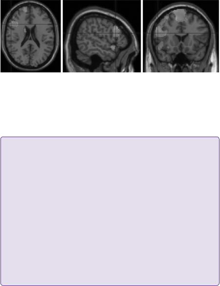
T2-weighted coronal MRI head scans demonstrated hyperintensity in a shrunken right hippocampus consistent with hippocampal sclerosis (Figure 5.1). Other associated findings may include loss of hippocampal head digitations, dilatation of the ipsilateral temporal horn of the lateral ventricle, and atrophy of the fornix and mammillary bodies. Hippocampal volumetric analysis was calculated by measuring the cross-sectional area from successive volumetric T1-weighted MRI coronal images. The right hippocampus was measured to be 40% smaller in comparison with the left.
46 |
Challenging concepts in neurosurgery |
1 2
3
Figure 5.1 T2-weighted coronal MRI head (3 Tesla) demonstrating severe diffuse right hippocampal sclerosis (arrow). Structure 1: cornu ammonis (CA) 1–4, structure 2: subiculum, structure 3: parahippocampal gyrus.
The patient was admitted for a period of video telemetry.
 Learning point
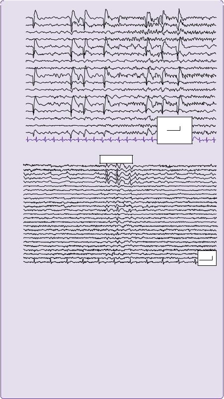
The EEG measures changes in voltage over the scalp resulting from summed post-synaptic potentials in cortical neurons. The international 10–20 electrode placement (Figure 5.2) allows standardization and reproducibility of electrode placement and naming. The ‘10’ and ‘20’ refer to the fact that the actual distances between adjacent electrodes are either 10 or 20% of the total nasion–inion distance of the
10%
|
|
NASION |
|
20% |
|
|
|
|
|
|
|
|
Fp1 |
|
Fp2 |
|
|
F7 |
F3 |
Fz |
F4 |
F8 |
20% |
T3 |
C3 |
Cz |
C4 |
T4 |
|
T5 |
P3 |
Pz |
P4 |
T6 |
20% |
|
O1 |
|
O2 |
|
|
|
|
INION |
|
20% |
|
|
|
|
|
||
|
|
|
10% |
|
|
Figure 5.2 Electrode placement according to the 10–20 international system. ‘Double banana’
longitudinal bipolar montage represented in purple.
(continued)
Case 5 Surgery for temporal lobe epilepsy |
47 |
|
|
|
|
Fp2-F8 |
|
|
F8-T4 |
|
|
T4-T6 |
|
|
T6-O2 |
|
|
Fp1-F7 |
|
|
F7-T3 |
|
|
T3-T5 |
|
|
T5-O1 |
|
|
Fp2-F4 |
|
|
F4-C4 |
|
|
C4-P4 |
|
|
P4-O2 |
|
|
Fp1-F3 |
|
|
F3-C3 |
|
|
C3-P3 |
70 µV |
|
P3-O1 |
||
|
||
Fz-Cz |
1000ms |
|
ECG |
||
|
||
(a) |
|
|
|
Sharp waves |
|
Fp2-AV |
|
|
Fp1-AV |
|
|
F12-AV |
|
|
RSph-AV |
|
|
F8-AV |
|
|
F4-AV |
|
|
Fz-AV |
|
|
F3-AV |
|
|
F7-AV |
|
|
LSph-AV |
|
|
F11-AV |
|
|
T4-AV |
|
|
C4-AV |
|
|
Cz-AV |
|
|
C3-AV |
|
|
T3-AV |
|
|
P12-AV |
|
|
T6-AV |
|
|
P4-AV |
|
|
P3-AV |
|
|
T5-AV |
|
|
P11-AV |
70 µV |
|
O2-AV |
||
O1-AV |
|
|
ECG |
1000ms |
(b)
Figure 5.3 (a) Ictal EEG with longitudinal bipolar ‘double banana’ montage demonstrating epileptiform discharges over the right frontal and temporal areas with spread to left fronto-temporal regions. (b) Interictal awake EEG with average reference montage demonstrates a run of sharp waves seen maximally over the right anterior inferior temporal and frontal areas extending into the left temporal and frontal area.
Courtesy of Dr B. Diehl, Department of Neurophysiology, NHNN, London, UK.
skull. The letter describes anatomical location (Fp = frontopolar, F = frontal, T = temporal, P = parietal, O = occipital, C = central). Electrodes placed over the right hemisphere are represented by even numbers, over the left hemisphere by odd numbers, and midline represented by ‘z’ (zero). Lower numbers indicate more medial placement, while higher numbers indicate more lateral placement. Additional electrodes may be useful, e.g. superficial sphenoidal electrodes when recording from the anterior or mesial temporal lobe.
The display montage represents the way the electrodes are connected and referenced. In the ‘longitudinal bipolar’ montage, also known as the ‘double banana’, each channel represents the difference between two adjacent electrodes (Figure 5.3). Whereas in the ‘average reference’ montage, the outputs of all of the electrodes are summed and averaged, and this averaged signal is used as the common reference for each channel.
Sharp waves 70–200msec and spikes lasting less than 70ms duration indicate epileptiform activity.
48 |
Challenging concepts in neurosurgery |
Figure 5.4 Language fMRI paradigms for verbal fluency (orange) and verb generation (yellow) provided evidence for right-sided language dominance (for colour version of this figure please see online edition).
Courtesy of Dr C. Micallef & Mr J. Stretton Department of Neuroradiology, NHNN, London, UK.
In this left-handed patient with right-sided hippocampal sclerosis and post-ictal dysphasia functional MRI (fMRI) was performed with language paradigms to lateralize language dominance (Figure 5.4).
 Learning point
The incidence of language hemisphere dominance has been investigated using fMRI studies in healthy populations. In the right-hand dominant group, 94% were considered left hemisphere language dominant, with 6% demonstrating bilateral language representation [5]. A similar study in a non-right- hand dominant group demonstrated only that 8% were right hemisphere language dominant, with 78% demonstrating left hemisphere language dominance [6].
The language system is variable in location across individuals, influenced by handedness, and in patients with TLE may reorganize with bilateral representation or transfer to the contralateral hemisphere [5].
Anterior temporal lobe resection (ATLR) may impair both language and memory functions. Dominant hemisphere ATLR may impair naming and verbal memory, while non-dominant hemisphere ATLR may impair visual memory. Precise pre-operative assessment of language and memory lateralization is essential prior to surgery, to predict risk of post-operative deficits allowing informed consent of the patient.
The Wada test has been, and in some centres still is, considered to be the Gold standard investigation of both language and memory functions. Reversible pharmacological deactivation of one cerebral hemisphere is achieved by injection of the barbiturate sodium amobarbital into the internal carotid artery, while the patient is awake, allowing evaluation of functions in the opposite hemisphere. The inherent risks of this invasive procedure and the emergence of non-invasive alternatives, particularly fMRI has led to replacement in many centres [7–9].
To summarize, this 30-year-old, left-handed woman had presented with complex motor seizures since the age of 18 years. MRI demonstrated right hippocampal sclerosis. Inter-ictal EEG showed right anterior temporal spikes and the ictal EEG showed a right hemisphere seizure with maximal activation in the temporal region. Semiology indicated right hemisphere lateralizing signs of seizure onset including:
●Head deviation to the right at seizure onset.
●Left tonic fist posturing.
●Post-ictal nose rubbing with right hand.
●A left Todd’s paresis.
●Post-ictal speech difficulty, supporting right hemisphere language dominance.

Case 5 Surgery for temporal lobe epilepsy |
49 |
Table 5.3 Specific operative risks of anterior temporal lobe resection (ATLR) depending on hemisphere dominance:
ATLR dominance |
Risk |
Dominant |
Impairment of language, particularly naming and verbal memory (20–30%) |
Non-dominant |
Impairment of visual memory (10%) |
Both |
Infection (<3%) |
|
Hemiparesis and speech disturbance (<1%) |
|
Visual field loss (10%), visual field deficit resulting in driving ban (5%) |
|
Neuropsychiatric deficit: mood disturbances, including emotional lability, |
|
depression, and anxiety may occur on up to 1:5 patients. Rarely (1%) acute |
|
psychosis can occur.* |
|
|
Neuropsychological assessment is consistent with dominant cortical hemisphere dysfunction. fMRI demonstrated right hemisphere language localization. Importantly, she had no psychiatric contraindications to surgery.
This data was presented in a multidisciplinary epilepsy surgery planning meeting, which concluded that the findings were concordant with a presumed epileptogenic zone in the right temporal lobe secondary to right hippocampal sclerosis. A success rate of seizure freedom was predicted at 60%t, with an additional 20% of significant improvement in seizure frequency following a standard right-sided ATLR. Preand peri-operative counselling is essential for all patients undergoing epilepsy surgery. Risk–benefit discussions with the patient and their family should be performed and documented, and given to the patient in written format. The standard operative risks and specific risks of a dominant hemisphere ATLR were discussed with the patient (see Table 5.3).
Temporal lobe resection for epilepsy
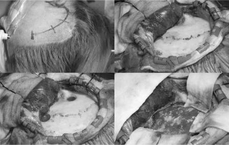
The patient was placed supine in the Mayfield head clamp. The head was rotated 90 degrees, with the zygoma parallel to the floor. A temporal question mark skin incision (Figure 5.5a) and myocutaneous flap were made (Figure 5.5b). Two burr holes were made, one over the pterion and the other above the root of the zygoma (Figure 5.5c), allowing a craniotomy (Figure 5.5d). The craniotomy should be made as low as possible to allow access to the floor of the middle cranial fossa with minimal retraction. The dura was opened in a C-shape to expose the Sylvian fissure.
ATLR involved resection of the lateral neocortex, facilitated by identification of the temporal horn of the lateral ventricle, followed by resection of the mesial temporal structures (amygdala, hippocampus, parahippocampal gyrus).
To achieve the neocortical resection, 4cm was measured from the temporal pole over the long axis of the middle temporal gyrus and the posterior border marked on the cortical surface using irrigating bipolar forceps. This was extended along the superior temporal gyrus approximately 5mm below the Sylvian vessels creating an ‘L shape’. The arachnoid of the L shape was divided with microscissors and subpial dissection performed using a Rhoton dissector.
Using intra-operative navigation, the temporal horn of the lateral ventricle was located. The approach to the ventricle was along the middle fossa floor, identifying the collateral sulcus. Dissection perpendicular to the floor, following the collateral
50 |
Challenging concepts in neurosurgery |
Figure 5.5 Right anterior temporal lobe exposure.
sulcus, enabled the ventricle to be entered inferiorly. This reduced the risk of damage to the optic radiation, in particular Meyer’s loop (which is lateral and superior to the ventricle). A cottonoid patty was inserted into the temporal horn of the ventricle to prevent CSF loss, and blood entering the ventricular system as the temporal pole was removed.
The temporal stem was disconnected by passing a blunt hook from the tip of the temporal horn towards the previously dissected superior temporal gyrus, allowing removal of the lateral temporal neocortex.
The mesial temporal structures were then resected. The uncus and amydala were removed using a low-powered Cavitron Ultrasonic Aspirator (CUSA). On removal of the uncus along its pial boundary, the third cranial nerve, posterior communicating artery and the edge of the tentorium could be visualized. The anterior choroid point is an important anatomical landmark as it represents the most anterior aspect of the choroid plexus, localizes entry of the anterior choroidal artery, and demarcates the posterior part of the hippocampal head.
The amygdala can be resected anteriorly and lateral to an imaginary line drawn from this anterior choroid point to the Sylvian fissure. Care must be taken not to go too deep during amygdala resection to avoid entering the frontal lobe.
A blunt hook was used to gently separate the choroid plexus from the hippocampus to reveal the fimbria. Gentle dissection of the fimbria off the underlying arachnoid revealed the hippocampal artery arcade lying within the hippocampal sulcus, which supplies the hippocampal head. The vessels were cauterized and the hippocampus resected. The parahippocampal gyrus and pes were finally resected, taking care not to damage the underlying adjacent cerebral peduncle. The craniotomy was closed and all samples sent to pathology.
Post-operatively, the patient continued her AEDs, experienced no neurological deficit, and was seizure-free at 1 year follow-up.

Case 5 Surgery for temporal lobe epilepsy |
51 |
 Clinical tip Anterior temporal lobe resection
●When positioning, the neck must be both extended and flexed to allow the ventricular approach along the middle temporal fossa.
●When positioning the patient, care should be taken that the neck is not over-flexed, reducing the risk of brachial plexus injury. All bony prominences must be well padded to avoid pressure sores.
●During the craniotomy, if the mastoid air cells are opened, these must be packed off with bone wax or biological sealant to prevent CSF leak or encephalocoele.
●The vein of Labbe is located over the dominant posterior temporal lobe with variable anatomy. Dissection should proceed anterior to the vein as damage to this vessel may produce venous infarction in cortical and subcortical speech areas.
●Subpial dissection of the superior temporal gyrus exposes the arachnoid overlying the insular cortex and branches of the middle cerebral artery. These landmarks can facilitate dissection of the temporal stem, but care must be taken not to penetrate the arachnoid layer and enter the Sylvian Fissure.
●The CUSA is used on low power to ensure no breech of the pial boundary into the basal cisterns. If blood enters these cisterns chemical meningitis may ensue, resulting in headache, neurological irritation, and a prolonged hospital stay.
●Meyer’s loop lies lateral and superior to the temporal horn of the lateral ventricle and damage will result in superior temporal visual field defect.
●When resecting the amygdala, care must be taken, particularly at the superior medial aspect, as there are no clear anatomical landmarks. Amygdala resection below an imaginary line taken from the anterior choroidal point to the anterior superior arachnoid of the limen insulae and uncus is deemed safe. Above this line lies the globus pallidus and branches of the middle cerebral artery en route to the basal ganglia, which if damaged may result in hemiplegia and hemianopia.
Discussion
The estimated incidence of epilepsy is 50 per 100,000 in the general population [10] and 20% are estimated to become refractory to medication [11]. A UK study reported that the commonest surgical procedure for epilepsy was temporal resection for hippocampal sclerosis [12]. Brain tumours represent a common finding in patients with intractable focal epilepsies. Approximately 60% are dysembryoplastic neuroepithelial tumours and gangliomas [13]. Other lesions include glioma, focal cortical dysplasia, heterotopia, arteriovenous malformation, cavernoma, infarct, granuloma (e.g. secondary to cysticercosis, tuberculosis), and traumatic intraparenchymal brain injury. Dual pathology exists in up to 30% of surgical specimens of patients operated on for TLE, with the most common extrahippocampal lesion abnormality reported as cortical dysplasia. The pathogenesis of dual pathology is poorly understood [14]. Cryptogenic or paradoxical TLE refers to patients in whom no pathology is identified in the resected temporal lobe and constitutes less than 10% of cases [15].
 Expert comment Hippocampal sclerosis
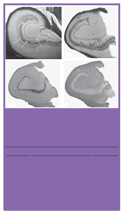
The pathological diagnosis of HS is characterized by gliosis and neuronal loss (especially pyramidal neurons) predominantly in CA1, but also in the CA3 and CA4 subfields. Typically, there is an accompanying loss of excitatory mossy cells, neuropeptide Y-secreting neurons and GABAergic neurons [16]. There may be associated gliosis and similar neuronal loss in the ipsilateral amygdala and entorhinal cortex (Figure 5.6). A recent classification of Hippocampal Sclerosis (HS) suggests that a non-classical histological pattern (although occurring less frequently) has a worse outcome in relation
(continued)
52 |
Challenging concepts in neurosurgery |
|
CA3 |
|
CA4 |
|
|
|
|
|
CA2 |
|
CA1 |
|
CA1 |
Dentate |
|
|
|
||
|
|
gyrus |
|
|
|
|
|
(a) |
|
|
(b) LFB/CV |
(c) NeuN |
(d) GFAP |
Figure 5.6 Sections demonstrating (a) healthy hippocampus; (b, c, d) classical hippocampal sclerosis. Sections are stained with: (a) Luxol Fast Blue (LFB); (b) LFB and cresyl violet (CV). (c) Neuronal nuclei (NeuN) stained section demonstrating neuronal loss mainly in CA1 and CA4. (d) Glial fibrillary acidic protein (GFAP) demonstrating the diffuse fibrillary gliosis in CA4 and CA1, (for colour version of this figure please see online edition).
Images courtesy of Dr Thom, Department of Neuropathology, Institute of Neurology, London.
Table 5.4 Characteristics of classical and non-classical features of HS and post-operative seizure freedom
|
Percentage in a |
Neuronal loss |
Post-operative seizure |
|
series of 178 patients |
|
freedom at 1 year (%) |
No Mesial temporal |
19 |
Nil |
59 |
sclerosis (MTS) |
|
|
|
MTS 1a (classical) |
19 |
CA1 predominantly with |
72 |
|
|
some neuronal loss in all |
|
|
|
other subfields excluding CA2 |
|
MTS 1b (classical) |
53 |
All hippocampal subfields |
73 |
MTS 2 (non-classical) |
6 |
CA1 |
67 |
MTS 3 (non-classical) |
4 |
End folium sclerosis in hilar area |
29 |
|
|
|
|
Summarized from Blümcke I, Pauli E, Clusmann H et al. A new clinico-pathological classification system for mesial temporal sclerosis. Acta Neuropathol 2007; 113: 235–44. Distributed under the terms of the Creative Commons Attribution License 2.0 [17].
to seizure freedom 1 year post-operatively (Table 5.4) [17]. Although HS was identified nearly 200 years ago [18], it is still an area of active research, particularly with regard to identifying the cause and mechanism of seizure generation in this region of mesial neuronal loss.

Case 5 Surgery for temporal lobe epilepsy |
53 |
 Learning point
A MDT and systematic approach to investigations is essential in evaluating candidates for epilepsy surgery [19].
Clinical evaluation
Birth and developmental history, epilepsy history, current seizure pattern, drug history (failure of two AEDs), co-morbidities preventing surgery, clinical examination (with emphasis on neurological, cardiovascular, skin systems (stigmata of epilepsy syndromes), ECG.
Initial investigations
MRI head, prolonged inter-ictal EEG, video-EEG, neuropsychology, and neuropsychiatry.
Multi-disciplinary meeting
If concordant data: surgical planning is required—fMRI for language/motor function, visual fields, if surgical resection is in proximity to these areas.
If discordant, but a potential surgical candidate, further imaging techniques can be used to derive the target for intracranial EEG using depth electrode, subdural grids, and strips.
It is imperative that patients and their families are given realistic expectations and associated risks of surgery, and to ensure that long-term post-operative follow-up is maintained.
Neurophysiology
The aim of prolonged scalp inter-ictal and ictal EEG in pre-surgical work-up for resective epilepsy procedures includes:
●Demonstration of an electrophysiological-clinical seizure: up to 10% of candidates undergoing pre-surgical work up for epilepsy surgery have psychogenic non-epileptic attacks.
●Characterization of electro-clinical seizures to ensure concordance that all seizures are arising from the same location.
●If there is discordance due to other epileptogenic areas (e.g. dual pathology), this will reduce the chance of seizure freedom.
 Expert comment If dual epileptogenic areas are present, invasive EEG can be considered
Depth electrodes are multiple contact wires inserted under stereotactic MRI guidance and are most useful for investigating deep epileptogenic zone. Their disadvantage is they sample only small areas of brain. Subdural grid and strip electrodes record from large superficial cortical areas and are inserted via a craniotomy. Electrical stimulation to map function can also be performed through both types of electrodes.
Neuroimaging in temporal lobe epilepsy
A 3-Tesla MRI brain scan consisting of T1and T2-weighted, and FLAIR imaging in axial and coronal planes, can reveal lesions that were not identified on previous imaging. Gradient-echo imaging may be useful to detect a vascular abnormality. Hippocampal volumetric analysis can provide evidence of HS. If imaging does not yield a structural cause for the epilepsy, or there is discordance between clinical

54 Challenging concepts in neurosurgery
and EEG data, functional imaging can be considered: positron emission tomography (PET), single-photon emission CT (SPECT) with ictal–inter-ictal subtraction, simultaneous EEG and fMRI [20] and magneto-encephalography (MEG). Cortical regions can be highly variable in terms of size and location, and stereotactic atlases fail to take into account this intrinsic variability. Eloquent functions can be mapped with fMRI, using language, memory, primary motor, and somatosensory paradigms with diffusion tensor imaging for white matter tracts [21]. Co-registration of multi-modal imaging can facilitate surgical planning and intra-operative MRI can aid surgical resection.
 Learning point
ATLR for TLE may be complicated by a visual field deficit (VFD), typically a contralateral superior quadrantinopia caused by damage to the anterior optic radiation (Meyer’s loop). Despite being seizure free, post-operative patients may have significant VFD to prevent driving [22].
There is significant anatomical variability in Meyer’s loop and integration of tractography of optic radiation and real-time neuronavigation is currently being characterized [23].
Neuropsychological assessment
Pre-operative neuropsychological assessment is performed using the Wechsler Adult Intelligence Scale. The full-scale intelligence quotient (IQ) is made up from scores obtained by testing verbal IQ comprising verbal comprehension and working memory, and performance IQ comprising perceptual organization, and processing speed. The results may aid localization and lateralization of cerebral dysfunction— impaired memory functions suggest localization to the temporal lobes. Comparison of visual IQ to performance IQ can help with lateralization, impaired verbal memory lateralizes to the dominant temporal lobe (usually the left), whereas impaired visual memory lateralizes to the non-dominant temporal lobe. fMRI memory paradigms are now also providing information that helps predict post-operative cognitive deficit [8,9]. The extent of any post-operative deficit depends both on the functionality of the removed tissue and the functional reserve of the non-resected structures. Pre-operative assessment can help predict the risk for post-surgical cognitive decline, allowing appropriate pre-operative counselling and post-operative support. Assessment of the cognitive baseline is useful, as this may be deleteriously affected by the seizures themselves, as part of the spectrum of epilepsy syndromes, AED, and is of paramount importance to ensure the patient has a high enough IQ for informed consent.
Neuropsychiatry assessment
Psychiatric morbidity (particularly depression, anxiety, and psychosis) is common with lifetime prevalence rates of up to 50% in TLE. 25% of patients post-surgery for TLE, report transient mood disturbances, including emotional lability, depression, and anxiety during the first 3 months with 10% experiencing more persistent symptoms. Post-operative de novo psychosis has been reported to occur in 1% of patients. The risk factors for developing post-operative psychiatric disorders include pre-operative psychiatric history, pre-operative bilateral epileptiform discharges, and poor post-operative seizure control [24].
Case 5 Surgery for temporal lobe epilepsy |
55 |
Surgery for temporal lobe epilepsy
Sir Victor Horsley (National Hospital of Neurology and Neurosurgery, London, UK) was one of the pioneers of epilepsy surgery in the 1800s, correlating clinical observation and seizure semiology to plan lesionectomy. With the invention of the EEG by Hans Berger in 1924, preand intra-operative recordings allowed resection to include areas that gave rise to epileptiform activity, even though the area to be removed may have appeared grossly normal. Wilder Penfield (Montreal Neurological Institute, Canada) first identified that, for patients with TLE, the temporal lobe, hippocampus, and amygdala could be removed safely and with good result in terms of reduction in seizure frequency. In the 1950s, Murray Falconer (Maudsley Hospital, London, UK) pioneered the en bloc anatomical temporal lobectomy. However, this led to significant deficits especially with regard to neuropsychological and superior quadrantanopia visual field deficits. Alternative procedures aiming to reduce the size of the neocortical resection include the Spencer-type technique and the selective amygdalohippocampectomy [25,26]. The Spencer technique was developed to preserve the function of the lateral temporal cortex and to access the medial temporal structures through a temporal corridor. The selective amygdalohippocampectomy can be approached transcortically, transylvian, and subtemporally with the perceived advantage that the adjacent temporal neocortex remains intact. It has been difficult to compare the various surgical techniques, as there has been a lack of standardized operative outcome criteria, and the optimal size of temporal resection is still controversial and requires further study [27,28].
In the late 1990’s, a Canadian trial randomly assigned 80 patients with medically refractory TLE, to surgery or 1 year of AED therapy [29]. The primary outcome was freedom of seizures that impair awareness of self and surroundings. Wiebe et al. demonstrated that surgery afforded 58% seizure freedom in comparison with 8% in the medical therapy arm. Similarly, a cohort study of consecutive patients undergoing anterior temporal lobe resections or temporal lesionectomies for TLE at a single UK centre, demonstrated that approximately 60% of 537 patients were seizure free (excluding simple partial seizures) at 5 years [30].
A RCT was recently started to compare the outcome of standard surgical temporal lobe resection with the results of gamma knife radiosurgery of the same surgical target. The main study hypothesis of the Radiosurgery or Open Surgery for Epilepsy (ROSE) trial was that radiosurgery (which requires no hospital stay), is as safe and effective as open surgery in treating patients with seizures arising from the medial temporal lobe. The study was abandoned in 2012 due to difficulty in patient recruitment.
Palliative procedures exist for medically refractory patients that are either not suitable for resective surgery, or who do not improve following resective surgery. These adjunctive therapies include continuous stimulation with deep-brain stimulation of the anterior thalamus (DBSANT; Medtronic Inc.), intermittent stimulation with vagal nerve stimulation (VNS; Cyberonics Inc.), and responsive neurostimulation (RNS; Neuropace Inc.). To date, only VNS is licensed for use in both Europe and the USA.
VNS provides a third of patients with greater than 50% reduction in seizures, a third experience less than 50% reduction, and no improvement is seen in the remaining third [31]. Spiral-shaped bipolar electrodes are wrapped around the left vagal nerve, the distal ends are tunnelled over the clavicle into a subcutaneous
56 Challenging concepts in neurosurgery
pocket above the pectoral fascia in which an implantable pulse generator (IPG) is sited. Stimulation parameters can be adjusted via a programming device, using radio-frequency signals. Typically, patients slowly have their stimulation parameters increased until individual optimal seizure control and side effect tolerance is reached. The patient can also trigger the stimulator by resting a magnet (often worn as a bracelet) over it, allowing extra stimulation during an aura, which may arrest progression into a seizure.
 Learning point
The left vagus nerve is stimulated due to concerns that bradycardias or arrhythmias may be induced on stimulating the right vagal nerve.
The vagus is approximately 80% afferent and composed of unmylinated fibres. The fibres project to the nucleus tractus solitarius in the brainstem and then on to other structures including hypothalamus, thalamus, and cortex. The exact mechanism of action for seizure reduction is unknown.
Insertion and stimulation may be associated with a hoarse voice, cough, throat pain, or paraesthesia and dyspnoea.
A final word from the expert
●
●
●
●
●
Operative safety may be improved with the development of surgical image guidance systems, allowing reliable integration of structural and functional data available in real time as surgery progresses. Neuromodulation is a rapidly evolving field. DBS of different targets are being investigated, e.g. centromedian thalamic nucleus [32]. Seizure prediction devices are also under development. Implantable electrodes communicate to a hand-held device, indicating the likelihood of seizure activity in the coming hours and allow direct electrical stimulation to prevent or reduce severity of the seizure. Electrically-active drug release implants may allow direct access of drugs to epileptogenic areas avoiding systemic metabolism, and impermeability of the blood–brain barrier.
Optogenetic tools comprise a variety of different light-sensitive proteins that, via a viral vector, can be expressed in mammalian neurons to effectively control their excitability. Expression of the inhibitory halorhodopsin in hippocampal principal cells has recently been used as a tool to effectively control chemicallyand electrically-induced epileptiform activity in animal slice preparations [33]. Optogenetic tools represent a novel way of manipulating neural networks at a molecular level and may offer a future strategy in epilepsy treatment [34].
Cell transplantation therapies with either mature cells or stem cells derived from foetal tissue or genetically-engineered cells that enhance synaptic inhibition, and so reduce seizures or protect neurons from damage, are being investigated in animal models [35].
References
1.Commission on Classification, International League Against Epilepsy. Proposed provisions of clinical and electroencephalographical classification of epileptic seizures. Epilepsia 1981; 22: 489–501.
2.Berg AT, Berkovic SF, Brodie MJ, et al. Revised terminology and concepts for organization of seizures and epilepsies: report of the ILAE Commission on Classification and Terminology, 2005–2009. Epilepsia 2010; 51: 676–85.
3.Annegers JF, Hauser WA, Shirts SB, et al. Factors prognostic of unprovoked seizures after febrile convulsions. N Engl J Med 1987; 316: 493–8.
4.Brodie MJ, Kwan P. Staged approach to epilepsy management. Neurology 2002; 58: S2–8.
Case 5 Surgery for temporal lobe epilepsy |
57 |
5.Springer JA, Binder JR, Hammeke TA, et al. Language dominance in neurologically normal and epilepsy subjects. A functional MRI study. Brain 1999; 122: 2033–45.
6.Szaflarski JP, Binder JR, Possing ET, et al. Language lateralization in left-handed and ambidextrous people fMRI data. Neurology 2002; 59: 238–44.
7.Arora J, Pugh K, Westerveld M, et al. Language lateralization in epilepsy patients: fMRI validated with the Wada procedure. Epilepsia 2009; 50: 2225–41.
8.Bonelli SB, Powell RHW, Yogarajah M, et al. Imaging memory in temporal lobe epilepsy: predicting the effects of temporal lobe resection. Brain 2010; 133: 1186–99.
9.Bonelli SB, Thompson PJ, Yogarajah M, et al. Imaging language networks before and after anterior temporal lobe resection: results of a longitudinal fMRI study. Epilepsia 2012; 53: 639–50.
10.Sander JWAS. Some aspects of prognosis in the epilepsies: a review. Epilepsia 1993; 34: 1007–16.
11.Hart YM.Shorvon SD. The nature of epilepsy in the general population: I. Characteristics of patients receiving medication for epilepsy. Epilepsy Res 1995; 21: 43–9.
12.Lhatoo SD, Soloman JK, McEvoy AW, et al. A prospective study of the requirement for and the provision of epilepsy surgery in the United Kingdom. Epilepsia 2003; 44: 673–6.
13.Blümcke I. Epilepsy-associated brain tumors. Handb Clin Neurol. 2012; 108: 559–68.
14.Harroud A, Bouthillier A, Weil AG, et al. Temporal lobe epilepsy surgery failures: a review. Epilepsy Res Treatment 2012. Available at: http://dx.doi.org/10.1155/2012/201651
15.Thom M. Hippocampal sclerosis: progress since Sommer. Brain Pathol 2008; 19: 565–72.
16.Thom M, Sisodiya SM, Najm I. Epilepsy. In: S Love, DN Lewis, DW Ellison (eds), Greenfield’s neuropathology 8th edn. (pp. 833–87). London: Hodder-Arnold, 2008.
17.Blümcke I, Pauli E, Clusmann H, et al. A new clinico-pathological classification system for mesial temporal sclerosis. Acta Neuropathol 2007; 113: 235–44.
18.Bouchet C, Cazauvieilh C.A. De l’ épilepsie considerée dans ses rapports avec aliénation mentale. Recherche sur la natur et le siège de ces maladies. Arch Gén Méd 1825; 9: 510–42.
19.Duncan JS. Selecting patients for epilepsy surgery: synthesis of data. Epilepsy Behav 2011; 20: 230–2.
20.Vulliemoz S, Thornton R, Rodionov R, et al. The spatio-temporal mapping of epileptic networks: combination of EEG-fMRI and EEG source imaging. Neuroimage 2009; 46: 834–43.
21.Duncan JS. Imaging in the surgical treatment of epilepsy. Nat Rev Neurol 2010; 6: 537–50.
22.Yogarajah M.Focke NK, Bonelli S, et al. Defining Meyer’s loop—temporal lobe resections, visual field deficits and diffusion tensor tractography. Brain 2009; 132: 1656–68.
23.Winston GP. Epilepsy surgery, vision, and driving: what has surgery taught us and could modern imaging reduce the risk of visual deficits? Epilepsia 2013; 54: 1877–88.
24.Foong J, Flugel D. Psychiatric outcome of surgery for temporal lobe epilepsy and presurgical considerations. Epilepsy Res 2007; 75: 84–96.
25.Harkness W. Temporal lobe resections. Childs Nerv Syst 2006; 22: 936–44.
26.Campero A, Tróccoli G, Martins C, et al. Microsurgical approaches to the medial temporal region: an anatomical study. Neurosurgery 2006; 59: 279–308.
27.Schramm J. Temporal lobe epilepsy surgery and the quest for optimal extent of resection: a review. Epilepsia 2008; 49: 1296–307.
28.Al-Otaibi F, Baeesa SS, Parrent AG, et al. Surgical techniques for the treatment for temporal lobe epilepsy. Epilepsy Res Treat 2012; 2012: 374848.
29.Wiebe S, Blume WT, Girvin JP, et al. A randomized controlled trial of surgery for temporal lobe epilepsy. N Engl J Med 2001; 345: 311–18.
30.De Tisi J, Bell, GS, Peacock JL, et al. The long-term outcome of adult epilepsy surgery, patterns of seizure remission, and relapse: a cohort study. Lancet 2011; 378: 1388–95.
31.Janszky J, Hoppe M, Behne F, et al. Vagus nerve stimulation: predictors of seizure freedom. J Neurol Neurosurg Psychiat 2005; 76: 384–9.
58 |
Challenging concepts in neurosurgery |
|
|
32. |
Velasco F, Velasco AL, Velasco M, et al. Deep brain stimulation for treatment of the |
|
|
epilepsies: the centromedian thalamic target. Acta Neurochir Suppl. 2007; 97: 337–42. |
|
33. |
Tønnesen J.Sørensen AT, Deisseroth K, et al. Optogenetic control of epileptiform activity. |
|
|
Proc Natl Acad Sci 2009; 106: 12162–7. |
|
34. |
Kokaia M.Andersson M.Ledri M. An optogenetic approach to epilepsy. |
|
|
Neuropharmacology 2013; 69: 89–95. |
|
35. |
Boison D. Cell and gene therapies for refractory epilepsy. Curr Neuropharmacol 2007; 5: |
|
|
115–25. |