


CASE
20Spontaneous intracerebral haemorrhage
Peter Bodkin
 Expert commentary Patrick Statham
Case history
A 68-year-old man presented to his local emergency department with a left hemiparesis of sudden onset. His background medical history included poorly-controlled hypertension, ischaemic heart disease, and previous TIAs. He was medicated on amlodipine 5mg, aspirin 75mg, dypiridamole 100mg tds, and simvastatin 20mg. Blood parameters, including clotting, were within normal range.
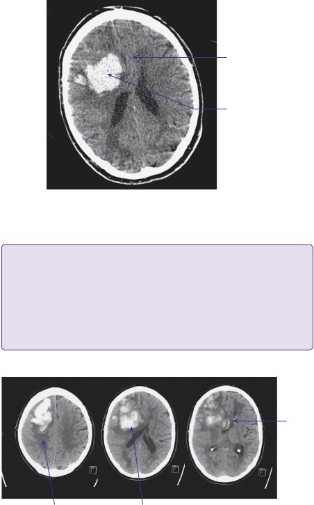
On admission, his conscious level was normal, but he had MRC grade 3/5 weakness of the left upper and lower limb. Cranial nerves were normal apart from a left upper motor neurone facial palsy. His blood pressure was 210/105 in the emergency department. A CT brain scan (Figure 20.1) revealed a right frontal intracerebral haematoma (41 × 53 × 39mm, approximately 42mL; see Learning point: estimation of volume of intracranial haemorrhage). Antiplatelet agents were immediately stopped.
 Learning point Estimation of volume of intracranial haemorrhage
An ellipsoid can be described by its Cartesian coordinates of:
●The largest cross-sectional diameter.
●A second diameter drawn at right angles to the first.
●The height of the ellipsoid [1].
Volume = (4π/3) × (a/2) × (b/2) × (c/2)
If π is approximated to 3 the equation simplifies to:
Volume = (a × b × c)/2
In practice, the axial image with the largest cross-sectional area of clot should be chosen, and a and b values measured in centimetres. The c value should be worked out by counting the number of slices the intracranial haemorrhage (ICH) is visible on and multiplied by the slice thickness in centimetres. A value in millilitres will be given. There has been proven to be a very close correlation between this method of volume estimation and computer-assisted planimetric image analysis [2].
He was initially managed conservatively with careful control of his blood pressure by labetolol infusion with a target systolic blood pressure of below 180mmHg.
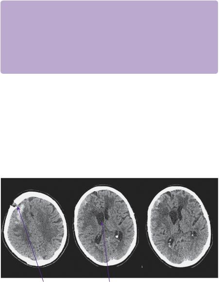
Between days 1 and 5 he remained neurologically stable. On day 6 he became increasingly drowsy and confused, with eye opening to speech and speech limited to occasional words (E3 V3 M6). His eyes were noted to be deviated to the right at rest. A repeat CT brain showed haematoma expansion causing increased mass effect (Figure 20.2). There were no systemic factors contributing to his deterioration.
190 |
Challenging concepts in neurosurgery |
Midline shift
Right frontal intracerebral haematoma
Figure 20.1 Patient’s initial CT. Non-contrasted axial image showing haematoma in the right frontal lobe with effacement of the right frontal horn and 4mm of midline shift.
 Learning point Frontal eye fields [3]
Horizontal conjugate gaze deviation towards the side of a lesion (Prévost or Vulpian sign) may be a result of damage to the frontal eye fields (FEFs). These are located in the posterior part of the middle frontal gyrus and adjacent precentral sulcus (Brodman areas 6 and 4). The FEFs are involved in complex neural pathways, modulating responses from visual and other stimuli for horizontal saccadic and pursuit movements, ultimately via the abducens nucleus. For pursuit movements, the dorsolateral pontine nucleus, cerebellum, and vestibular nucleus are intermediaries; for saccades the superior colliculus and paramedian pontine reticular formation (PPRF) play important roles. Oculocephalic manoeuvres and caloric stimulation can typically override the gaze palsy. A seizure focus in the region of the FEF will cause deviation of gaze away from the side of origin.
Midline shift
Peri-haematoma oedema |
Right frontal haematoma |
Figure 20.2 Preoperative CT. Non-contrasted axial slices demonstrating significantly larger right frontal haematoma with mid-line shift now of 1cm.
Case 20 Spontaneous intracerebral haemorrhage |
191 |
The balance of risk versus benefit at this point favoured surgical evacuation, which was discussed with his family. A right frontal burr hole was created under general anaesthesia, and a Dandy cannula was used to aspirate the clot. This was done with a freehand technique. The most appropriate entry point and target depth had been planned based on measurements in axial, sagittal, and coronal planes. 30mL of dark blood clot was aspirated. The brain was seen to relax after clot aspiration.
 Clinical tips Burr hole aspiration of intracerebral clot
Minimally-invasive techniques for clot aspiration are an attractive treatment option to avoid the inevitable trauma of craniotomy with corticotomy, brain retraction, etc. Aspiration may be most accurately performed with stereotactic methods using the same equipment as for tumour biopsy or shunt placement. Alternatively, modern digital imaging software usually provides measuring tools so that a target can be localized from anatomical landmarks using reconstructed 3D images. Given that the clot was large and in a relatively non-eloquent region of the brain, we felt that this method was appropriate. Adjunctive methods of dissolution of the clot may also be used (see Discussion). In this case, we were fortunate that the clot had largely liquefied, given that it was 6 days old.
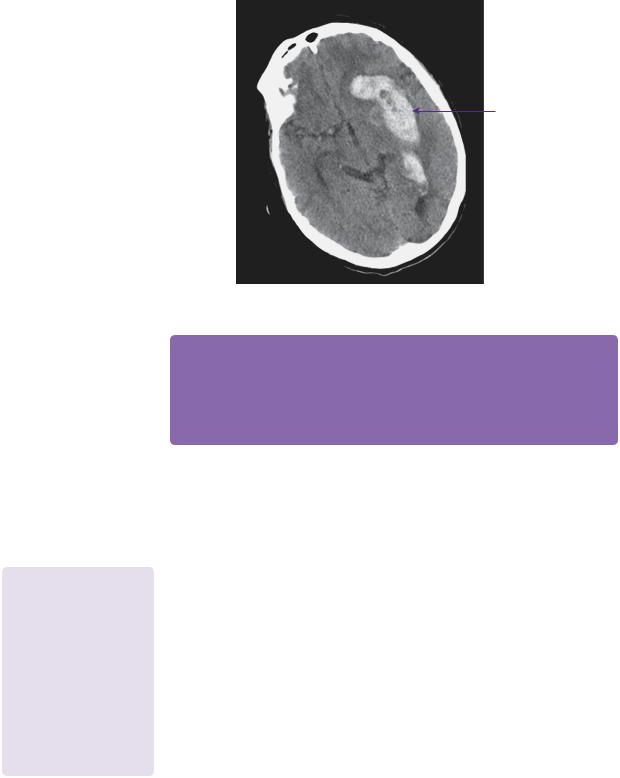
Following surgery, he recovered to being mildly confused (GCS E4 V4 M6). His hemiparesis (MRC 3/5) persisted. A post-operative CT showed markedly improved appearances (Figure 20.3. He was discharged to a rehabilitation unit, where his neurological deficit improved such that he was mobile with a Zimmer frame and independent of most activities of daily living.)
Unfortunately, he presented again 3 months later with a left-sided basal ganglia bleed with intraventricular extension (Figure 20.4). This may well relate to his being recommenced on antiplatelet agents 2 weeks previously. Further surgery was not undertaken at that point.
Right frontal burr hole |
Right lateral ventricular horn |
Figure 20.3 Post-operative CT 2 days following surgery. Non-contrasted axial slices showing minimal residual haematoma and resolution of mid-line shift.
192 |
Challenging concepts in neurosurgery |
Let basal ganglia haematoma
Figure 20.4 3 months later - CT non-contrasted axial image showing extensive left basal ganglia and intraventricular haemorrhage.
 Expert comment
Primary intracerebral haematoma (PICH) represents the end stage of a longstanding disease. Removing a haematoma may reduce the immediate risk to life, but at the cost of residual disability. Careful discussion with the patient and relatives is vital to prevent misunderstandings in the weeks and months following intervention. This should start before referral for ‘urgent’ neurosurgery. Secondary intracerebral haematoma is a very different surgical problem, which has to be considered on the merits of the underlying disease.
 Learning point Anticoagulation following intracranial haemorrhage
There is some evidence to suggest that deep nuclear ICH patients
at high risk of thromboembolic disease, that is, those with atrial fibrillation (AF) or pulmonary embolism (PE), may benefit from treatment with anticoagulation [4]. For those with lobar haemorrhages, however, the risks of anticoagulation outweigh any benefits as their risk of recurrence is higher (4.4 versus 2.1% per patient-year) [5].
Discussion
In 1888, the Glasgow surgeon Sir William Macewen described the first operation to evacuate an ICH [6]. Since then, the surgeon’s role in the treatment of haemorrhagic stroke has been highly controversial. Deciding on the best management of these patients in relation to operative versus non-operative treatment, management of hypertension, choosing between different operative techniques and timing of surgery is a challenge faced by neurosurgeons on a daily basis.
Spontaneous ICH may be classified as primary or secondary. Secondary ICH includes those due to:
●Trauma.
●Structural lesions, such as aneurysms, AVMs, cavernomas, and neoplasms.
●Haemorrhage into arterial or venous infarctions.
Neoplasms that have a particular tendency to bleed include melanoma, choriocarcinoma, thyroid, and renal cell carcinoma. However, due to its high prevalence, the commonest metastasis to cause haemorrhage is from carcinoma of the lung. Primary ICH is related to hypertension, amyloid angiopathy, and coagulopathy, and accounts for 78–85% of all cases. The incidence is 24.6 per 100,000 person years, more than twice that of SAH [7]. It affects in the order of 2 million people worldwide per annum.

Case 20 Spontaneous intracerebral haemorrhage |
193 |
 Learning point The causes of intracerebral haemorrhage
Table 20.1 is from a review of 1013 cases between 2005 and 2010 in Helsinki. The mnemonic
SMASH-U is useful to remember the causes of spontaneous ICH [8].
Table 20.1 A review of 1013 cases of ICH between 2005 and 2010 in Helsinki
Aetiology |
Percentage of total |
Structural vascular lesion (cavernoma, AVM) |
5 |
Medication (anticoagulation) |
14 |
Amyloid angiopathy |
20 |
Systemic disease (liver cirrhosis, |
5 |
thrombocytopenia, other rare conditions) |
|
Hypertension |
35 |
Undetermined |
21 |
|
|
Masotti L, Di Napoli M, Godoy DA, et al. The practical management of intracerebral hemorrhage associated with oral anticoagulant therapy. Int J Stroke 2011:6;228–240
Risk factors fall into non-modifiable and modifiable categories. Race and age are important non-modifiable factors. The incidence of ICH is slightly higher in men [9] and is higher in Japanese, Chinese, and African American populations. Age plays a significant role, with risk doubling every decade after 35. Those under 45 are much more likely to have an underlying lesion. The most significant modifiable risk factor is hypertension, which is present in 75% of cases [10]. The increasingly effective management of hypertension has reduced the incidence of ICH in some populations over the last few decades [11]. Conversely, ICH may be a direct consequence of anticoagulation treatment. Long-term anticoagulation increases the risk of ICH eightto eleven-fold, and causes haemorrhages twice the volume, on average, compared with non-anticoagulated patients. The mortality is also significantly increased (60–65%). Most bleeds occur in the first 6 months of treatment [12]. Aspirin therapy also increases the risk, but by much less (0.7% compared with 0.37% with placebo). Thrombolysis for myocardial infarction and ischaemic stroke also carries a significant risk (0.4–1.3% [13] and as much as 11%, respectively [14]). Alcohol consumption may also contribute, probably due to its adverse effect on clotting and a direct effect on blood vessel walls. Intake of more than two standard units of alcohol per day doubles the risk of ICH. Smoking has only a modest effect, if any.
 Learning point Dabigatran—a safer alternative to warfarin?
Dabigatran (Pradaxa®) is an orally-administered direct thrombin inhibitor. It does not require any coagulation monitoring, and has no food or major drug interaction. The RE-LY trial (Randomized Evaluation of Long-Term Anticoagulation Therapy) [15] showed similar efficacy compared with warfarin in preventing stroke in patients with AF. A dose of 110mg had a lower risk of major bleeding events than a comparable dose of warfarin. Most pertinently, the incidence of haemorrhagic stroke was almost 75% lower in the dabigatran group. Dabigatran is now licenced in the UK. Methods of reversing it are not yet available, however, and more time is needed to see whether it should replace warfarin [16].
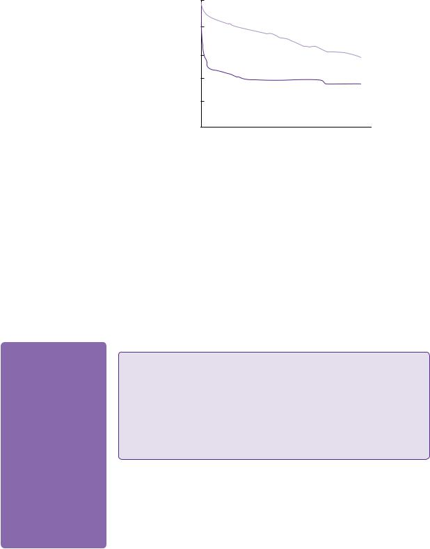
Primary ICH makes up 10–20% of all strokes, but has a far higher mortality rate, up to 40–45% in the first month compared with ischaemic stroke (Figure 20.5) [17].
194 |
Challenging concepts in neurosurgery |
|
100 |
Ischaemic stroke
surviving% |
50 |
|
PICH
0 0 |
|
|
|
|
|
|
|
|
|
|
|
|
|
|
|
|
|
1 |
2 |
3 |
4 |
|||||
|
|
|
Years |
|
|
|
|
|
Figure 20.5 Kaplan–Meier plot showing long-term survival following first-ever stroke due to PICH or cerebral infarction.
Source: Dennis MS, Burn JPS, Sandercock PAG, BamfordJM, Wade DT, Warlow CP: Long-term survival after first-ever stroke: The Oxfordshire Community Stroke Project. Stroke 1993;24:796–800.
 Expert comment
A number of factors have been identified that predict outcome. The functional outcome risk stratification scale (FUNC) score brings together the most important predictors (ICH volume, age, ICH location, GCS, pre-ICH cognitive impairment) to give a likelihood of functional independence 90 days after PICH [19]. Some studies have shown absolute values for
clot size predictive of death or survival, for instance, all patients in a cohort study with haematomas greater than 85cm3died with or without
surgery, whereas all patients with haematoma less than 26cm3 survived without surgery [24].
At 1 year, only around 25% are independent in activities of daily living with 10–15% alive, but dependent. The new-onset seizure rate is 4.6–8.2% [18].
Common sites for ICH include the deep structures of the periventricular white matter, caudate nucleus, globus pallidus, putamen, internal capsule, and thalamus (50%), in the gray matter or subcortical white matter (lobar (35%), cerebellum (10%), and brainstem (6%) [20]). On a histological level it has been shown that degenerative changes within the cerebral blood vessels, often attributable to chronic hypertension, are responsible for leakage of blood into the brain parenchyma. This is mainly seen at or near the bifurcation of small arteries, 50–700μm in diameter [21]. Charcot and Bouchard described micro-aneurysms, sites of fibrinoid necrosis of the subendothelium, which have been implicated in underlying PICH. It has been difficult to establish conclusively, however, whether these lesions are the source of hypertensive ICH.
 Learning point Cerebral amyloid angiopathy
Lobar or subcortical haemorrhages, especially in the elderly, are often caused by amyloid angiopathy. This results from the deposition of beta-amyloid in the media and adventitia of small cerebral arteries and capillaries. There is loss of smooth muscle cells, vessel wall thickening, luminal narrowing, concentric splitting of the vessel wall, and micro-aneurysm formation. The result is cortical and subcortical micro-infarctions and microhaemorrhages, some visible on DWI and gradient echo sequence MRI. Although individually clinically silent, their gradual accumulation over some years results in significant cognitive impairment. There is also some overlap with deposition of betaamyloid in the parenchyma (Alzheimer’s disease). Both are underpinned by abnormalities in the genes responsible for apolipoprotein E production [22].
There is evidence that many haematomas expand slowly over the first few hours after the initial bleed. Some sequential imaging studies have shown up to 73% demonstrate some enlargement of haematoma in the first 24 hours, the majority of this occurring within the first 4 hours [23]. This corresponds with the typical clinical presentation with a progression of neurological deficit in the early period with a minority initially presenting with dense deficits. Haematoma expansion and the development of intraventricular haemorrhage (IVH) are independent risk factors for poor outcome [24].
Case 20 Spontaneous intracerebral haemorrhage |
195 |
0–60 min
Neuronal and glial Haematoma 
 mechanical disruption,
ogligaemia or ischaemia
Glutamate release
0–4 h
|
Neuronal and glial |
|
|
|
|
|
Calcium influx, |
|
|
|
|
|
|
Sodium accumulation, |
|
|
|
|
||||||||||
|
mechanical stretch |
|
|
|
|
mitochondrial failure |
|
|
|
|
|
cytotoxic oedema, necrosis |
|
|
|
|
||||||||||||
|
|
|
|
|
|
|
|
|
|
|
|
|
|
|
|
|
|
|
|
|
|
|
|
|
|
|
|
|
|
|
|
|
|
|
|
|
|
|
|
|
|
|
|
|
|
|
|
|
|
|
|
|
|
|
|
|
|
|
4 h to 7 days |
|
|
|
|
|
|
|
|
|
|
|
|
|
|
|
|
|
|
|
|
|
|
|
|
|||
|
|
|
|
|
|
|
|
|
|
|
|
|
|
Oxygen free |
|
|
|
|
|
|
|
|
|
|
|
Increased BBB |
|
|
|
|
|
|
|
|
|
|
|
|
|
|
|
|
radicals |
|
|
|
|
|
|
|
|
|
permeability, |
|
|||
|
|
|
|
|
|
|
|
|
|
|
|
|
|
|
|
|
|
|
|
|
|
|
|
|
|
|||
|
|
|
|
|
|
|
|
|
|
|
|
|
|
|
|
|
|
|
|
|
|
AQ-4 expression in |
|
|
|
vasogenic |
|
|
|
|
|
|
|
|
|
|
|
|
|
|
|
|
MMP |
|
|
|
|
|
|
astrocytes, breakdown |
|
|
|
oedema |
|
||
|
Thrombin, ferrous |
|
|
|
|
|
|
|
|
|
|
|
|
|
|
|
|
|
of connective tissue |
|
|
|
|
|
||||
|
iron, haemin, |
|
|
Microglial |
|
|
|
|
|
|
|
|
|
|
|
|
|
in BBB, expression of |
|
|
|
|
|
|||||
|
halotransferrin |
|
|
|
activation |
|
|
|
|
|
Complement |
|
|
|
|
|
|
adhesion molecules |
|
|
|
Recruitment of |
|
|||||
|
release |
|
|
|
|
|
|
|
|
|
|
factors |
|
|
|
|
|
|
|
|
|
|
|
PMNs and |
|
|||
|
|
|
|
|
|
|
|
|
|
|
|
|
|
|
|
TFNK- |
|
|
macrophages |
|
||||||||
|
|
|
|
|
|
|
|
|
|
|
|
|
|
|
|
|
|
|
|
|
|
|
||||||
|
|
|
|
|
|
|
|
|
|
|
|
|
|
|
|
|
|
|
|
|
|
|
|
|
|
|
|
|
|
|
|
|
|
|
|
|
|
|
|
|
|
|
TNF |
|
|
|
|
Caspase activation, |
|
|
|
|
|
||||
|
|
|
|
|
|
|
|
|
|
|
|
|
|
interleukin 1 |
|
|
|
|
apoptosis in neurons |
|
|
|
|
|
||||
|
|
|
|
|
|
|
|
|
|
|
|
|
|
|
|
|
|
|
|
|
|
|
|
|||||
|
|
|
|
|
|
|
|
|
|
|
|
|
|
|
|
|
|
|
|
|
|
and glia |
|
|
|
|
|
|
|
|
|
|
|
|
|
|
|
|
|
|
|
|
Cytochrome C |
|
|
|
|
|
|
|
|
|
|||||
|
|
|
|
|
|
|
|
|
|
|
|
|
|
|
|
|
|
|
||||||||||
|
|
|
|
|
|
|
|
|
|
|
|
|
|
|
|
|
|
|
|
|
|
|
|
|
|
|
|
|
Figure 20.6 Sequence of neural damage initiated by intracerebral haemorrhage. Note that in the initial 4 hours injury is caused by the mass effect and subsequent damage by release of breakdown products.
BBB = blood–brain barrier; MMP = matrix metallopeptidase; TNF = tumour necrosis factor; PMN = polymorphonuclear cells [1]
Qureshi AI, Mendelow AD, Hanley DF. Intracerebral haemorrhage. Lancet 2009; 373(9675): 1632–44.
Following haemorrhage, subsequent secondary damage may occur through a cascade of pathological processes, including cytotoxicity of blood products, hypermetabolism, excitotoxicity, spreading depression, oxidative stress, and inflammation (Figure 20.6) [25]. Resulting peri-haematoma oedema peaks around 5–6 days and lasts up to 14 days.
The optimal medical management of these patients requires co-ordinated input from prehospital care practitioners, general practitioners, A&E staff, stroke physicians, neurosurgeons, and geriatricians. Specialist stroke unit care [26], as well as neurosciences intensive care unit [27,28] has been shown to improve outcome.
Blood pressure management is a controversial, but important issue to address early on in the management of these often medically complex patients (see Evidence base: blood pressure control after primary intracranial haemorrhage).
 Evidence base Blood pressure control after primary intracranial haemorrhage
Seventy-five percent of patients admitted with ICH have systolic blood pressure over 140mmHg, and 20% present with systolic blood pressure over 180mmHg [29].
Although instinctively logical, a causal link has not been firmly established between ICH and hypertension. Blood pressure may be affected by parenchymal haemorrhage itself through activation of neuroendocrine systems and by alterations in ICP. Some studies report an inconsistent
(continued)
196 |
Challenging concepts in neurosurgery |
|
|
|
|
relationship between blood pressure and haematoma expansion. There are also theoretical arguments about the balance between permissive elevation of blood pressure to maintain viability of the peri-haematoma penumbra versus lowering the blood pressure to reduce the risk of haematoma expansion.
Two recent trials have sought to clarify some of these questions. The Intensive Blood Pressure Reduction in Acute Cerebral Haemorrhage Trial (INTERACT) [30] randomized 404 Chinese patients between a target systolic blood pressure of 140 and 180mmHg. There was a weak association with reduced haemorrhagic expansion, however, secondary clinical outcomes were statistically unchanged. The Antihypertensive Treatment of Acute Cerebral Haemorrhage (ATACH) [31] had three cohorts of patients with a target blood pressure of 170–200, 140–170, or 110–140. The study did not show any difference in haematoma expansion, peri-haematoma oedema, and 3-month
outcome between the groups, but with only sixty patients, the study was somewhat underpowered. Follow-up studies are under way and may advance our understanding. American and European guidelines based on Class 3 (IIb) evidence are shown (Table 20.2), but have been shown to be poorly adhered to [32].
Table 20.2 Options for urgent warfarin reversal [37] |
|
||
|
|
|
|
Agent |
Pros |
Cons |
Usefulness |
|
|
|
for urgent |
|
|
|
reversal |
Vitamin K1 |
Widely available; inexpensive; |
|
directly reverses warfarin effect; |
|
small volume infused; low |
|
infective and thrombotic risk |
Fresh-frozen |
Widely available; contains |
plasma |
all coagulation factors; low |
|
thrombotic risk |
Prothrombin |
Rapid onset; small volume |
complex |
infused; low infective risk |
concentrate |
|
Recombinant |
Rapid onset; small volume |
activated |
infused; thrombin burst; low |
factor VII |
infective risk |
TRALI: transfusion-related acute lung injury.
The most recent ASA/AHA guidelines [33] state:
Slow onset of action; possible |
Poor |
allergy |
|
Large volumes usually needed; |
Fair |
requires cross-matching and |
|
thawing; slow onset of action; |
|
not negligible infective risk, |
|
possible TRALI |
|
Expensive; variable factor |
Good |
concentrations in different |
|
preparations; no negligible |
|
thrombotic risk |
|
Very expensive; acts directly |
Good |
on only a single factor; INR |
|
correction may be ‘lab artefact’; |
|
off label use |
|
|
|
Until ongoing clinical trials of blood pressure intervention for ICH are completed, physicians must manage blood pressure on the basis of incomplete evidence.
●If SBP is >200mmHg or MAP is >150mmHg, then consider aggressive reduction of blood pressure with continuous intravenous infusion, with frequent blood pressure monitoring every 5 minutes.
●If SBP is >180mmHg or MAP is >130mmHg, and there is the possibility of elevated ICP, then
consider monitoring ICP and reducing blood pressure using intermittent or continuous iv medications, while maintaining a cerebral perfusion pressure ≥60mmHg.
●If SBP is >180mmHg or MAP is >130mmHg, and there is no evidence of elevated ICP, then consider a modest reduction of blood pressure (e.g. MAP of 110mmHg or target blood pressure of 160/90mmHg), using intermittent or continuous iv medications to control blood pressure, and clinically re-examine the patient every 15 minutes.
(continued)

Case 20 Spontaneous intracerebral haemorrhage |
197 |
|
|
|
|
According to the European Guidelines [34], routine blood pressure lowering is not recommended. Treatment is recommended if blood pressure is elevated above the following levels, confirmed by repeat measurements (class IV evidence):
●Patients with a known history of hypertension or signs (ECG, retina) of chronic hypertension: systolic blood pressure >180mmHg and/or diastolic blood pressure >105mmHg. If treated, target blood pressure should be 179/100mmHg (or a MAP of 125mmHg).
●Patients without known hypertension: systolic blood pressure >160mmHg and/or diastolic blood pressure > 95mmHg. If treated, target blood pressure should be 150/90 mmHg (or a MAP of 110mmHg).
●A reduction of MAP by > 20% should be avoided.
●These limits and targets should be adapted to higher values in patients undergoing monitoring if increased ICP, to guarantee a sufficient CPP>70mmHg.
●Recommended drugs for blood pressure treatment: iv labetalol or urapidil, iv sodium nitroprusside or nitroglycerine, and captopril (per os). Avoid oral nifedipine and any drastic blood pressure decrease.
Non-contrasted brain CT is the first line imaging modality. Gradient echo MRI can be used to detect hyperacute haemorrhage and microhaemorrhages. Ancillary studies, e.g. angiography may be used when aneurysmal/AVM haemorrhage is suspected, or delayed MRI to look for underlying cavernomas or tumours.
Activated recombinant factor VII (fVIIa) has been investigated in its capacity to limit haematoma expansion. A phase II trial showed significantly reduced haematoma volume (from 29 to 11–16%) and 90-day mortality (29% compared with 18%) [35]. A phase III trial (the FAST trial) [36] had rather less encouraging results, however. Although the effect on limiting haematoma volume was confirmed, at 3 months the percentage who were dead or disabled in the placebo group was 24%, compared with 26% and 29% in the 20μg/kg and 80μg/kg groups, respectively. In patients who develop ICH on oral anticoagulation fVIIa is an option, although an expensive one. Rapid reversal of anticoagulants not only prevents ongoing bleeding, but also allows the possibility of surgical intervention. Normalization of INR within 2 hours from hospital admission is associated with low rates of haematoma enlargement and is achieved in the majority of patients (84%) treated with prothrombin complex concentrates, while fresh frozen plasma (FFP) infusions show only a partial effect in reducing haematoma enlargement (39%) and vitamin K1 has no effects [37]. Vitamin K, however, must be given to avoid a rebound in coagulopathy.
Surgical intervention
The use of surgery for ICH is highly variable across the world with reports of 50% in German and Japanese literature, whereas other countries report 2–20% [38].
The concept of ischaemic penumbra has been used as an argument for early surgical intervention. DWI, perfusion-weighted imaging (PWI), and positron emission tomography (PET) studies do not consistently demonstrate the existence of ischaemic change in the peri-haematoma region, however [40]. Nonetheless, damage may be occurring through mitochondrial dysfunctional, leading to impaired oxidative metabolism. Raised glutamate, lactate, glycerol, and lactate/pyruvate ratio point to a biochemical, rather than ischaemic crisis. These metabolic changes are seen to reverse 24–48 hours following clot evacuation [41].
 Expert comment
The rationale for surgical evacuation of ICH has a sound theoretical basis. It directly addresses the damaging mass effect, prevents release of inflammatory blood breakdown products, and may improve perfusion of the vulnerable penumbra. A wide variety
of surgical strategies are available—open craniotomy, frameless stereotactic aspiration, or endoscopic evacuation with or without thrombolysis [39].
198 |
Challenging concepts in neurosurgery |
 Evidence base Medical versus surgical treatment of intracranial haemorrhage
The investigation of the relative merits of medical versus surgical treatment of ICH has a long and illustrious history. Wylie McKissock published the first prospective randomized trial in neurosurgery on the subject in 1961 [49]. After randomizing 180 patients diagnosed by catheter angiogram
and air ventriculography, surgical intervention produced no obvious advantage. The conclusion of this landmark paper was: ‘We have clearly made no contribution to the treatment of primary intracerebral haemorrhage by surgery.’ Most subsequent studies have supported this conclusion. The largest trial to date is the International Surgical Trial in Intracerebral Haemorrhage (STICH) led by Mendelow from Newcastle, UK and published in 2005 [50] in the Lancet. In summary, 1033 patients from twenty-seven countries were randomized either to early surgery (within 24 hours of randomization) or medical treatment. The results showed that 26% of surgically-treated patients
had a favourable outcome compared with 24% treated medically. However, this was not statistically significant and the overall conclusion was that there was ‘no benefit from early surgery compared with initial conservative treatment’. Indeed, for some groups, especially those patients presenting in a coma (where risk was increased by 8%), surgery seemed to be harmful. When one examines the Forest plots of the twelve meaningful trials (including STICH) there may, however, be a suggestion of overall benefit for surgery (odds ratio of 0.85, CI 0.71, 1.03), when the unfavourable outcome was death (Figure 20.7).
Review: |
Surgery in intracerebral haemorrhage |
|
|
|
|
|
|
|
|
|
|
|
|
|
|
|
|
|
|
|
|
|
|
|
|
|
|
|
|
|
|
|
|
|
|
|
|
|
|
|
|
|
|
|
|
Comperison: |
01 surgery v control |
|
|
|
|
|
|
|
|
|
|
|
|
|
|
|
|
|
|
|
|
|
|
|
|
|
|
|
|
|
|
|
|
|
|
|
|
|
|
|
|
|
|
|
|
Outcome: |
02 death |
|
|
|
|
|
|
|
|
|
|
|
|
|
|
|
|
|
|
|
|
|
|
|
|
|
|
|
|
|
|
|
|
|
|
|
|
|
|
|
|
|
|
|
|
Study |
|
Treatment |
Control |
|
|
|
|
|
|
|
|
|
|
|
Peto OR |
|
|
|
|
|
|
|
|
|
|
|
Peto OR |
||||||||||||||||||
or sub-category |
n/N |
n/N |
|
|
|
|
|
|
|
|
|
|
|
95% CI |
|
|
|
|
|
|
|
|
|
|
|
95% CI |
|||||||||||||||||||
|
|
|
|
|
|
|
|
|
|
|
|
|
|
|
|
|
|
|
|
|
|
|
|
|
|
|
|
|
|
|
|
|
|
|
|
|
|
|
|
|
|
|
|
|
|
McKissock (1961) |
58/89 |
46/91 |
|
|
|
|
|
|
|
|
|
|
|
|
|
|
|
|
|
|
|
|
|
|
|
|
|
|
|
|
|
|
|
|
|
|
|
|
|
|
|
1.81 (1.01, 3.27) |
|||
|
|
|
|
|
|
|
|
|
|
|
|
|
|
|
|
|
|
|
|
|
|
|
|
|
|
|
|
|
|
|
|
|
|
|
|
|
|
|
|||||||
Auer (1989) |
|
21/50 |
35/50 |
|
|
|
|
|
|
|
|
|
|
|
|
|
|
|
|
|
|
|
|
|
|
|
|
|
|
|
|
|
|
|
|
|
|
|
|
|
|
|
0.32 (0.15, 0.71) |
||
|
|
|
|
|
|
|
|
|
|
|
|
|
|
|
|
|
|
|
|
|
|
|
|
|
|
|
|
|
|
|
|
|
|
|
|
|
|
|
|
||||||
Juvela (1989) |
|
12/26 |
10/26 |
|
|
|
|
|
|
|
|
|
|
|
|
|
|
|
|
|
|
|
|
|
|
|
|
|
|
|
|
|
|
|
|
|
|
|
|
|
|
|
1.36 (0.46, 4.05) |
||
|
|
|
|
|
|
|
|
|
|
|
|
|
|
|
|
|
|
|
|
|
|
|
|
|
|
|
|
|
|
|
|
|
|
|
|
|
|
|
|
||||||
Batjer (1990) |
|
4/8 |
11/13 |
|
|
|
|
|
|
|
|
|
|
|
|
|
|
|
|
|
|
|
|
|
|
|
|
|
|
|
|
|
|
|
|
|
|
|
|
|
|
|
0.20 (0.03, 1.33) |
||
|
|
|
|
|
|
|
|
|
|
|
|
|
|
|
|
|
|
|
|
|
|
|
|
|
|
|
|
|
|
|
|
|
|
|
|
|
|
|
|
||||||
Chen (1992) |
|
15/64 |
11/63 |
|
|
|
|
|
|
|
|
|
|
|
|
|
|
|
|
|
|
|
|
|
|
|
|
|
|
|
|
|
|
|
|
|
|
|
|
|
|
|
1.44 (0.61, 3.40) |
||
|
|
|
|
|
|
|
|
|
|
|
|
|
|
|
|
|
|
|
|
|
|
|
|
|
|
|
|
|
|
|
|
|
|
|
|
|
|
|
|
||||||
Morgenstern (1998) |
3/17 |
4/17 |
|
|
|
|
|
|
|
|
|
|
|
|
|
|
|
|
|
|
|
|
|
|
|
|
|
|
|
|
|
|
|
|
|
|
|
|
|
|
|
0.71 (0.14, 3.63) |
|||
|
|
|
|
|
|
|
|
|
|
|
|
|
|
|
|
|
|
|
|
|
|
|
|
|
|
|
|
|
|
|
|
|
|
|
|
|
|
|
|||||||
Zuccarello (1999) |
2/9 |
3/11 |
|
|
|
|
|
|
|
|
|
|
|
|
|
|
|
|
|
|
|
|
|
|
|
|
|
|
|
|
|
|
|
|
|
|
|
|
|
|
|
0.77 (0.11, 5.62) |
|||
|
|
|
|
|
|
|
|
|
|
|
|
|
|
|
|
|
|
|
|
|
|
|
|
|
|
|
|
|
|
|
|
|
|
|
|
|
|
|
|||||||
Cheng 2001 |
|
26/266 |
34/234 |
|
|
|
|
|
|
|
|
|
|
|
|
|
|
|
|
|
|
|
|
|
|
|
|
|
|
|
|
|
|
|
|
|
|
|
|
|
|
|
0.64 (0.37, 1.09) |
||
|
|
|
|
|
|
|
|
|
|
|
|
|
|
|
|
|
|
|
|
|
|
|
|
|
|
|
|
|
|
|
|
|
|
|
|
|
|
|
|
||||||
Teernstra (2001) |
20/36 |
20/34 |
|
|
|
|
|
|
|
|
|
|
|
|
|
|
|
|
|
|
|
|
|
|
|
|
|
|
|
|
|
|
|
|
|
|
|
|
|
|
|
0.88 (0.34, 2.25) |
|||
|
|
|
|
|
|
|
|
|
|
|
|
|
|
|
|
|
|
|
|
|
|
|
|
|
|
|
|
|
|
|
|
|
|
|
|
|
|
|
|||||||
Hosseini 2003 |
|
3/20 |
9/17 |
|
|
|
|
|
|
|
|
|
|
|
|
|
|
|
|
|
|
|
|
|
|
|
|
|
|
|
|
|
|
|
|
|
|
|
|
|
|
|
0.19 (0.05, 0.72) |
||
|
|
|
|
|
|
|
|
|
|
|
|
|
|
|
|
|
|
|
|
|
|
|
|
|
|
|
|
|
|
|
|
|
|
|
|
|
|
|
|
||||||
Hattori (2004) |
|
9/121 |
20/121 |
|
|
|
|
|
|
|
|
|
|
|
|
|
|
|
|
|
|
|
|
|
|
|
|
|
|
|
|
|
|
|
|
|
|
|
|
|
|
|
0.42 (0.20, 0.92) |
||
|
|
|
|
|
|
|
|
|
|
|
|
|
|
|
|
|
|
|
|
|
|
|
|
|
|
|
|
|
|
|
|
|
|
|
|
|
|
|
|
||||||
Mendelow (2005) |
173/477 |
189/505 |
|
|
|
|
|
|
|
|
|
|
|
|
|
|
|
|
|
|
|
|
|
|
|
|
|
|
|
|
|
|
|
|
|
|
|
|
|
|
|
0.95 (0.73, 1.23) |
|||
|
|
|
|
|
|
|
|
|
|
|
|
|
|
|
|
|
|
|
|
|
|
|
|
|
|
|
|
|
|
|
|
|
|
|
|
|
|
|
|||||||
Total (95% CI) |
|
1183 |
1182 |
|
|
|
|
|
|
|
|
|
|
|
|
|
|
|
|
|
|
|
|
|
|
|
|
|
|
|
|
|
|
|
|
|
|
|
|
|
|
|
0.85 (0.71, 1.02) |
||
|
|
|
|
|
|
|
|
|
|
|
|
|
|
|
|
|
|
|
|
|
|
|
|
|
|
|
|
|
|
|
|
|
|
|
|
|
|
|
|
||||||
Total events: 346 (treatment), 392 (control) |
|
|
|
|
|
|
|
|
|
|
|
|
|
|
|
|
|
|
|
|
|
|
|
|
|
|
|
|
|
|
|
|
|
|
|
|
|
|
|
|
|
|
|
||
Test for heterogeneity: Chi2 = 26.29, df = 11 (p = 0.006), I2 = 58.2% |
|
|
|
|
|
|
|
|
|
|
|
|
|
|
|
|
|
|
|
|
|
|
|
|
|
|
|
|
|
|
|
|
|
|
|
|
|
|
|||||||
Test for overall effect: z = 1.73 (p = 0.08) |
|
|
|
|
|
|
|
|
|
|
|
|
|
|
|
|
|
|
|
|
|
|
|
|
|
|
|
|
|
|
|
|
|
|
|
|
|
|
|
|
|
|
|
||
|
|
|
|
|
|
|
|
|
|
|
|
|
|
|
|
|
|
|
|
|
|
|
|
|
|
|
|
|
|
|
|
|
|
|
|
|
|
|
|
|
|
|
|
|
|
|
|
|
0.1 |
|
|
0.2 |
0.5 |
|
|
|
|
|
1 |
|
|
|
|
|
|
2 |
|
5 |
10 |
||||||||||||||||||||||
|
|
|
Favours treatment |
|
|
|
|
|
|
|
|
|
Favours control |
||||||||||||||||||||||||||||||||
Figure 20.7 Forrest plots of twelve ICH trials [1].
Source: Mendelow AD, Gregson BA, Mitchell PM, Murray GD, Rowan EN, Gholkar AR. Surgical trial in lobar intracerebral haemorrhage (STICH II) protocol. Trials 2011:12:124. Distributed under the terms of the Creative Commons Attribution License 2.0
When subgroups are analysed, it appears that there is quite a difference in outcome in patients with IVH (42%) (favourable outcome of only 15% compared with 31% in those without IVH). A metaanalysis of purely lobar haemorrhages seems to favour surgical evacuation (Figure 20.8). Given the results of this post hoc analysis, the STICH collaborators have pointed out that the overall conclusion should be that, for certain patients with ICH, surgery can be justified. A further ongoing study (STICH II) is looking prospectively at the subgroups of patients most likely to benefit from surgery. The following lists are the criteria for this to date unpublished study (http://research.ncl.ac.uk/stich):
Inclusion criteria
●Evidence of a spontaneous lobar ICH on CT scan (1cm or less from the cortex surface of the brain).
●Patient within 48 hours of ictus.
(continued)

|
|
|
Case 20 Spontaneous intracerebral haemorrhage |
199 |
|||||||||||||||||||
|
|
|
|
|
|
|
|
|
|
|
|
|
|
|
|
|
|
|
|
|
|
|
|
|
|
|
|
|
|
|
|
|
|
|
|
|
|
|
|
|
|
|
|
|
|
|
|
|
|
Surgery |
Control |
|
|
|
|
|
Peto OR |
|
|
|
|
Peto OR |
|
||||||||
|
|
n/N |
n/N |
|
|
|
|
|
|
95% CI |
|
|
|
|
95% CI |
|
|||||||
|
|
|
|
|
|
|
|
|
|
|
|
|
|
|
|
|
|
|
|
|
|
|
|
|
Auer (1989) |
11/24 |
15/21 |
|
|
|
|
|
|
|
|
|
|
|
|
|
|
|
|
|
0.36 (0.11–1.16) |
|
|
|
|
|
|
|
|
|
|
|
|
|
|
|
|
|
|
|
|
|
|||||
|
Teernstra (2001) |
12/16 |
7/9 |
|
|
|
|
|
|
|
|
|
|
|
|
|
|
|
|
|
0.86 (0.13–5.63) |
|
|
|
|
|
|
|
|
|
|
|
|
|
|
|
|
|
|
|
|
|
|||||
|
Mendelow (2004) |
56/110 |
71/113 |
|
|
|
|
|
|
|
|
|
|
|
|
|
|
|
|
|
0.62 (0.36–1.05) |
|
|
|
|
|
|
|
|
|
|
|
|
|
|
|
|
|
|
|
|
|
|||||
|
Total (95% CI) |
150 |
143 |
|
|
|
|
|
|
|
|
|
|
|
|
|
|
|
|
|
0.58 (0.36–0.92) |
|
|
|
Total events: 79 (surgery), 93 (control) |
|
|
|
|
|
|
|
|
|
|
|
|
|
|
|
|
|
|
|
|
|
|
|
Test for heterogeneity: 2 = 0.87, df = 2 (p = 0.65), I2 = 0% |
|
|
|
|
|
|
|
|
|
|
|
|
|
|
|
|
|
|
|
|||
|
Test for overall effect: z = 2.30 (p = 0.02) |
|
|
|
|
|
|
|
|
|
|
|
|
|
|
|
|
|
|
|
|
|
|
|
|
|
|
|
|
|
|
|
|
|
|
|
|
|
|
|
|
|
|
|
|
|
|
|
|
|
|
|
|
|
|
|
|
|
|
|
|
|
|
|
|
|
|
|
|
|
|
|
|
|
0.1 |
0.2 |
0.5 |
|
|
1 |
|
2 |
5 |
10 |
|
|
|||||||||
|
|
|
Favours treatment |
|
|
|
Favours control |
|
|||||||||||||||
Figure 20.8 Forrest plot of lobar haematomas without IVH [1].
Source: Mendelow AD, Gregson BA, Mitchell PM, Murray GD, Rowan EN, Gholkar AR. Surgical trial in lobar intracerebral haemorrhage (STICH II) protocol. Trials 2011:12:124. Distributed under the terms of the Creative Commons Attribution License 2.0
●Best motor score on the GCS of 5 or 6, and best eye opening score on the GCS of 2 or more.
●Volume of haematoma between 10 and 100mL (calculated using (a × b × c)/2 method).
Exclusion criteria
●Clear evidence that the haemorrhage is due to an aneurysm or angiographically-proven arteriovenous malformation.
●IVH of any sort.
●ICH secondary to tumour or trauma.
●Basal ganglia, thalamic, cerebellar, or brainstem haemorrhage or extension of a lobar haemorrhage into any of these regions.
●Severe pre-existing physical or mental disability, or severe co-morbidity that might interfere with assessment of outcome.
●If surgery cannot be performed within 12 hours.
●If the haematological effects of any previous anticoagulants are not completely reversed.
STICH II aims to recruit 600 patients by May 2012, efforts are ongoing. There are other important trials ongoing in the subject. Clot lysis evaluating accelerated resolution on intraventricular haemorrhage (CLEAR III) is a phase III clinical trial examining how recombinant tissue plasminogen activator (rtPA), placed through EVD changes, modified Rankin score at 12 months compared with EVD alone. minimally invasive surgery plus rtPA for intracerebral haemorrhage evacuation (MISTIE) also looks at tPA, this time its effect after being placed within the clot itself. Currently (February 2012), CLEAR III is still ongoing and MISTIE has finished recruiting, but is waiting for follow-up data.
Surgical methods of intracranial haemorrhage treatment
Traditional craniotomy provides a relatively large access to the haematoma with direct visibility of the cavity wall. This allows bipolar cautery of the haematoma bed, placement of haemostatic agents, and visualization of any underlying abnormalities such as AVMs, tumours, etc. It is, of course, an invasive method with increased risk of causing damage to the cortex and white matter tracts, whilst accessing the clot. This is obviously most significant in deep-seated basal ganglia and thalamic bleeds. Neuronavigation may be usefully applied to plan the safest trajectory. Intra-operative ultrasound has also proven to be a simple and effective localization technique [42].
Less invasive methods have been applied. The simplest of these is aspiration via a burr hole, sometimes undertaken under local anaesthesia. Stereotactic aspiration in 175 patients achieved at least 50% clot removal in 75%. By contrast, 7.4 % had

200 Challenging concepts in neurosurgery
post-operative bleeding [43]. Fifty-two percent achieved independent outcomes at 6 months. This method may also allow the delivery of fibrinolytics to the haematoma. Catheter systems may be left in situ to allow infusion of rtPA or urokinase following aspiration of the clot. A Japanese study found only 58% of patients had evacuation of 50% of haematoma by initial aspiration, but after the urokinase infusion, the haematoma was 80% removed in more than 70% of patients. The mortality rate was 3% [44]. It should be noted, however, that no correlation between degree of clot removal and neurological outcome has been proven [45].
The endoscope has also been applied to ICH, providing a view of the haematoma cavity, so that clot can be identified and removed, allowing greater total clot removal and direct coagulation of visualized bleeding points. This is reflected in the clinical results with 96% removal in the putaminal group, 86% in the thalamic group, and 98% in the subcortical group [46].
Other mechanical devices have been described to aid haematoma evacuation, such as ‘Archimedes screw’ type devices, ultrasonic aspirators, and oscillating cutters.
Hemicraniectomy (without primary clot evacuation) has also been used as a treatment method. A case series of twelve patients with evacuation and hemicraniectomy had a survival rate of 92% and good functional outcome in 55% [47].
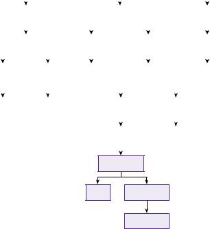
 Learning box Cerebellar haemorrhage [48]
Cerebellar haemorrhage accounts for 5–13% of spontaneous ICH and has a high mortality rate (20–75%). It can be an uncommon complication of supratentorial surgery. Kirollos [48] published one of the few prospective studies in cerebellar haemorrhage. The configuration of the fourth ventricle was used as a measure of mass effect and a guide to treatment:
● Grade I: fourth ventricle was not effaced or displaced from the midline. ● Grade II: partial effacement or shift to one side.
● Grade III: total effacement or brainstem distortion. The following treatment protocol was used.
Results (good outcome)
Grade III with GCS<8 (0%), Grade III with GCS>8 (38%), Grade II with GCS<8 (57%), Grade II with
GCS>8 (58%), Grade I (100%).
The importance of urgent surgical treatment of the Grade III patients before conscious level deteriorates (when outcome is universally poor) was emphasized.
A protocol for the management of infratentorial is shown in Figure 20.9 .
A final word from the expert
For most patients with PICH, surgery is not the best option, and attention to the best medical treatment is vital, with management in a dedicated stroke unit likely to confer an advantage. Where there is uncertainty, or clinical equipoise, it is most helpful to enter the patient into a prospective randomized trial, such as STICH II. This will elucidate whether the subgroup of patients who made a better recovery after surgery, such as lobar haemorrhage, did so because of surgery or from confounding factors.
|
|
|
|
|
|
|
|
|
|
|
|
|
|
Case 20 Spontaneous intracerebral haemorrhage |
201 |
|||||||||||||||
|
|
|
|
|
|
|
|
|
|
|
|
|
|
|
|
|
|
|
|
|
|
|
|
|
|
|||||
|
|
|
|
|
|
|
|
|
|
|
|
IVth ventricular grade |
|
|
|
|
|
|
|
|
|
|||||||||
|
|
|
|
|
|
|
|
|
|
|
|
|
|
|
|
|
|
|
|
|
|
|
|
|
|
|
|
|
|
|
|
|
|
|
|
|
|
|
|
|
|
|
|
|
|
|
|
|
|
|
|
|
|
|
|
|
|
|
|
|
|
|
|
|
|
|
|
|
|
|
|
|
|
|
|
|
|
|
|
|
|
|
|
|
|
|
|
|
|
|
|
|
|
|
|
|
|
|
I |
|
|
|
|
|
|
|
|
II |
|
|
|
|
|
|
|
|
III |
|
|
|
|||
|
|
|
|
|
|
|
|
|
|
|
|
|
|
|
|
|
|
|
|
|
|
|
|
|
|
|||||
|
|
|
|
|
|
|
|
|
|
|
|
|
|
|
|
|
|
|
|
|||||||||||
|
|
|
|
|
|
|
|
|
|
|
|
|
|
|
|
|
|
|
|
|
|
|
|
|
|
|
|
|
|
|
|
|
|
|
|
|
|
|
|
|
|
|
|
|
|
|
|
|
|
|
|
|
|
|
|
|
|
|
|
|
|
|
|
|
GCS (<13) |
|
|
GCS >13 |
|
|
|
|
GCS |
|
Any GCS |
|
|
|||||||||||||||
|
|
|
|
|
|
|
|
|
|
|
|
|
|
|
|
|
|
|
|
|
|
|
|
|
|
|
|
|
|
|
|
|
|
|
|
|
|
|
|
|
|
|
|
|
|
|
|
|
|
|
|
|
|
|
|
|
|
|
|
|
|
|
|
|
|
|
|
|
|
|
|
|
|
|
|
|
|
|
|
|
|
|
|
|
|
|
|
|
|
|
|
|
|
|
|
|
|
|
|
|
|
|
|
|
|
|
|
|
|
|
|
|
|
|
|
|
|
|
|
|
|
|
|
|
No |
|
|
|
|
Yes |
|
Conservative |
|
|
Hydrocephalus |
|
|
Evacuate clot |
|
|
||||||||||||||
|
|
|
|
|
|
|
|
|
+ CSF-D |
|
|
|||||||||||||||||||
|
|
|
|
|
|
|
|
|
|
|
|
|
|
|
|
|
|
|
|
|
|
|
|
|
|
|
||||
|
|
|
|
|
|
|
|
|
|
|
|
|
|
|
|
|
|
|
|
|
|
|
|
|
|
|
|
|
|
|
|
|
|
|
|
|
|
|
|
|
|
|
|
|
|
|
|
|
|
|
|
|
|
|
|
|
|
|
|
|
|
|
|
|
|
|
|
|
|
|
|
|
|
|
|
|
|
|
|
|
|
|
|
|
|
|
|
|
|
|
|
|
Conservative |
|
|
CSF-D |
|
|
|
|
|
Yes |
|
|
|
No |
|
|
|
|
|
|
|||||||||||
|
|
|
|
|
|
|
|
|
|
|
|
|
|
|
|
|
|
|
|
|
|
|
|
|
|
|
|
|
|
|
|
|
|
|
|
|
|
|
|
|
|
|
|
|
|
|
|
|
|
|
|
|
|
|
|
|
|
|
|
|
|
|
|
|
|
|
|
|
|
|
|
|
|
|
|
|
|
|
|
|
|
|
|
|
|
|
|
|
|
|
|
|
|
|
|
|
|
|
|
|
|
|
|
|
|
|
|
CSF-D |
|
|
|
Evacuate clot |
|
|
|
|
|
||||||
|
|
|
|
|
|
|
|
|
|
|
|
|
|
|
|
|
|
|
|
|
|
|
|
|
|
|
|
|
|
|
|
|
|
|
|
|
|
|
|
|
|
|
|
|
|
|
|
|
|
|
|
|
|
|
|
|
|
|
|
|
|
Improvement
Yes No
Evacuate clot
Figure 20.9 Protocol for infratentorial ICH.
Source: lvi Kirollos RW, Tyagi AK, Ross SA, van Hille PT, Marks PV. Management of spontaneous cerebellar hematomas: a prospective treatment protocol. Neurosurgery 2001:49(6):1378–1387
References
1.Newman GC. Clarification of the abc/2 rule for ICH volume. Stroke 2007; 38: 862.
2.Kothari RU, Brott T, Broderick JP, et al. The ABCs of measuring intracerebral hemorrhage volume. Stroke 1996; 27: 1304–5.
3.Brazis PW, Masdeu JC, Biller J. Gaze palsy. Localization in clinical neurology. Philadelphia: Lippincott, Williams and Wilkins, 2007.
4.Eckman MH, Rosand J, Knudsen KA, et al. Can patients be anticoagulated after intracerebral hemorrhage? A decision analysis. Stroke 2003; 34: 1710–16.
5.Bailey RD, Hart RG, Benavente O, et al. Recurrent brain hemorrhage is more frequent than ischemic stroke after intracranial hemorrhage. Neurology 2001; 56: 773–77.
6.Singh RVP, Prusmack CJ, Morcos JJ. Spontaneous intra cerebral hemorrhage: non arteriovenous malformation, non aneurysm. In HR Winn (ed.), Youmans Neurological Surgery, 5th edn (pp. 1753–5). Philadelphia, PA: Saunders, 2004.
7.van Asch CJJ, Luitse MJA, Rinkel GJE, et al. Incidence, case fatality, and functional outcome of intracerebral haemorrhage over time, according to age, sex, and ethnic origin: a systematic review and meta-analysis. Lancet Neurology 2010; 9: 167–76.
8.Meretoja A, Strbian D, Putaala J,. SMASH-U: a proposal for etiologic classification of intracerebral hemorrhage. Stroke 2012 43(10): 2592–7.
9.Gupta RK, Jamjoom AAB, Nikkar-Esfahani A, et al. Spontaneous intracerebral haemorrhage: a clinical review. British Journal of Hospital Medicine 2010; 71: 499–504.
10.Butcher K, Laidlaw J. Current intracerebral hemorrhage management. Journal of Clinical Neuroscience 2003; 10(2): 158–67.
202 |
Challenging concepts in neurosurgery |
|
|
11. |
Lovelock CE, Molyneux AJ, Rothwell PM. Change in incidence and aetiology of intracere- |
|
|
bral haemorrhage in Oxfordshire, UK, between 1981 and 2006: a population-based study. |
|
|
Lancet Neurology 2007; 6(6): 487–93. |
|
12. |
Singh RVP, Prusmack CJ, Morcos JJ. Spontaneous intra cerebral hemorrhage: non arterio- |
|
|
venous malformation, non aneurysm. In: HR Winn (ed.), Youmans Neurological Surgery, |
|
|
5th edn (pp. 1753–5). Philadelphia, PA: Saunders, 2004. |
|
13. |
Camerlingo M, Casto L, Censori B, et al. Immediate anticoagulation with heparin for |
|
|
first—ever ischaemic stroke in the carotid artery territories observed within 5 hours of |
|
|
onset. Archives of Neurology 1994; 51: 462–7. |
|
14. |
Pessin MS, Del Zoppo GJ, Estol CJ. Thrombolytic agents in the treatment of stroke. |
|
|
Clinical Neuropharmacology 1990; 13: 271–89. |
|
15. |
Connolly SJ, Ezekowitz MD, Yusuf S, et al. RE-LY steering committee and investigators |
|
|
dabigatran versus warfarin in patients with atrial fibrillation. New England Journal of |
|
|
Medicine 2009; 361(12): 1139–51. |
|
16. |
Rahme RJ, Bernstein R, Batjer HH, et al. Is it time to abandon warfarin and embrace |
|
|
oral direct thrombin inhibitors to prevent stroke in patients with atrial fibrillation? |
|
|
Neurosurgery 2011; 68(2): N16–17. |
|
17. |
Dennis MS. Outcome after brain haemorrhage. Cerebrovascular Disease 2003; |
|
|
16(Suppl. 1): 9–13. |
|
18. |
Yang TM, Lin WC, Chang WN, et al. Predictors and outcomes of seizures after spontane- |
|
|
ous intracerebral hemorrhage. Journal of Neurosurgery 2009; 111: 87–93. |
|
19. |
Rost NS, Smith EE, Chang Y, et al. Prediction of functional outcome in patients with |
|
|
primary intracerebral hemorrhage: the FUNC score. Stroke 2008; 39(8): 2304–9. |
|
20. |
Flaherty ML, Woo D, Haverbusch M, et al. Racial variations in location and risk of |
|
|
intracerebral hemorrhage. Stroke 2005: 36; 934–7. |
|
21. |
Qureshi AI, Mendelow AD, Hanley DF. Intracerebral haemorrhage. Lancet 2009; |
|
|
373(9675): 1632–44. |
|
22. |
Viswanathan A, Greenberg SM. Cerebral amyloid angiopathy in the elderly. Annals of |
|
|
Neurology 2011; 70(6): 871–80. |
|
23. |
Brott T, Broderick J, Kothari R, et al. Early hemorrhage growth in patients with intracer- |
|
|
ebral hemorrhage. Stroke 1997; 28: 1–5. |
|
24. |
Davis SM, Broderick J, Hennerici M, et al. Hematoma growth is a determinant of mortal- |
|
|
ity and poor outcome after intracerebral hemorrhage. Neurology 2006; 66: 1175–81. |
|
25. |
Aronowski J, Zhao X. Molecular pathophysiology of cerebral hemorrhage: secondary |
|
|
brain injury. Stroke 2011; 42(6): 1781–6. |
|
26. |
Candelise L, Gattinoni M, Bersano A. Stroke-unit care for acute stroke patients: an obser- |
|
|
vational follow-up study. Lancet 2007; 369(9558): 299–305. |
|
27. |
Diringer MN, Edwards DF. Admission to a neurologic/neurosurgical intensive care unit |
|
|
is associated with reduced mortality rate after intracerebral hemorrhage. Critical Care |
|
|
Medicine 2001; 29: 635–40. |
|
28. |
Mirski MA, Chang CW, Cowan R. Impact of a neuroscience intensive care unit on neu- |
|
|
rosurgical patient outcome and cost of care: evidence-based support for an intensivist- |
|
|
directed speciality ICU model of care. Journal of Neurosurgical Anesthesiology 2001; 13: |
|
|
83–92. |
|
29. |
Qureshi AJ, Ezzedine MA, Nasar A, et al. Prevalence of elevated blood pressure in |
|
|
563,704 adults with stroke presenting to the ED in the United States. American Journal of |
|
|
Emergency Medicine 2007; 25: 32–8. |
|
30. |
Anderson CS, Huang Y, Wang JG, et al. Intensive blood pressure reduction in acute cer- |
|
|
ebral haemorrhage trial (INTERACT): a randomised pilot trial. Lancet Neurology 2008; 7: |
|
|
391–9. |
|
31. |
Antihypertensive Treatment of Acute Cerebral Haemorrhage (ATACH) investigators. |
|
|
Antihypertensive treatment of acute cerebral haemorrhage. Critical Care Medicine 2010; |
|
|
38(2): 637–48. |
Case 20 Spontaneous intracerebral haemorrhage |
203 |
32.Manawadu D, Jeerakatil T, Roy A, et al. Blood pressure management in acute intraerebral haemorrhage. Guidelines are poorly implemented in clinical practice. Clinical Neurology and Neurosurgery 2010; 112: 858–64.
33.Morgenstern LB, Hemphill JC, Anderson C, et al. Guidelines for the management of spontaneous intracerebral hemorrhage: a guideline for healthcare professionals from the American Heart Association/American Stroke Association. Stroke 2010; 41: 2108–29.
34.Steiner T, Kaste M, Forsting M, et al. Recommendations for the management of intracranial haemorrhage. The European Stroke Initiative Writing Committee and the Writing Committee for the EUSI Executive Committee. Cerebrovascular Disease 2006; 22: 294–316.
35.Mayer SA, Brun NC, Begtrup K, et al. Recombinant activated factor VII for acute intracerebral hemorrhage. New England Journal of Medicine 2005; 352: 777–85.
36.Mayer SA, Davis SM, Begtrup K, et al. Subgroup analysis in the FAST trial: a subset of intracerebral hemorrhage patients that benefit from recombinant activated factor VII. Stroke 2008; 39: 528.
37.Masotti L, Di Napoli M, Godoy DA, et al. The practical management of intracerebral hemorrhage associated with oral anticoagulant therapy. International Journal of Stroke 2011; 6: 228–40.
38.Prasad K, Mendelowe AD, Gregson B. Surgery for primary supratentorial intracerebral haemorrhage. Cochrane Database System Review 2008; 8: C D000200.
39.Dubourg J, Messerer M. State of the art in managing nontraumatic intracerebral hemorrhage. Neurosurgery Focus 2011; 30(6): E22.
40.Kirkman MA. Debate—does an ischaemic penumbra exist in intracerebral haemorrhage. British Journal of Neurosurgery 2011; 25: 523–5.
41.Nilsson OG, Polito A, Saveland H, et al. Are primary supratentorial intracerebral hemorrhages surrounded by a biochemical penumbra? A microdialysis study. Neurosurgery 2006; 59(3): 521–8.
42.Lee J-K., Lee J-H. Ultrasound-guided evacuation of spontaneous intracerebal hematoma in the basal ganglia. Journal of Clinical Neuroscience 2005; 12: 553–6.
43.Niizuma H, Shimizu Y, Yonemitsu T, et al. Results of stereotactic aspiration in 175 cases of putaminal hemorrhage. Neurosurgery 1989; 24: 814–19.
44.Niizuma H, Otsuki T, Johkura H, et al. CT-guided stereotactic aspiration of intracerebral hematoma—result of a hematoma-lysis method using urokinase. Applied Neurophysiology 1985; 48: 427–30.
45.Choy DKS, Wu PH, Tan D, et al. Correlation of the long-term neurological outcomes with completeness of surgical evacuation in spontaneous supratentorial intracerebral hemorrhage: a retrospective study. Singapore Medical Journal 2010; 51(4): 320.
46.Kuo LT, Chen CM, Li CH, et al. Early endoscope-assisted hematoma evacuation in patients with supratentorial intracerebral hemorrhage: case selection, surgical technique, and long-term results. Neurosurgery Focus 2011; 30(4): E9.
47.Murthy JM, Chowdary GV, Murthy TV, et al. Decompressive craniectomy with clot evacuation in large hemispheric hypertensive intracerebral hemorrhage. Neurocritical Care 2005; 2: 258–62.
48.Kirollos RW, Tyagi AK, Ross SA, et al. Management of spontaneous cerebellar hematomas: a prospective treatment protocol. Neurosurgery 2001; 49(6): 1378–87.
49.Mckissock W, Richardson A, & Taylor J. Primary intracerebral haemorrhage: a controlled trial of surgical and conservative treatment in 180 unselected cases. The Lancet 1961; 278(7196): 221–226.
50.Mendelow AD, Gregson BA, Fernandes HM, et al. Early surgery versus initial conservative treatment in patients with spontaneous supratentorial intracerebral haematomas in the International Surgical Trial in Intracerebral Haemorrhage (STICH): a randomised trial. The Lancet 2005; 365(9457): 387–397.