


CASE
1The management of chronic subdural haematoma
Nick Borg and Angelos G. Kolias
 Expert commentary Thomas Santarius and Peter J. Hutchinson
Case history
A 78-year-old retired solicitor was admitted to the Emergency Department with a 1-week history of worsening confusion. His wife had initially noted occasional short episodes of confusion, when he would appear wandering aimlessly around the house, but for the previous two days he had been unable to hold a coherent conversation. In addition, there had been a marked deterioration in his gait, with his right foot catching the edge of a carpet and causing a number of falls over the preceding 2 weeks. Although there was no clear history of significant head trauma, his wife thought he had become progressively more unsteady with every fall.
Prior to this he had been in good physical health, and was able to walk the dogs 2 miles a day and play bowls at the village club. His medical co-morbidities included well-controlled hypertension and atrial fibrillation with one episode of transient ischaemic attack, subsequent to which he was prescribed lifelong anticoagulation with warfarin.
On admission, he was drowsy, but opening his eyes to verbal commands (E3); he was confused and slightly dysphasic (V4), and was obeying commands (M6), giving a Glasgow Coma Score (GCS) of 13. Limb examination revealed a right-sided pronator drift. His gait was unsteady, with a tendency to fall over to the right. General systemic examination confirmed rate-controlled atrial fibrillation, but he was otherwise unremarkable.
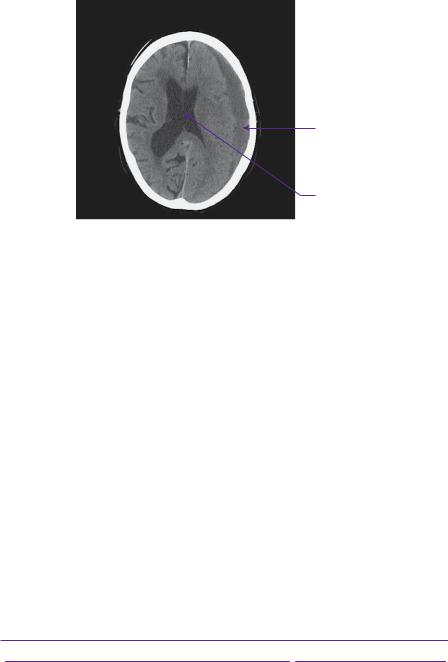
Admission blood tests were normal, apart from an international normalized ratio (INR) of 2.7. In view of his age and anticoagulation regime, he was referred for a plain CT head scan, which showed a left-sided chronic subdural haematoma (see Figure 1.1).
In view of his symptomatology, pre-morbid functional status, and the mass effect from the haematoma, surgical evacuation was advised. The risks and benefits of surgery were discussed with the patient and his wife. After liaising with the haematologist, he was given 10mg of vitamin K intravenously, followed by 1000 units (15 units/kg [1]) of Beriplex immediately prior to transfer to the operating theatre.
Under general anaesthetic, he was positioned supine with a sandbag under his left shoulder and his head in a horseshoe. The haematoma was evacuated using two burr holes (frontal and parietal), and irrigating the subdural space with warm saline until the effluent was clear. At the end of the procedure a soft subdural drain was inserted through the frontal burr hole and directed anteriorly.
His post-operative recovery was unremarkable. The next day he was more alert and orientated, and his dysphasia had completely resolved. On the second
2 |
Challenging concepts in neurosurgery |
Chronic subdural haematoma
Midline shift
Figure 1.1 Plain axial head CT showing a 12-mm crescent fluid collection overlying the left hemisphere, exerting enough mass effect to shift the midline 6mm to the right. The attenuation of haematoma older than about a week becomes lower than the underlying cortex as blood products are hydrolysed into smaller and more radiolucent molecules. The collection is seen to cross suture lines, but not dural attachments.
post-operative day, the drain was removed, with about 200mL drainage fluid in the bag. Prophylactic low molecular weight heparin (40mg enoxaparin) was commenced. After a further 2 days of physiotherapy he was discharged home.
A follow-up appointment was arranged for 3 months following discharge and he was advised to contact the Driver and Vehicle Licensing Agency (DVLA) regarding his driving licence. Given his clinical improvement, no post-operative imaging was organized. When seen at his follow-up appointment he was very happy with his progress and had returned to his hobbies.
Discussion
Epidemiology and pathophysiology
Chronic subdural haematoma is one of the most common conditions in general neurosurgical practice. Its incidence in the general population is about 5 per 100,000/year [2], with increasing incidence linked to age. Therefore, it is expected to become more common as the population ages. There is a strong male preponderance, with a male- to-female ratio of 3:1 [3]. It presents with a wide variety of symptoms (see Table 1.1) and accounts for approximately 1% of all hospital admissions with acute confusion [4].
Table 1.1 Most common presenting symptoms of chronic subdural haematoma [3]
Symptom |
Frequency |
Gait disturbance or falls |
57% |
Mental deterioration |
35% |
Limb weakness |
35% |
Acute confusion |
33% |
Headache |
18% |
Drowsiness or coma |
10% |
Speech impairment |
6% |
|
|
Reprinted from The Lancet, 374:9695, Santarius, T, et al., Use of drains versus no drains after burr-hole evacuation of chronic subdural haematoma - a randomised controlled trial 1067–73., Copyright (2009), with permission from Elsevier
Case 1 Management of chronic subdural haematoma |
3 |
 Expert comment
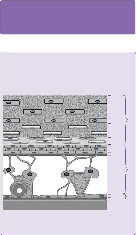
It is now widely accepted that subdural haematomas (SDH) result from the rupture of a dural bridging vein into the weakly adherent dural border cell layer, allowing blood to collect between the dura and the arachnoid mater (see Figure 1.2). As a consequence of cerebral atrophy in elderly patients, head trauma results in a greater displacement of brain in relation to dura. Bridging veins are subjected to a greater degree of stretch and, thus, SDHs may develop after relatively minor head injuries.
 Learning point Microarchitecture of the dura mater
The dura mater is composed of fibroblasts and a large amount of collagen. The arachnoid barrier cells are supported by a basement membrane (black) and bound together by numerous tight junctions (red). The dural border cells layer is formed by flattened fibroblasts, with no tight junctions and no intercellular collagen. It is, therefore, a relatively loose layer positioned between firm dura mater and arachnoid. The subdural space is a potential space that can form within the dural border cell layer. (Figure 1.2).
Periosteal |
dura mater |
|
mater |
Meningeal |
dura |
mater |
Dura |
Dural |
border |
cells |
|
Arachnoid |
barrier |
cells |
|
Arachnoid |
trabeculae |
|
Arachnoid |
Brain Pia |
mater |
|
Pia mater |
Figure 1.2 Schematic section through the dura and arachnoid mater; it is the dural border cell layer that separates in CSDH [5].
Santarius, T, et al. (2010), ‘Working toward rational and evidence-based treatment of cSDH’, Clinical
Neurosurgery, 57.

4 |
Challenging concepts in neurosurgery |
 Evidence base Pathogenesis of chronic subdural haematoma
The presence of blood in the subdural space elicits a complex inflammatory cascade involving proliferation of dural border cells, migration of macrophages, formation of granulation tissue, and angiogenesis [5]. In the majority of cases, this process ultimately results in resorption of the haematoma, but should this fail, the haematoma may grow and become symptomatic.
Chronic subdural haematoma (CSDH) often presents in patients whose acute subdural haematoma (ASDH) was initially not symptomatic enough for the patient to seek medical attention. Many groups have studied the mechanisms underlying the evolution of ASDH into CSDH and it is likely to involve an interplay of multiple pathways, leading to an increase in the haematoma fluid volume and, consequently, mass effect. Traditionally, it was thought that the hydrolysis of acute blood products into smaller molecules increased the oncotic pressure of the haematoma, thereby drawing in water by osmosis [6]. This hypothesis fell out of favour following the publication of Markwalder’s
landmark paper, which first demonstrated that CSDH fluid osmolality is the same as that of blood and cerebrospinal fluid (CSF) [7].
Rebleeding is one of the mechanisms that may contribute to haematoma growth. There is an abundance of coagulation inhibitors and fibrinolytic factors in the subdural fluid. High levels of tissue plasminogen activator (tPA) have been found in the subdural fluid and its concentration is predictive of recurrence [8]. Vascular endothelial growth factor (VEGF) is also found at higher concentrations in the subdural fluid [9]. VEGF is a pro-angiogenic factor and is also known to increase the ‘leakiness’ of capillary junctions.
The hypothesis of rebleeding is supported by the frequent observation of mixed attenuation blood on CT and mixed consistency haematoma intra-operatively. Furthermore, it is hypothesized that the serial dilution of anticoagulant and fibrinolytic factors by thorough lavage may be responsible for at least some of the therapeutic efficacy of burr hole drainage.
Indications and techniques for surgical intervention
Given the relatively low morbidity and mortality associated with evacuation of CSDH, symptomatic presentation merits strong consideration for surgical evacuation. Nonsurgical management is reserved for cases at both extremes in the spectrum of severity of their clinical presentation. At one end of the scale, an asymptomatic collection with minimal mass effect may be managed expectantly. At the other end, patients who are otherwise very unwell or moribund may be offered palliation.
Notably, there is a considerable variety of surgical and anaesthetic techniques that can be employed to evacuate CSDH, allowing clinicians to customize treatment to the characteristics of their patient. In the simplest of circumstances, where the patient is fit enough, general anaesthetic and burr hole evacuation is the most common technique used in most UK units. Either one or two burr holes can be used and, although there is no clear evidence supporting one over the other [5], the general consensus is that, where practicable, two burr holes allow a more complete evacuation. General anaesthetic appears to be more comfortable to patients and surgeons. It allows a higher standard of surgical technique in terms of asepsis, retention of subdural air, drain placement, wound closure, to be achieved, etc.

Case 1 Management of chronic subdural haematoma |
5 |
 Expert comment One versus two burr holes
While individual surgeons may have their own preference, it is generally agreed that two burr holes allow more thorough evacuation and irrigation, which in itself is probably associated with a better outcome [10]. Taussky et al. demonstrated a reduction in the incidence of recurrence where two burr holes were used [11]. Conversely, Han et al. found a 2% (n = 51) recurrence rate for one burr hole, compared with 7% (n = 129) where two burr holes were used [12]. Crucially, both of these studies were retrospective, such that there was no randomization process or equipoise. The marked disparity between them is, therefore, more likely to reflect differences in the conditions and patients being treated, rather than the technique employed.
A recent systematic review has found no difference in outcome between the use of one and two burr holes [13]. As a treatment of choice we use two burr holes. One burr hole may be considered if the CSDH is more localized or the procedure is performed under local anaesthetic.
 Clinical tip
The position of burr holes should be based on CT, in order to span as much of the haematoma as possible and allow conversion to craniotomy if required.
Copious lavage with warm isotonic solution should be used until the effluent is clear. Some surgeons use a Jaques catheter to irrigate in different directions and aid complete evacuation.
Over-enthusiastic advancement of the catheter into remote parts of the subdural space may result in bleeding. Irrigation with Jaques catheter alone may significantly prolong the length of the operation and it may be prudent to omit this step in high-risk surgical candidates, in whom a shorter operating time may be preferable.
Closing the dependent (usually parietal) burr hole first in a strictly watertight fashion allows the subdural space to be filled with irrigation fluid, reducing the volume of pneumocephalus and the risk of recurrence.
Patient positioning is important. Sandbags under the ipsilateral shoulder allow the side of the head to be almost horizontal without placing too much strain on the neck. Strapping the patient to the operating table allows safe tilting of the table, to bring the frontal burr hole to the highest point of the head prior to closure.
Using a high-speed drill enables the creation of a tangential frontal burr hole, which enables passing of the drain at an angle closer to parallel than perpendicular in relation to the brain surface. This may be relevant, especially in cases with a thick skull.
In instances where the patient is unfit for general anaesthetic, but generally cooperative, infiltration with local anaesthetic and scalp block can be used. In this case, the shorter operating time of a single burr hole may be preferable.
A second surgical option for evacuation is twist drill craniostomy (TDC) and closed-system drainage. Here, a small hole is drilled, 1cm anterior to the coronal suture, above the superior temporal line or over the maximum thickness of the subdural collection. Although morbidity and mortality is similar to burr hole evacuation (apart from a higher risk of recurrence with TDC), it can be performed at the bedside under local anaesthetic, providing a safe treatment modality in unfit patients, while reducing the costs of running an operating theatre [14].
6 |
Challenging concepts in neurosurgery |
|
Finally, craniotomy remains an option at the surgeon’s disposal in selected cases. |
|
Craniotomy was the treatment of choice until the publication of a paper in 1964, com- |
|
paring craniotomy to burr hole evacuation in sixty-nine patients [15], which showed |
|
improved functional outcome and lower recurrence rate following burr hole evacua- |
|
tion. These findings were subsequently confirmed in a number of other studies over |
|
the following two decades. However, mini-craniotomies remain useful, particularly |
|
in the context of multiple subdural membranes, solid haematoma, re-accumulation, |
|
or failure of brain expansion. Modern minicraniotomy probably has a similar risk |
|
and benefit profile as burr hole evacuation, but thus far a direct comparison of these |
|
two techniques has not been reported in the literature. |
 Expert comment Outcomes in modern chronic subdural haematoma surgery
Surgical treatment of symptomatic CSDH results in a rapid improvement of patient symptoms and a favourable outcome in excess of 80% of patients [16]. However, there are a number
of rare, but recognized early complications, including acute subdural haematoma, tension pneumocephalus, and cerebral infarction (Table 1.2). Recurrence rates in various series are approximately between 10 and 20% [5,17], but some papers have reported rates between 5 and 30%. Post-operative seizures occur in 3–10% of patients, but there is no evidence to support prophylactic anticonvulsant use [18].
Table 1.2 Intracranial complications of CSDH drainage [32]. The overall rate of intracranial complications in this series of 500 consecutive cases was 4.6%. Recurrence is considered separately.
Complication |
Rate |
Acute subdural haematoma |
2.6% |
Tension pneumocephalus |
0.8% |
Cerebral infarction |
0.4% |
Intracerebral haemorrhage |
0.2% |
Extradural haematoma |
0.2% |
Subdural empyema |
0.2% |
|
|
Mori, K. and Maeda, M. (2001), ‘Surgical treatment of chronic subdural hematoma in 500 consecutive cases: clinical characteristics, surgical outcome, complications, and recurrence rate’, Neurologia medico-chirurgica, 41 (8), 371-81.
 Learning point Non-operative management
Recognition of biochemical cascades producing a localized procoagulant and angiogenic state raises the possibility of using anti-inflammatory drugs, such as corticosteroids, as an alternative or adjuvant to surgery. Steroids have been shown to inhibit tPA activity [19] and VEGF expression [20] among others. Despite multiple reports of steroid use in CSDH management [21,22], there is a distinct lack of good quality clinical studies showing any therapeutic efficacy in CSDH, and the rationale for their use is largely theoretical. At present, the further elucidation of biochemical pathways, with the promise of potential pharmaceutical targets, remains an area of important academic interest.
Anticoagulation and anti-platelet agents in CSDH
Anticoagulation with warfarin and other drugs has been associated with both occurrence [23] and recurrence of CSDH. As a consequence of widespread use among elderly patients with cardiovascular co-morbidities, therapeutic anticoagulation is frequently encountered in patients presenting with CSDH and, therefore, merits a thorough understanding and effective management.

Case 1 Management of chronic subdural haematoma
Table 1.3 Pharmacokinetic properties of a commercially-available prothrombin complex concentrate as derived from healthy volunteers.
Component |
Median half-life (h) |
Range (h) |
Factor II |
60 |
25–135 |
Factor VII |
4 |
2–9 |
Factor IX |
17 |
10–127 |
Factor X |
31 |
17–44 |
Protein C |
47 |
9–122 |
Protein S |
49 |
33–83 |
|
|
|
Source data from: www.medicines.org.uk
Warfarin inhibits vitamin K-dependent synthesis of coagulation factors II, VII, IX, and X in the liver, which in turn blocks the extrinsic coagulation cascade, thus prolonging prothrombin time (PT) and INR. The desired degree of anticoagulation is determined by the risk of thromboembolism from the underlying condition.
The principle behind reversing anticoagulation with warfarin is to restore normal circulating concentrations of coagulation factors, which can broadly be achieved in two ways. The first is to directly transfuse clotting factors, with the dose of products depending on body weight and degree of anticoagulation. This first method is quick, but expensive and its effect is short-lived (Table 1.3). The second is to supplement vitamin K, enterally or intravenously. This allows the liver to resume synthesis of vitamin K-dependent clotting factors, a process that requires hours to days. By using a combination of blood products and vitamin K, a normal coagulation profile can be achieved throughout the entire periand post-operative periods, allowing safe surgical intervention.
For atrial fibrillation, the risk of thromboembolic events is 2.03% per year in the absence of therapeutic anticoagulation, falling to 1.15% for patients taking warfarin [24]. Here, the target INR is 2.5 (2.0–3.0). In contrast, the risk from prosthetic heart valves may be as high as 22% [25] and the target INR is accordingly higher—3.5 (3.0–4.0). While there is no doubt that anticoagulation increases the risk of chronic subdural haematoma [5,16], there is a distinct lack of data to quantify the risks resulting from restarting anticoagulation and its timing. A recent systematic review by Chari et al summarizes the relevant evidence [26].
 Expert comment
Antiplatelet agents, such as aspirin, clopidogrel, and dipyridamole are another important consideration in the management of CSDH [16]. While there is clear evidence that they promote occurrence, their effect on recurrence is less clear. In addition, there are no studies to determine the effect of aspirin on perioperative bleeding in intracranial surgery, but a recent survey showed neurosurgeons prefer to discontinue it’s use, on average, 7 days before an elective procedure [27]. On this basis, two general principles apply. First, antiplatelet agents should be stopped the moment CSDH is diagnosed, whether the patient is likely to be a candidate for surgery or not. Secondly, if neurological status is stable, one might consider postponing surgery. In instances where early surgical intervention is required, we prefer to transfuse one pool of platelets immediately prior to surgery, with the possibility of further transfusions in the initial postoperative days.
7
 Expert comment
The decision as to whether to resume anticoagulant or anticoagulation therapy after
evacuation is more challenging. A multi-disciplinary discussion between the neurosurgeon, general practitioner, and possibly cardiologist should consider
the patient’s clinical status and indication for anticoagulation. It is vital that the patient understands the pros and cons of starting and withholding the anticoagulation treatment.

8 |
Challenging concepts in neurosurgery |
Use of subdural drains
The reduction in pressure and/or mass effect following surgical evacuation allows the brain to gradually re-expand and fill up the space occupied by the haematoma. Filling that space with irrigation fluid reduces the amount of air trapped in the space. Fluid drained via a dependent drain facilitates brain re-expansion. The drain acts as a valve, where the forces moving the fluid out of the intracranial cavity are systolic brain expansion and the syphoning effect of the dependent drain. Both, but especially the latter are much diminished if air is trapped in the subdural space. The amount of air in the subdural space has been shown to be associated with recurrence [28–30].
Subdural drains permit continuing drainage of blood and irrigation fluid after surgical treatment. They are left in situ for an arbitrary 48 hours, which is thought to balance the risk of recurrence from inadequate brain re-expansion against the potential for infection. There is class I evidence that they reduce the incidence of recurrence and 6-month mortality, while improving functional status at discharge [3].
 Clinical tip Inserting subdural drains
Where drains are used, they should be inserted via the frontal burr hole and directed anteriorly, as this is an area in which the collection persists the longest. Placement of the drain via the frontal burr hole has been associated with a lower risk of recurrence [31]. It is important to direct the drain parallel to the inside of the calvarium in order to avoid inadvertent parenchymal insertion and intracerebral bleeding. Drilling the burr hole tangentially with a high-speed drill, rather than a perforator will
help achieve this aim. While a dedicated subdural drain is yet to be developed, the softest and most flexible drain available should be used.
Always check that drains are working at the end of the procedure and later on the ward. Drainage bags should be placed in a dependent position, and it is important to ensure that nursing staff are aware of the importance of continually maintaining dependency of the drain.
A final word from the expert
As more patients survive into their ninth and tenth decades, not only will the incidence of CSDH continue to rise, but surgeons will be faced with a patient population with
an increasingly complex profile of medical co-morbidities. In particular, the issue of anticoagulation is likely to become more pertinent. Further research should be directed towards establishing evidence-based guidelines for resuming anticoagulation and antiplatelet medication after CSDH surgery. Surgery will remain the mainstay of treatment of patients with CSDH, but further work is also needed to understand the rationale efficacy of various aspects of the surgical technique, and to refine indications for the different surgical techniques used, especially for craniotomy and twist-drill craniostomy.
Case 1 Management of chronic subdural haematoma |
9 |
References
1.Evans G, Luddington R, Baglin T. Beriplex P/N reverses severe warfarin-induced overanticoagulation immediately and completely in patients presenting with major bleeding. Br J Haematol 2001; 115(4): 998–1001.
2.Santarius T, Hutchinson PJ. Chronic subdural haematoma: time to rationalize treatment? Br J Neurosurg 2004; 18(4): 328–32.
3.Santarius T, Kirkpatrick PJ, Ganesan D, et al. Use of drains versus no drains after burrhole evacuation of chronic subdural haematoma—a randomised controlled trial. Lancet 2009; 374(9695): 1067–73.
4.George J, Bleasdale S, Singleton SJ.Causes and prognosis of delirium in elderly patients admitted to a DGH. Age Ageing 1997; 26: 423–7.
5.Santarius T, Kirkpatrick PJ, Kolias AG, et al. Working toward rational and evidencebased treatment of chronic subdural hematoma. Clin Neurosurg 2010; 57: 112–22.
6.Zollinger R, Gross RE. Traumatic subdural hematoma, an explanation of the late onset of pressure symptoms. JAMA 1934; 103: 245–9.
7.Markwalder TM, Steinsiepe KF, Rohner M, et al. The course of chronic subdural hematomas after burr-hole craniostomy and closed-system drainage. J Neurosurg 1981; 55(3): 390–6.
8.Katano H, Kamiya K, Mase M, et al. Tissue plasminogen activator in chronic subdural hematomas as a predictor of recurrence. J Neurosurg 2006; 104(1): 79–84.
9.Hohenstein A, Erber R, Schilling L, et al. Increased mRNA expression of VEGF within the hematoma and imbalance of angiopoietin-1 and -2 mRNA within the neomembranes of chronic subdural hematoma. J Neurotrauma 2005; 22(5): 518–28.
10.Matsumoto K, Akagi K, Abekura M, et al. Recurrence factors for chronic subdural hematomas after burr-hole craniostomy and closed system drainage. Neurol Res 1999; 21(3): 277–80.
11.Taussky P, Fandino J, Landolt H. Number of burr holes as independent predictor of postoperative recurrence in chronic subdural haematoma. Br J Neurosurg 2008; 22(2): 279–82.
12.Han, H. J., Park CW, Kim EY, et al. One vs. two burr hole craniostomy in surgical treatment of chronic subdural hematoma. J Korean Neurosurg Soc 2009; 46(2): 87–92.
13.Smith, M.D., Kishikova, L., & Norris, J.M., Surgical management of chronic subdural haematoma: one hole or two? Int J Surg (London, England), 2012; 10(9): 450–2.
14.Chari, A., Kolias, A.G., Santarius T, et al., 2014b Twist-drill craniostomy with hollow screws for evacuation of chronic subdural hematoma. J Neurosurg 2014; 121: 176–83.
15.Svien SJ, Gelety JE. On the surgical management of encapsulated chronic subdural hematoma: a comparison of the results of membranectomy and simple evacuation. J Neurosurg 1964; 21: 172–7.
16.Ducruet AF, Grobelny BT, Zacharia BE, et al. The surgical management of chronic subdural hematoma. Neurosurg Rev 2012; 35(2): 155–69; discussion 169.
17.Weigel R, Schmiedek P, Krauss JK. Outcome of contemporary surgery for chronic subdural haematoma: evidence based review. J Neurol Neurosurg Psychiat 2003; 74(7): 937–43.
18.Ratilal B, Costa J, Sampaio C. Anticonvulsants for preventing seizures in patients with chronic subdural haematoma. Cochrane Database Syst Rev 2005 Jul 20;(3): CD004893.
19.Coleman PL, Patel PD, Cwikel BJ, et al. Characterization of the dexamethasone-induced inhibitor of plasminogen activator in HTC hepatoma cells. J Biol Chem 1986; 261(9): 4352–7.
20.Gao T, Lin Z, Jin X. Hydrocortisone suppression of the expression of VEGF may relate to toll-like receptor (TLR) 2 and 4. Curr Eye Res 2009; 34(9): 777–84.
10 |
Challenging concepts in neurosurgery |
|
|
21. |
Delgado-Lopez PD, Martín-Velasco V, Castilla-Díez JM, et al. Dexamethasone treatment |
|
|
in chronic subdural haematoma. Neurocirugia (Astur) 2009; 20(4): 346–59. |
|
22. |
Berghauser Pont LME, et al., Clinical factors associated with outcome in chronic sub- |
|
|
dural hematoma: a retrospective cohort study of patients on preoperative corticosteroid |
|
|
therapy. Neurosurgery 2012; 70(4): 873–80; discussion 880. |
|
23. |
Robinson RG. Chronic subdural hematoma: surgical management in 133 patients. |
|
|
J Neurosurg 1984; 61(2): 263–8. |
|
24. |
Go AS, Hylek EM, Chang Y, et al. Anticoagulation therapy for stroke prevention in atrial |
|
|
fibrillation: how well do randomized trials translate into clinical practice? JAMA 2003; |
|
|
290(20): 2685–92. |
|
25. |
Liebermann A, Hass W, Pinto R. Intracranial hemorrhage and infarction in anticoagu- |
|
|
lated patients with prosthetic heart valves. Stroke 1978; 9: 18–24. |
|
26. |
Chari, A., Clemente Morgado, T., & Rigamonti, D., Recommencement of anticoagulation |
|
|
in chronic subdural haematoma: a systematic review and meta-analysis. Br J Neurosurg |
|
|
2014; 28(1): 2–7. |
|
27. |
Korinth MC. Low-dose aspirin before intracranial surgery—results of a survey among |
|
|
neurosurgeons in Germany. Acta Neurochir 2006; 148(11): 1189–96; discussion 1196. |
|
28. |
Shiomi, N., Sasajima, H., & Mineura, K., [Relationship of postoperative residual air and |
|
|
recurrence in chronic subdural hematoma]. No shinkei geka. Neurolog Surg 2001; 29(1): |
|
|
39–44. |
|
29. |
Nakajima H, Yasui T, Nishikawa M, et al. The role of postoperative patient posture in the |
|
|
recurrence of chronic subdural hematoma: a prospective randomized trial. Surg Neurol |
|
|
2002; 58(6): 385–7; discussion 387. |
|
30. |
Ohba, S., Kinoshita Y, Nakagawa T, et al., 2013. The risk factors for recurrence of chronic |
|
|
subdural hematoma. Neurosurg Rev 2013; 36(1): 145–9; discussion 149–50. |
|
31. |
Nakaguchi H, Tanishima T, Yoshimasu N. Relationship between drainage catheter loca- |
|
|
tion and postoperative recurrence of chronic subdural hematoma after burr-hole irriga- |
|
|
tion and closed-system drainage. J Neurosurg 2000; 93(5): 791–5. |
|
32. |
Mori K, Maeda M. Surgical treatment of chronic subdural hematoma in 500 consecutive |
|
|
cases: clinical characteristics, surgical outcome, complications, and recurrence rate. |
|
|
Neurol medico-chir 2001; 41(8): 371–81. |