


CASE
16The surgical management of the rheumatoid spine
Robin Bhatia
 Expert commentary Adrian Casey
Case history
A 52-year-old woman was referred to the spinal outpatients’ clinic with a 5-year history of neck pain, loss of hand dexterity, and gait deterioration. The previous medical history was remarkable for rheumatoid arthritis (RA) affecting the hand and knee joints symmetrically, and pulmonary nodules diagnosed on chest radiography. She had previous multiple small joint replacements in her hands and a hip replacement.
The loss of hand dexterity manifested itself as increasing difficulty fastening buttons and using cutlery to eat. In the year prior to presentation, the patient’s walking distance had progressively deteriorated, and she required a zimmer frame to mobilize. She taking methotrexate (5mg/week) and NSAIDs. The visual analogue score of neck pain at presentation was 7/10 (where 0/10 corresponded to no pain, and 10/10 was the worst pain imaginable), and her sleep was disturbed nightly.
Examination revealed bilateral hand deformities consistent with rheumatoid disease, with a gross grip strength of 3/5 (MRC grading). Proximal upper limb power was limited by neck pain and shoulder girdle rigidity. Power was globally 4/5 in the lower limbs with marked hypertonia, and exaggerated knee and ankle reflexes. Plantar reflexes were bilaterally up-going, with 3 beats of clonus at the right ankle. Her pre-operative Ranawat class was IIIa, based on the clinical findings. The Myelopathy Disability Index (MDI) was 85%, and Neck Disability Index (NDI) was 75%.
 Learning point Functional measures in rheumatoid cervical spine disease
The Ranawat Class classifies neurological function in rheumatoid spine disease into four broad categories, as shown in Table 16.1. This measure is simple to use and has been widely accepted. In the original publication of 1979, Ranawat et al. applied the classification scheme to 33 patients
Table 16.1 Classification of neurological function in rheumatoid disease according to Ranawat et al. [1]
Ranawat Class |
Description |
I |
No neurological deficit |
II |
Subjective weakness/dysaesthesia +/–hyperreflexia |
IIIa |
Objective weakness with long tract signs and ambulatory |
IIIb |
Quadraparetic and non-ambulatory |
Data from: Ranawat CS, O'Leary P, Pellicci P, Tsairis P, Marchisello P, Dorr L: Cervical spine |
|
fusion in rheumatoid arthritis. J Bone Joint Surg Am 1979;61:1003–1010 |
(continued) |
|
150
 Expert comment
This is a fairly classical presentation in a patient with rheumatoid cervical myelopathy. It typically presents after 15–20 years disease duration. Neurological examination can be difficult in the presence
of a painful deforming arthritis. The Ranawat classes are the most widely used gradation system with most patients fitting into Class II, symptomatic, but no objective signs, or IIIa, with objective neurological signs, but still able to walk.
Challenging concepts in neurosurgery
with rheumatoid cervical spine disease, including atlanto-axial subluxation, superior odontoid peg migration and subaxial subluxation. Two patients improved from Class III to II after spinal fusion, five patients improved within Class III, i.e. they became ambulatory, but the remaining patients showed no change in class or deterioration post-operatively [1].
The Myelopathy Disability Index (MDI), as developed by Casey et al. in 1996, described a functional scale of myelopathic disability based on an abridged form of the Stanford Health Assessment Questionnaire (HAQ). Responses to ten questions relating to rising, eating, walking, self-hygiene, gripping, and getting into and out of a car, were scored in terms of level of difficulty, with the overall score out of 30 converted to a percentage. MDI showed excellent correlation (r = 0.98) to the original Stanford HAQ, and was an accurate predictor of functional outcome in 194 rheumatoid spine patients operated on in comparison to the Ranawat and Steinbrocker grades [2].
The Neck Disability Index (NDI) was developed by Howard Vernon in 1989, as a modification of the Oswestry Low Back Pain Disability Index. The NDI primarily tests for pain-related functional deficits in categories such as personal care, lifting, driving, reading, sleeping, work, and recreation. The overall score is out of 50 converted to a percentage. The NDI showed moderate to high correlations (r = 0.6 and r = 0.7) to the visual analogue score and McGill Pain Questionnaire respectively, in small groups of conservatively-managed whiplash-injured patients [3].
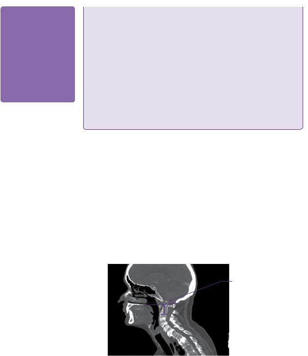
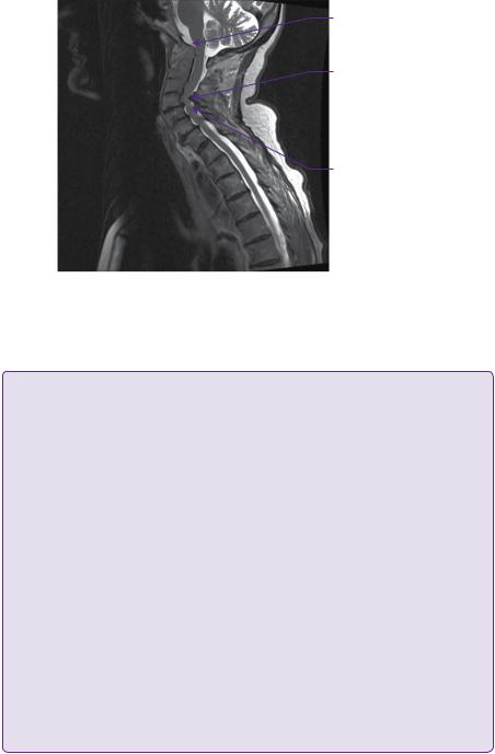
Plain lateral radiographs in flexion and extension revealed atlanto-axial subluxation with atlanto-dental interval (ADI) of 5mm and posterior atlanto-dental interval (PADI) of 15.5mm. CT of the craniocervical junction and upper thoracic spine revealed basilar impression with a Redlund-Johnell measurement of 25mm, and odontoid bony erosion with generalized osteopaenia (Figure 16.1). MRI imaging confirmed basilar impression, but without overt brainstem compression, and cervical canal stenosis at C4 and, to a lesser extent, at C5 due to anterior disc-osteophyte bars and posterior buckling of ligamentum flavum. There was intramedullary signal change of the spinal cord at the C4 and C5 levels on the T2-weighted imaging (Figure 16.2).
Basilar invagination of the odontoid peg
Figure 16.1 Sagittal CT image through the lower skull and cervicothoracic spine. The markedly degenerate and osteopenic rheumatoid spine shows basilar invagination, cervical canal stenosis, and subaxial cervical disease. The Redlund-Johnell measurement (purple arrow: 25mm) and the posterior atlanto-dental interval (white arrow: 15mm) are demonstrated.
Case 16 Surgical management of the rheumatoid spine |
151 |
Basilar invagination of the odontoid peg
Subaxial cervical canal stenosis
Intramedullary cord signal change
Figure 16.2 Sagittal T2-weighted MRI demonstrating spinal cord compression in the subaxial region particularly C4 /C5, with intramedullary cord signal change. The abnormally high position and proximity of the odontoid tip to the cervicomedullary junction is noted.
 Learning point Measuring basilar migration
The terms basilar invagination and basilar impression are more or less used interchangeably with ‘upward migration’, ‘vertical translocation’, and ‘cranial settling’. Multiple eponymous measurements were developed to describe the degree of basilar impression/invagination on radiographic imaging. One reason for this was the unreliability of eroded bony landmarks as a consequence of the disease process.
In 1939, Chamberlain stated that if the odontoid peg protruded more than 3mm above a line between the hard palate and the opisthion, then this was diagnostic of invagination [4]. McGregor, in 1948, defined a line between posterior hard palate and the most inferior point of the occipital curve (over 4.5mm odontoid projection was positive for basilar invagination) [5]. Wackenheim
in 1974 defined invagination if there was odontoid protusion posterior to a line drawn along the superior surface of the clivus [6]. Ranawat described a line from the midpoint of the odontoid peg to a transverse line through the atlas (positive if <15mm in males or <13mm in females) [1]. RedlundJohnell and Peterson drew a line from the midpoint of the caudal surface of C2 body to McGregor’s line, defining significant basilar invagination if the distance was <34mm in males and <29mm in females, as shown in Figure 16.2 [7]. Clark’s stations refer to the division of the sagittally-imaged odontoid peg into three equal parts; if the ring of the atlas was level with the middle or caudal third, then basilar invagination was diagnosed [8].
The modern era of early MRI has made many of these measurements obsolete, since clinicians can now view ligamentous integrity and the direct impact of basilar migration on the craniocervical junction. Riew et al. examined cervical radiographs in 131 rheumatoid patients and found that the only three measurements with over 90% sensitivity were the Redlund-Johnell, Clark’s stations, and the Ranawat measurement, with a combined sensitivity of 94% and negative predictive value of 91% [9].
Given the severity of her presenting symptomology, and clinical progression, the decision was made to proceed to posterior occipito-cervicothoracic fixation and C4/C5 laminectomy. Methotrexate was stopped 2 weeks prior to surgery. After induction, the patient was positioned prone in Mayfield head pins, and
152 |
Challenging concepts in neurosurgery |
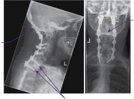
Cross-link of OCF
construct
Occipital plate of OCF construct
T1 and T2 pedicle screws at lower end of OCF construct
Figure 16.3 Lateral and AP radiographs immediately post-occipito-cervicothoracic fixation with rigid occipital plate/rod construct.
intra-operative X-ray imaging revealed satisfactory atlanto-axial and subaxial cervical alignment, with a neutral head position in order to maintain horizontal vision. Intravenous third generation cephalosporin (1.5g cefuroxime) and steroid (8mg dexamethasone) were administered on induction. Somatosensoryand motor-evoked potential spinal cord monitoring was used throughout the operation. A midline incision was made extending between the external occipital protuberance (EOP) and T4 posterior spinous process. After bilateral subperiosteal paraspinal muscle strips (extending laterally enough to reveal the facet joints), bilateral C3, C4, and C5 poly-axial lateral mass screws, and bilateral T1 and T2 pedicle screws were inserted under intra-operative image guidance. An occipital plate was applied by drilling two holes into the midline keel of the occiput beneath the EOP, and affixed using bicortically purchasing screws. C4 and C5 laminectomies were then carried out. The occipital plate was attached to bilateral lordotic titanium rods, positioned and torque-affixed into the heads of the cervical and thoracic screws. A cross-link was also affixed to the rods between C3 and C4. Morcelized own-bone fragments from the laminectomies and demineralized synthetic bone graft were applied lateral to the rods, and the wound was closed in layers of vicryl with clips to skin.
Immediate post-operative assessment revealed that the patient’s neurological deficit was unchanged, and the patient was once again mobilizing on day 2 with her zimmer frame. Plain X-rays revealed satisfactory placement of intrumentation and occipitocervical alignment (Figure 16.3). The patient was discharged from hospital after 12 days. Follow-up at 6 months revealed the persistence of long tract signs and MRC 4/5 global lower limb power (Ranawat IIIa, still mobilizing with a zimmer frame). However, NDI had significantly improved to 45% (with particular improvements in sleeping, eating, and reading), and MDI had improved to 50%. Her post-operative visual analogue score of neck pain was 4/10, with appropriate reductions in her analgesia. Repeat X-ray imaging did not reveal evidence of bony fusion at this stage. She recommenced methotrexate 4 weeks postoperatively, and remained without exacerbation of her rheumatoid disease in this period.

Case 16 Surgical management of the rheumatoid spine |
153 |
 Expert comment
The results of surgery are generally good for the ambulatory patient. They are correlated with preoperative neurological grade. However, for Ranawat IIIb the non-ambulatory myelopath the results are very poor, with mortality as poor as a patient with a malignant glioma. Surgical morbidity is also closely correlated with the patient’s pre-operative neurological class [10]. This includes wound healing issues, fusion success, and most importantly general complications, e.g. chest or urine infection. The operation is successful for pain relief in most patients. Typically, the pain is in the C2 distribution. It is caused by direct trauma in the area of inflammatory mediators.
Spinal fixation for RA has evolved from on-lay bone grafts with poor fusion and fixation rates, to wire and loop systems (e.g. Ransford loop), and now rigid screw rod systems that use the midline occiput, C2 pedicles and subaxial lateral masses [11,12]. For more limited involvement of the atlanto-axial joint, trans-articular or C1 C2 pedicle screws can be very effective [13].
Fusion rates can be also improved by the off-label use of bone morphogenic protein (BMP). This is potentially useful as many patients will be on immunosupressant drugs, e.g. methotrexate.
Discussion
RA affects approximately 1% of the population and, of those affected, approximately 50% will have cervical spine involvement [14]. Conlon et al. reported that 295/333 (88%) rheumatoidpatientscomplainedofcervicalsymptoms[15],butonly2/609(0.3%)inastudy by da Silva et al. required cervical spine fusion between 1955 and 1995 [16]. Therefore, there appears to be a significant difference at present in the numbers of rheumatoid spine patients managed by rheumatologists and those managed by spinal surgeons.
There is a female predominance of RA, with F:M ratio of approximately 3:1. RA is an autoimmune disease, the target of which is primarily joint synovium leading ultimately to articular cartilage and bone destruction with the development of synovial cysts and ligamentous laxity; atlanto-axial instability follows transverse ligament laxity, and basilar invagination is secondary to alar ligament and occipital condyle destruction. Cardiovascular, respiratory, skin, renal, vascular, and ocular systems may be affected by extra-articular rheumatoid disease. In general, the longer the disease, the more extensive and severe the articular and systemic involvement, although in recent years disease-modifying anti-rheumatoid drugs (DMARDs) have positively influenced disease progression [17].
The spinal neurosurgeon needs to address the following fundamental questions when managing the rheumatoid cervical spine.
What are the pathologies commonly encountered?
Broadly and clearly, these commonly co-exist and comprise atlanto-axial (horizontal) subluxation, vertical translocation giving rise to basilar invagination, pannusrelated craniocervical stenosis/ligamento-osseous erosion, and subaxial instability/ stenosis, as shown in Figure 16.4.
|
|
Atlantoaxial |
|
|
|
|
(’horizontal’) |
|
|
|
|
subluxation |
|
|
Vertical |
|
|
|
Subaxial |
|
|
|
||
translocation |
|
|
|
stenosis/instability |
|
|
Odontoid pannus |
|
|
|
|
|
|
|
|
|
|
|
|
Figure 16.4 Basic concepts in rheumatoid cervical spine pathology. There is a relationship and interplay between different pathologies.

154 |
Challenging concepts in neurosurgery |
 Expert comment
RA affects the synovial joints of the atlanto-dens joint. The inflammatory process will cause laxity or frank rupture of the transverse ligament. This allows anterior horizontal atlanto-axial subluxation. Posterior subluxation is a more dangerous subtype. It implies that the dens is fractured. Pathological fracture of the dens secondary to RA erosions is not an uncommon radiological finding. This pathology is probably responsible for unexplained death in RA.
As the destructive process continues, the lateral masses of the atlas (C1) and to a lesser degree the axis (C2) cause vertical translocation (cranial settling) to occur. Concomitant with the development of vertical translocation the atlanto-dens interval decreases, potentially giving a false sense of security. Vertical translocation is an end-stage development, and is associated with increased mortality and progressive neurological problems.
At what point in the disease process (from asymptomatic to clinically severe brainstem and spinal cord compression) should we surgically intervene?
The natural history of cervical rheumatoid disease is not entirely known, and the decision to operate balances the risks of intervention with the likelihood of preventing/improving neurological and functional decline. Moreover, using neurological examination parameters to guide operative decision-making reliably is difficult due to the peripheral joint and nervous involvement of RA.
The development of surgical techniques to manage the rheumatoid cervical spine was mirrored by the development of instrumentation. Although this case highlights the use of occipitocervical fixation for severe disease, spinal surgeons may also chose to carry out ‘segment-saving surgery’, or C1–C2 fixation.
 Learning point C1–C2 fixation
Atlanto-axial subluxation presents earlier in the disease process of rheumatoid cervical spine disease, with a prevalence range of 10–25% at a mean of 3.9 years after the diagnosis of RA [18]. There is now common acceptance for early surgical intervention to help neck pain and occipital neuralgia, prevent (occasionally catastrophic) spinal cord damage due to instability, and possibly to slow down disease progression, i.e. the development of vertical translocation at the craniocervical junction. However, there is no clear consensus on the relationship between the ADI and/or PADI, and the absolute requirement for surgical intervention.
Historically, the Gallie fusion described in 1939, and later modified by Brookes and Jenkins, employed sublaminar wires passed under the C1 arch and around the C2 spinous process [19], or under the
C2 lamina with bone blocks between C1 and C2 [20]. There were risks of accidental durotomy and spinal cord damage on sublaminar wire passage, however, and commonly patients were subsequently managed in a halo brace.
The 1980s saw the rise of interlaminar clamps, such as the Halifax clamp described by Tucker [21], with improved stability in rotation and extension. However, there was a high incidence of posterior element fracture, MRI artefact, and pseudoarthrosis with these devices.
In 1987, Magerl described C1–C2 fixation using posterior trans-articular screws [22]. This result enhanced rigidity, particularly in rotation, since the screws are passed through the articulating surfaces between C1 and C2. Added rigidity in flexion and extension can be obtained by Gallie-type C1–C2 wiring, although one major advantage of the Magerl C1–C2 fixation was the opportunity afforded to posteriorly decompress the craniocervical junction without affecting the construct. However, despite high reported fusion rates with this procedure (90–98% [22], the variable anatomy of the vertebral
(continued)

Case 16 Surgical management of the rheumatoid spine |
155 |
|
|
|
|
artery can sometimes preclude safe screw placement; indeed, pre-operative vascular imaging, such as CT angiography, is required therefore.
An alternative to the two screw fixation described by Magerl was reported by Goel in 1994 (and later modified by Harms in 2001) using four screws to fix the lateral masses of C1 to the pedicles of C2 [23,24]. The screws are secured by rods bilaterally. The development of poly-axial screws greatly aided this operative procedure. The Goel–Harms procedure is particularly advantageous when the atlanto-axial subluxation will not reduce, precluding the passage of trans-articular screws, when there is aberrant vertebral anatomy, or when the posterior elements are fractured or required to be decompressed. If C2 pedicle screws are not feasible due to bony anatomy, then C2 pars or
translaminar screws can be placed. The C2 nerve root is an important landmark, and there is a debate about whether to section or retract the nerve root during the insertion of the C1 lateral mass screw; ‘long shank’ screws avoid screw thread irritation of the nerve in this regard.
Does early C1–C2 fixation alter the pathological process in the rheumatoid spine? This question at present remains definitively unanswered, but there is a school of thought that early C1–C2 fixation can arrest the progression to vertical or basilar translocation, as well as reduce pannus formation [25]. However, it may also exacerbate the development of subaxial cervical disease [18].
What are the systemic issues related to the underlying diagnosis of RA, and how do these impact on peri-operative morbidity and mortality?
The peri-operative management of the patient with RA requires a multidisciplinary approach, and sufficient time pre-operatively to optimize the patient’s general medical status.
Cardiovascular system abnormalities include pericarditis (30–80%), arrhythmia, mitral valvular disease (30–80%), coronary vasculitis, and cardiomyopathy [26]. Hakala reported a poor prognosis in rheumatoid patients admitted with interstitial lung fibrosis [27]. Generalized osteopenia may compromise bony fusion and is compounded by chronic steroid use.
Rheumatoid patients also have more fragile skin making them susceptible to pressure sores, and poor wound healing. Rheumatoid patients undergoing cervical surgery often require post-operative management on the High Dependency Unit, given their poor pre-operative status and limited physiological reserve.
 Expert comment
The risk benefit profile for the RA patient is best for those with pain alone or minimal neurology. Careful attention to tissue handling, patient warming, prophylactic antibiotics, and pre-operative optimization of medication (stop steroids and DMARDs) and nutrition will potentially reduce complications. Rheumatoid pulmonary interstitial fibrosis and a higher apparent incidence of bronchiectasis are challenges for the anaesthetist, particularly in the prone position. Tracheostomy is rarely needed, even in those patients who need trans-oral decompression of the odontoid process. Mouth opening may be limited in the presence of temporomandibular (synovial joint) rheumatoid disease. Awake fibre-optic intubation is often required in the presence of the combined airway issues and an unstable cervical spine.
What has been the impact of disease-modifying drugs on the natural history of rheumatoid spinal disease, and what are the risks associated with continued use during the peri-operative period?
Biologic disease-modifying antirheumatic drugs (bDMARDs) extend the treatment choices for RA patients with suboptimal response or intolerance to a conventional
156 |
Challenging concepts in neurosurgery |
|
DMARD, such as methotrexate. Adalimumab, etanercept, and infliximab are exam- |
|
ples of tumour necrosis factor (TNF) inhibitors, and are effective treatments com- |
|
pared with placebo in the control of symptoms, improving physical function, and |
|
slowing radiographic changes in joints [28]. Other DMARDs include interleukin-1 |
|
receptor antagonists (e.g. anakinra) and anti-CD20 monoclonal antibodies (e.g. |
|
rituximab). These drugs hold promise, but are relatively new and the long-term |
|
benefits, particularly with regards to alteration of the natural history of rheumatoid |
|
disease in the spine is unknown. Moreover, they are not free of side effects; there |
|
may be an increased risk of bacterial infection associated with TNF inhibitor ther- |
|
apy [29]. |
 Learning point Disease-modifying drugs in the perioperative period
Although the side-effects of NSAIDs and glucocorticoids in the peri-operative period are well-described in the literature, less is known about the effects of biologic response-modifying medications, such as methotrexate, tumour necrosis factor-inhibitors (e.g. infliximab), interleukin-1 receptor antagonists (e.g. anakinra), and anti-CD20 monoclonal antibodies (e.g. rituximab).
Sany et al. conducted a randomized unblinded prospective trial on sixty-four rheumatoid patients receiving methrotrexate therapy, half of whom stopped taking methotrexate pre-operatively. There were no infections in either group, with comparable numbers of delayed wound healing [30].
Carpenter et al., on the other hand, reported increased local infection rates in patients continuing methotrexate compared with those stopping, in a prospective study of thirty-two patients undergoing hip arthroplasty [31]. A larger prospective randomized study by Grennan et al (n = 388), failed to show any significant increase in infection rate or wound complications in those on methotrexate compared with those stopping in the peri-operative period (in fact, the rates of local sepsis were lower in those continuing methotrexate), with no differences in rates of rheumatoid flare-ups [32].
No formal trials have addressed the peri-operative complications associated with continuing other disease-modifying drugs. Based on the extrapolation of limited studies in rheumatoid patients on infliximab undergoing abdominal surgery, and animal studies, Rosandich et al. and Pieringer et al. tentatively proposed discontinuation of these medications 2 weeks pre-operatively and restarting after a similar period of time, although further clinical data is required [33,34].
What is the optimal way of decompressing and fusing the rheumatoid cervical spine, and when should we consider (two-stage) anterior and posterior decompression? What type of instrumentation should one use and what is the capability of the underlying bony substrate in this disease to support fusion?
The application of occipitocervical fixation (OCF) constructs is challenging because of the unique 3D anatomy of the craniocervical region. The major considerations include the varying thickness of the occipital bone and the unusual load requirements on the construct, which is in essence supporting the head [35]. Historically, occipitocervical fusion using on-lay morcelized or fibular strut grafting with halo placement [36] was superceded by the internal rod and sublaminar wire technique, popularized in the last two decades of the twentieth century [37,38]. Roy-Camille introduced the concept of rigid occipitocervical fusion with skull base screws, plates, and lateral mass screws in 1989 [39], and in 1991 Grob et al. reported a technique of rigid occipitocervical (OC) fixation using an occipital Y-plate and lateral mass

Case 16 Surgical management of the rheumatoid spine |
157 |
screws (including trans-articular atlanto-axial screws) for a variety of pathologies in fourteen patients, all of whom achieved solid bony fusion and satisfactory clinical outcomes [40]. Furthermore, biomechanical cadaveric studies were supportive of rigid OC fixation over semi-rigid rod and wire systems [41,42], although in clinical practice fusion rates were similar and neither technique requires post-operative halo fixation.
 Evidence base A systematic review of OCF
In 2010, Winegar et al. published an extensive literature review of occipitocervical fixation with regards to techniques and outcomes. Their search generated 799 adult patients who underwent OC fixation between 1969 and 2011. The mean age +/– SD of patients undergoing OCF was 54 +/– 17 years (range 18–87 years). The indications for OCF were classified into five groups: inflammatory diseases (of which RA was the most common), congenital abnormalities, tumour, trauma, and other, non-specific indications. There were six types of instrumentation employed:
●Posterior wires/rods.
●Screws/rods.
●Hooks/rods.
●Screws/plates.
●Wires/plates.
●Wire/on-lay grafts.
The highest rates of fusion were for systems employing wires/rods and screws/rods, although the latter technique was more effective in RA. Most occipitocervical fusions occurred over a period of 4 months, excepting tumours in which only 45% fused over that period of time. Fusion was associated with neurological improvement and improved patient satisfaction overall.
Although only 16% of articles in the review documented adverse complications, the overall rate was 52%, divided into instrumentation misplacement, vascular injury, thecal sac injury, anaesthetic complications, and unspecified. There was a 22% rate of intrumentation failure, posterior wiring notably was associated with 10% failure rate compared with 6.2% with screws [43].
One of the largest series of non-rigid OC fixations for RA specifically was published by Zygmunt et al., who reported 163 rheumatoid patients over a mean follow-up period of 54 months [44]. The most common presenting symptom in patients with RA and cervical subluxation was neck pain, often with associated occipital neuralgia, but a wide spectrum of symptoms and signs secondary to brainstem, cranial nerve, and cord or cervical root compression were also reported. The Brattstrom–Granholm method of occipital burr hole-to-C2 fixation using wires, C2 spinous process pin, and PMMA cement was employed. The indication for surgery was atlanto-axial subluxation (defined as ADI > 3mm). Seventy-four per cent of patients reported significant improvements in pain and neurological deficit. Complications in this large series included wire breakages in 21/163, eight of whom required revisional surgery, five wound infections, five cerebellar injuries related to occipital burr hole wire passage, nine patients had progressive postoperative myelopathy, two cases had progressive vertical subluxation of the odontoid peg. Thirty-seven of 163 patients developed progressive subaxial subluxation at the levels below fixation (typically C4/C5). Therefore, wire-based constructs do have the additional risks of wire breakage, bone abrasion, and accidental durotomy/neurological injury when being passed [35].

158
 Expert comment
Occipital fixation has evolved to maximize the midline keel of the occiput, where the bone is
thickest. The use of pre-contoured and/ or articulated rods facilitates easier rod placement, particularly when the anatomy is extreme in cases of basilar impression. Polyaxial screws using favoured angles allow easier rod approximation and minimize the bone screw interface, a potential point of weakness.
Challenging concepts in neurosurgery
 Clinical tip The optimal construct for occipital cervical fixation
According to Nockels et al., the optimal implant for OCF would allow for:
●Rigid fixation of the occiput to the subaxial cervical spine without the requirement for halo bracing.
●The ability to perform atlanto-axial screw fixation and posterior decompression.
●The ability of the construct to conform to the individual anatomy.
●Ease of use.
●Means to correct deformity.
●Overall low profile.
●The ability to tolerate biomechanical forces unique to the region over a long-term basis.
Nockels believed that rigid fixation systems best met these goals [35].
A controversial point is whether to operate on patients with severe cervical rheumatoid disease. The decision must weigh up the benefits and risks to the patient. Moskovich et al. reported that 40% of patients improved in Ranawat class, and a significant number had prolonged improvements in neck pain score in a group of 150 rheumatoid patients undergoing OCF using the Ransford loop and sublaminar wires. However, there was a 10% mortality rate and 11% re-operation rate due to disease progression and post-operative complications [38]. Rheumatoid patients with severe disease (Ranawat Class IIIb) at baseline have the worst post-operative prognosis. Casey et al. reported mortality rates up to 30%, with little improvement in disability in this subgroup, suggesting that OC fixation as a procedure to help patients is carried out ‘too little and too late’ [10]. On the other hand, it may be reasonable to observe closely rheumatoid patients who are asymptomatic with fixed basilar invagination and no spinal cord compression. In those with MRI T2-weighted signal change in the cord or syrinx surgery can be considered to prevent future neurological decline or sudden death. It is important that the surgery is carried out in specialist units with a high turnover of RA patients and by spinal surgeons who are experienced in the management of the rheumatoid spine.
A final word from the expert
The rheumatoid patient presents many challenges in diagnosis, pre-operative optimization, and selection of levels to instrument. The fixation and bony healing is in a challenging environment with immunosuppressive agents and often osteoporosis from the historical use of steroids. The results of surgery for pain relief and preservation of neurological function are good, provided that it is not left too late. If the patient is no longer ambulant, surgery may be too much, too late, with high complication rates. Surgical fixation for atlanto-axial fixation for those patients who can still walk has a lot to offer patients in terms of neurological protection and pain relief, and is a rewarding area.
References
1.Ranawat CS, O’Leary P, Pellicci P, et al. Cervical spine fusion in rheumatoid arthritis. Journal of Bone Joint Surgery American 1979; 61: 1003–10.
2.Casey AT, Bland JM, Crockard HA. Development of a functional scoring system for rheumatoid arthritis patients with cervical myelopathy. Annals of Rheumatic Diseases 1996; 55: 901–6.
Case 16 Surgical management of the rheumatoid spine |
159 |
3.Vernon H, Mior S. The Neck Disability Index: a study of reliability and validity. Journal of Manipulative Physiological Therapeutics 1991; 14: 409–15.
4.Chamberlain WE. basilar impression (platybasia): a bizarre developmental anomaly of the occipital bone and upper cervical spine with striking and misleading neurologic manifestations. Yale Journal of Biology and Medicine 1939; 11: 487–96.
5.McGreger M. The significance of certain measurements of the skull in the diagnosis of basilar impression. British Journal of Radiology 1948; 21: 171–81.
6.Wackenheim A. Roentgen diagnosis of the craniovertebral region. New York: Springer, 1974.
7.Redlund-Johnell I, Pettersson H. Radiographic measurements of the cranio-verte- bral region. Designed for evaluation of abnormalities in rheumatoid arthritis. Acta Radiologica. Diagnosis (Stockholm) 1984; 25: 23–8.
8.Clark CR, Goetz DD, Menezes AH. Arthrodesis of the cervical spine in rheumatoid arthritis. Journal of Bone Joint Surgery American 1989; 71: 381–92.
9.Riew KD, Hilibrand AS, Palumbo MA, et al. Diagnosing basilar invagination in the rheumatoid patient. The reliability of radiographic criteria. Journal of Bone Joint Surgery American 2001; 83-A: 194–200.
10.Casey AT, Crockard HA, Bland JM, et al. Surgery on the rheumatoid cervical spine for the non-ambulant myelopathic patient-too much, too late? Lancet 1996; 347: 1004–7.
11.Bhatia R, Haliasos N, Vergara P, et al. The surgical management of the rheumatoid spine: Has the evolution of surgical intervention changed outcomes? Journal of the Craniovertebral Junction and Spine 2014; 5 (1): 38–43.
12.Bhatia R, DeSouza, R, Bull J, et al. Rigid occipitocervical fixation: indications, outcomes, and complications in the modern era Journal of Neurosurgery of the Spine 2013; 18 (4): 333–9.
13.Vergara P, Singh J, Casey A, et al. C1–C2 posterior fixation: are 4 screws better than 2? Neurosurgery 2012; 71: ON S86–95.
14.Krauss WE, Bledsoe JM, Clarke MJ, et al. Rheumatoid arthritis of the craniovertebral junction. Neurosurgery 2010; 66: 83–95.
15.Conlon PW, Isdale IC, Rose BS. Rheumatoid arthritis of the cervical spine. An analysis of 333 cases. Annals of Rheumatic Diseases 1966; 25: 120–6.
16.da Silva E, Doran MF, Crowson CS, et al. Declining use of orthopedic surgery in patients with rheumatoid arthritis? Results of a long-term, population-based assessment. Arthritis & Rheumatology 2003; 49: 216–20.
17.Hamilton JD, Gordon MM, McInnes IB, et al. Improved medical and surgical management of cervical spine disease in patients with rheumatoid arthritis over 10 years. Annals of Rheumatic Diseases 2000; 59: 434–8.
18.Werle S, Ezzati A, El Saghir H, et al. Is inclusion of the occiput necessary in fusion
for C1–2 instability in rheumatoid arthritis? Journal of Neurosurgery of the Spine 2013; 18: 50–6.
19.Gallie WE. Skeletal traction in the treatment of fractures and dislocations of the cervical spine. Annals of Surgery 1937; 106: 770–6.
20.Brooks AL, Jenkins EB. Atlanto-axial arthrodesis by the wedge compression method. Journal of Bone Joint Surgery American 1978; 60: 279–84.
21.Tucker HH. Technical report: method of fixation of subluxed or dislocated cervical spine below C1–C2. Canadian Journal of Neurological Sciences 1975; 2: 381–2.
22.Magerl F, Sceman PS. Stable posterior fusion at the atlas and axis by trans-articular screw fixation. In: P Kehr, A Weidner,(eds for more than one name), Cervical spine (pp. 322–7). New York: Springer-Verlag, 1987.
23.Goel A, Laheri V. Plate and screw fixation for atlanto-axial subluxation. Acta Neurochirugia (Wien) 1994; 129: 47–53.
24.Harms J, Melcher RP. Posterior C1–C2 fusion with poly-axial screw and rod fixation.
Spine (Phila Pa 1976) 2001; 26: 2467–71.
160 |
Challenging concepts in neurosurgery |
|
|
25. |
Landi A, Marotta N, Morselli C, et al. Pannus regression after posterior decompression and |
|
|
occipito-cervical fixation in occipito-atlanto-axial instability due to rheumatoid arthritis: |
|
|
case report and literature review. Clinical Neurology and Neurosurgery 2013; 115: 111–16. |
|
26. |
Voskuyl AE. The heart and cardiovascular manifestations in rheumatoid arthritis. |
|
|
Rheumatology (Oxford) 2006; 45 (Suppl. 4): iv4–7. |
|
27. |
Hakala M. Poor prognosis in patients with rheumatoid arthritis hospitalized for intersti- |
|
|
tial lung fibrosis. Chest 1988; 93: 114–18. |
|
28. |
Chen YF, Jobanputra P, Barton P, et al. A systematic review of the effectiveness of adali- |
|
|
mumab, etanercept and infliximab for the treatment of rheumatoid arthritis in adults |
|
|
and an economic evaluation of their cost-effectiveness. Health Technology Assessment |
|
|
2006; 10: iii–iv, xi-xiii, 1–229. |
|
29. |
Nam JL, Winthrop KL, van Vollenhoven RF, et al. Current evidence for the management |
|
|
of rheumatoid arthritis with biological disease-modifying antirheumatic drugs: a system- |
|
|
atic literature review informing the EULAR recommendations for the management of RA. |
|
|
Annals of Rheumatic Diseases 2010; 69: 976–86. |
|
30. |
Sany J, Anaya JM, Canovas F, et al. Influence of methotrexate on the frequency of |
|
|
postoperative infectious complications in patients with rheumatoid arthritis. Journal of |
|
|
Rheumatology 1993; 20: 1129–32. |
|
31. |
Carpenter MT, West SG, Vogelgesang SA, et al. Postoperative joint infections in rheuma- |
|
|
toid arthritis patients on methotrexate therapy. Orthopedics 1996; 19: 207–10. |
|
32. |
Grennan DM, Gray J, Loudon J, et al. Methotrexate and early postoperative complications |
|
|
in patients with rheumatoid arthritis undergoing elective orthopaedic surgery. Annals of |
|
|
Rheumatic Diseases 2001; 60: 214–17. |
|
33. |
Rosandich PA, Kelley JT, 3rd, Conn DL. Perioperative management of patients with |
|
|
rheumatoid arthritis in the era of biologic response modifiers. Current Opinion |
|
|
Rheumatology 2004; 16: 192–8. |
|
34. |
Pieringer H, Stuby U, Biesenbach G. Patients with rheumatoid arthritis undergoing |
|
|
surgery: how should we deal with antirheumatic treatment? Seminars in Arthritis |
|
|
Rheumatics 2007; 36: 278–86. |
|
35. |
Nockels RP, Shaffrey CI, Kanter AS, et al. Occipitocervical fusion with rigid internal fix- |
|
|
ation: long-term follow-up data in 69 patients. Journal of Neurosurgery of the Spine 2007; |
|
|
7: 117–23. |
|
36. |
Newman P, Sweetnam R. Occipito-cervical fusion. An operative technique and its indica- |
|
|
tions. Journal of Bone Joint Surgery (British volume) 1969; 51: 423–31. |
|
37. |
Moskovich R, Crockard HA, Shott S, et al. Occipitocervical stabilization for myelopathy |
|
|
in patients with rheumatoid arthritis. Implications of not bone-grafting. Journal of Bone |
|
|
Joint Surgery American 2000; 82: 349–65. |
|
38. |
Chen HJ, Cheng MH, Lau YC. One-stage posterior decompression and fusion using a |
|
|
Luque rod for occipito-cervical instability and neural compression. Spinal Cord 2001; |
|
|
39: 101–8. |
|
39. |
Roy-Camille R, Saillant G, Mazel C. Internal fixation of the unstable spine by a posterior |
|
|
osteosynthesis with plate and screws. Cervical Spine Research Society 1989: 390–404. |
|
40. |
Grob D, Dvorak J, Panjabi M, et al. Posterior occipitocervical fusion. A preliminary |
|
|
report of a new technique. Spine (Phila Pa 1976) 1991; 16: S17–24. |
|
41. |
Sutterlin CE, 3rd, Bianchi JR, Kunz DN, et al. Biomechanical evaluation of occipitocervi- |
|
|
cal fixation devices. Journal of Spinal Disorders 2001; 14: 185–92. |
|
42. |
Oda I, Abumi K, Sell LC, et al. Biomechanical evaluation of five different occipito- |
|
|
atlanto-axial fixation techniques. Spine (Phila Pa 1976) 1999; 24: 2377–82. |
|
43. |
Winegar CD, Lawrence JP, Friel BC, et al. A systematic review of occipital cervical |
|
|
fusion: techniques and outcomes. Journal of Neurosurgery of the Spine 2010; 13: 5–16. |
|
44. |
Zygmunt SC, Christensson D, Saveland H, et al. Occipito-cervical fixation in rheumatoid |
|
|
arthritis-an analysis of surgical risk factors in 163 patients. Acta Neurochirugia (Wien) |
|
|
1995; 135: 25–31. |