

CASE
17Cervical spondylotic myelopathy
Ellie Broughton
 Expert commentary Nick Haden
Case history
A 60-year-old, right-handed male presented to his GP with a 1-year history of gradual decline in his gait and dexterity. In the last 6 months he had also been experiencing right-sided arm pain radiating into his hand. He was unable to perform fine motor tasks, such as doing up buttons, and had started feeling unsteady on his feet.
His past medical history included hypertension only. He was otherwise well and a retired accountant. Examination findings included right arm numbness in C5 distribution, and loss of co-ordination in his hands and feet. He was hyperreflexic throughout with up-going plantars. Power in his legs was reduced to 4+ in hip flexion bilaterally.
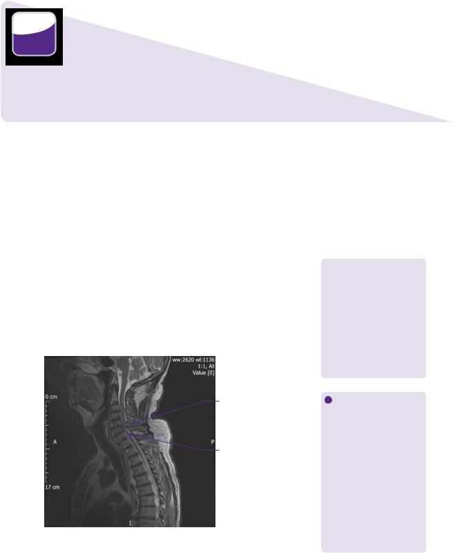
MRI of his cervical spine revealed marked degeneration at C5/6 with cord impingement and foraminal stenosis on the right. There was also less severe degenerative changes at C6/7. His overall spinal alignment was straight (see Figure 17.1).
Given this history and the findings on MRI, a diagnosis of CSM was made. The patient went on to have a two-level anterior cervical discectomy with fusion at the level C5/6 and C6/7.
Post-operatively the patient had resolution of his right-sided arm pain and some improvement in his right arm numbness. Immediate post-operative imaging revealed
 Learning point Clinical presentation of cervical spondylotic myelopathy
The course of cervical spondylotic myelopathy (CSM) is one of
slow and/or stepwise decline [1]. Symptoms include motor and sensory disturbance, diminished balance and dexterity, spasticity, paralysis and sphincter disturbance [2,3]. Additionally, there can be radicular symptoms from nerve root compression.
Cervical canal stenosis
Intramedullary cord signal change
Figure 17.1 T2-weighted MRI cervical spine of the patient showing canal stenosis at C5/6 and C6/7, with intramedullary cord signal change.
 Learning point Cord signal change
Increased signal on T2 and decreased signal on T1-weighted MRI is correlated with patients who are older, have a longer duration of disease, worse neurology, and worse long-term recovery [3,4]. It is suggested that these changes therefore indicate irreversible cord damage [3]. Focal T2 hyperintensity had better outcomes than multisegment
hyperintensity and T1 hypointensity [5,6]. Post-operative expansion of intramedullary high signal on T2 has also been observed and has a worse prognosis for recovery [3].

162 |
Challenging concepts in neurosurgery |
Cervical interbody cage |
Anterior cervical plate |
Figure 17.2 Post-operative imaging of 2 level ACDF.
satisfactory placement of instrumentation (Figure 17.2). At 6-month follow-up his walking improved. Follow-up imaging revealed no evidence of further kyphosis or instability in the spine, and fusion was obtained.
 Expert comment Principles of treating cervical spondylotic myelopathy
●Neurological decompression and alleviation of symptoms.
●Maintaining stability of the spine.
●Correcting or preserving deformity.
In addition to these key principles, some surgeons advocate maintaining motion
Discussion
CSM is a progressive degenerative process, resulting in motion abnormalities, loss of disc height, ligamentous redundancy, arthrosis of uncovertebral and facet joints, and formation of osteophytes [3]. This can result in circumferential spinal canal narrowing and nerve root compression [3]. Although this case focuses on the most common cause of myelopathy, degenerative spondylosis, compression can also result from other aetiologies, such as traumatic injuries, tumours, and ossification of the posterior longitudinal ligament [2].
Surgical intervention follows in the case of failed conservative management, or in patients with intractable pain and/or progressive neurology [7]. The prognosis is variable and, if stenosis has been present for many years, then demyelination and necrosis of gray and white matter can lead to irreversible deficits [1,7]. Matz et al. showed that mild CSM can be treated conservatively for up to 3 years with equivalent results to surgery, but it is a progressive disease and severe CSM always requires surgical management [8].
Surgical management of CSM is a challenging concept to neurosurgeons as there are many different factors to consider with each patient to determine the correct operative approach.
The options for treatment including the approach (anterior, posterior, or combined), the number of levels that should be treated and what should be done with the disc space (graft, arthroplasty, or nothing). These choices will be influenced by pathological and patient factors, in addition to the surgeon’s overall experience and skill set. It is also important to consider what is achievable, as the aim is to address the pathology while also limiting morbidity and optimizing long-term outcome [9].

Case 17 Cervical spondylotic myelopathy
The pathological factors that were relevant in this case were, first, that the disease was mostly anterior, the alignment was straight overall, and there were both radiculopathic and myelopathic symptoms. The patient factors to consider were his young age, no co-morbidities or neck deformity that might limit the approach and good overall bone quality.
Approach
The first aspect of treatment to consider is the approach.
 Learning point Techniques used in surgical treatment of cervical spondylotic myelopathy
Surgery can be anterior, posterior, or combined. There is no significant difference in outcome with any approach and they all are successful in the treatment of CSM [8,10–13]. The approaches include;
●Anterior cervical discectomy and fusion (ACDF).
●Anterior cervical corpectomy and fusion (ACCF).
●Anterior cervical discectomy and arthroplasty.
●Laminectomy and fusion: laminectomy has been associated with late deterioration with kyphosis and, therefore, is only recommended in combination with fusion [10,14].
●Laminoplasty: single or double-door.
The majority of spondylotic disease cases, as in this one, is located anterior and is easier to treat with an anterior approach, where the pathology can be directly visualized and removed without manipulating the spinal cord. This fulfils the first principle of neurological decompression and is the most common approach for CSM [7]. Anterior approaches show good neurological outcome, excellent stability, and carry a low morbidity [5]. When disease is at the extremes of the cervical spine, an anterior approach can become more challenging [7]. Dysphagia, odynophagia, and dysphonia are the most common complications in anterior surgery, but are almost always only in the early post-operative period [11].
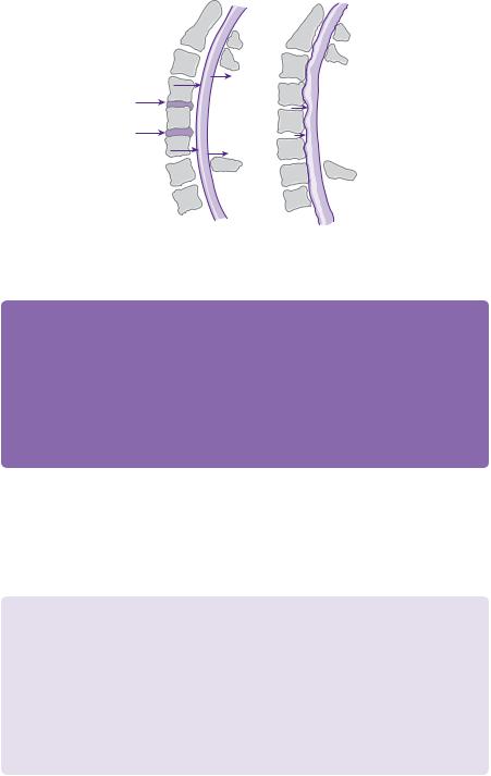
ACDF and ACCF are both suitable anterior approaches for decompression and with use of a plate have equivalent fusion rates, although corpectomy has higher rates of graft complication [2,10,15]. ACDF removes soft disc and bony osteophytes and is usually effective in decompressing the spinal canal [3]. Multiple levels can be treated in this way, but if there is disease behind the vertebral body, then ACCF may allow a more complete decompression [10]. This can be particularly important in kyphotic spines, where the cord is draped over osteophyte-disc bars which may not be fully removed with ACDF (see Figure 17.3)[7]. However, some authors report that not only can multilevel ACDF decompress, as well as ACCF, but that there is the additional benefit of improved restoration of lordosis due to increased distraction points and smaller inter-body grafts [16]. This is why multilevel discectomy was chosen in this case, although there is no denying that it can be more difficult and time-consuming than a corpectomy and, therefore, must be the surgeon’s choice.
163
 Clinical tip Factors in surgical decision making
Pathological factors
●Location of pathology.
●Overall and segmental alignment.
●Multilevel vs. single-level.
●Myelopathy vs. radiculopathy.
Patient factors
●Age, co-morbidities.
●Previous surgery.
●Pre-existing neck deformity.
●Bone quality (osteoporosis/ Ankylosing spondylitis (AS)).
 Expert comment The anterior approach
An anterior approach is excellent for decompressing the cord, thereby relieving myelopathic symptoms. This patient also had radicular symptoms and, therefore, wide foraminotomies with removal of uncinate spurs should be performed to alleviate this. Some consider it easier and safer (with direct visualization) to decompress the foramina with a posterior approach. However, in this patient, with a straight spine and anterior pathology, I would still advocate an anterior approach.
 Clinical tip Corpectomy
When performing corpectomy the space can be filled with an autoor allograft material. Allograft is normally bone harvested from the patient’s hip. In some centres titanium cages are preferred due to the potential risks of iliac bone harvesting, such as infection, haematoma, and the rare lateral femorocutaneous nerve neuralgia [5]. Some surgeons maintain
that autograft bone is superior, in particular tricortical iliac crest bone [16].
164 |
Challenging concepts in neurosurgery |
3 |
3 |
4 |
4 |
5 |
5 |
6 |
6 |
|
|
7 |
7 |
1 |
1 |
Figure 17.3 Normal lordosis of spine on left image with dorsal migration of cord following laminectomy. The right-hand picture shows a straighter spine with prevention of migration of cord and, therefore, persistent compression after laminectomy.
 Expert comment
Post-operative kyphosis is one of the most feared complications of cervical spine surgery; therefore, it is important to have an understanding of a patient’s spinal alignment and how surgery can affect it.
Sagittal balance can be assessed on MRI or CT and lateral flexion/extension X-rays will evaluate the degree of instability [17]. In straight or kyphotic spines, an anterior approach is advocated to allow proper distraction and extension, correcting alignment and improving lordosis [2,7,17]. Conversely, posterior approaches in straight or kyphotic spines can worsen the kyphosis, cause instability and restrict neurological improvement [2,7,17,18]. The latter occurs because the spinal cord is tented over anterior pathology and lordosis is required for dorsal migration of the cord away from anterior compression, which cannot occur in a kyphotic spine.
In this case, the spinal alignment was straight overall, with some loss of the natural lordosis of the cervical spine and the pathology was anterior; therefore, an anterior approach is ideal. If there is no anterior pathology then laminectomy with good instrumentation or laminoplasty can be performed in a kyphotic spine, with debate as to whether this increases risk of kyphosis and instability [2,17].
 Evidence base How approach affects kyphosis
In forty-eight CSM patients, where twenty-four were fixed anteriorly (corpectomy and cage) and twenty-four posteriorly (laminectomy and lateral mass screws), an initial improvement in lordosis of 8.8 degrees was seen in the anterior group and a decline of 6.5 degrees in the
posterior group. Over time, the lordotic correction declined in the anterior group, possibly due to subsidence of the cage before fusion has occurred [18]. Another study of forty-three patients with >10 degrees of kyphosis pre-operatively showed a significant decrease in kyphosis in twenty-eight patients fused anteriorly, compared with fifteen patients with en-bloc C3–7 posterior laminoplasty. However, at long-term follow-up (mean 3.3 years), there was no significant difference in neurological function [19]. Thus, posterior fixation leads to a more constant, but kyphotic alignment than anterior fixation, which may not affect clinical outcome in early follow-up.
The use of curved rods and screws with smaller heads, which can fit closer together, may help with posterior cervical surgery and sagittal balance [18]. However, ultimately anterior spondylotic osteophytes may prevent adequate distraction,

Case 17 Cervical spondylotic myelopathy
decompression, and realignment; in such cases, a combined approach can be considered [9,18,20]. Combined approaches are useful if there is a fixed kyphosis (where there is ankylosis of segments and kyphosis cannot be corrected posturally or with traction), and in cases where there is high risk of multicolumn instability [21].
 Learning point Fixed kyphosis
One review of sixteen patients demonstrated that combined approaches can correct an average deformity of +38 degrees (kyphosis) to –10 degrees (lordosis), rendering 75% lordotic and 25% straight post-operatively [21].
Combined approaches can be performed in oneor two-stage operations. They offer higher fusion rates for multilevel disease and lower complication rates, particularly in relation to anterior plate/fusion failure and graft dislodgement seen with anterior approach alone [9]. The latter
is probably due to the fact that the additional posterior fusion takes the load off the anterior system [20].
Posterior procedures allow dorsal migration of the cord, decreased axial tension and improved vascular perfusion leading to neurological improvement in 70–95% of patients [12,22]. The advantages over anterior surgery include adequate long-seg- ment decompression, wider decompression than anterior approaches (due to limit of vertebral arteries), and no complications of dysphagia or vocal cord palsy [23,24]. This makes them more attractive when performing multisegment surgery. However, the potentials of hardware failure, loss of alignment (kyphosis) or instability are recognized and feared complications in multilevel procedures [12,22].
There are different ways to approach laminectomy, and fusion and lateral mass fixation has been advocated as the procedure of choice, due to its biomechanical properties, simplicity, and low complication rate [23,25].
165
 Evidence base Combined approaches
One study of forty patients with anterior and posterior CSM, treated with a combined approach (corpectomy or
discectomy anteriorly then fusion +/– laminectomy posteriorly) showed relief of neurological symptoms in all patients at 1-year follow-up with 97.5% radiological fusion [20]. Another study of thirty-five patients with single-stage combined approach showed
no cases of pseudoarthrosis or graft/instrumentation related complications [9].
 Evidence base Laminectomy
Sekhon et al. looked at fifty patients with CSM, treated with wide posterior decompression (laminectomy) and lateral mass instrumentation with alignment maintained in 96% and fusion in >90% [23]. Kyphotic spines were excluded. The authors also suggest that posterior fusion can result in regression of spondylotic disease, and that there is a low rate of adjacent segment disease (2% at 25.6 months follow-up), due to posterior tension banding allowing micromotion anteriorly and shielding the intervertebral discs from stress of fusion [23].
Bapat et al. reviewed 129 CSM patients, comparing anterior, posterior, and combined approaches [11]. Overall, good outcome was achieved in 86% of patients, allowing a return to work or original levels of activity [11]. A further study of seventy CSM patients decompressed anteriorly showed that 94.2% had some improvement in functional status post-operatively, whilst 5.8% were unchanged and none worsened [5].
Laminoplasty is a good alternative to laminectomy for multilevel disease, with the aim to preserve lordosis and reduce post-op kyphosis [22,24,26]. Motion and stability are maintained and the clinical results at least match, and in some cases are better than that of laminectomy and anterior fusions [13,22,24,26]. It is, however, contraindicated in patients with a fixed kyphosis and anterior pathology due to the anterior tenting discussed previously [26]. Other criticisms are that it causes axial pain and limits the range of motion, although these have not been proven [22].
166 |
Challenging concepts in neurosurgery |
 Learning point What is laminoplasty?
This is a posterior approach, which expands the sagittal dimension of the canal, while maintaining the posterior elements and the tension band [22,24]. A mini-laminectomy and greenstick trough can be made on one (Figure 17.4) or both sides of lamina, then the lamina/spinous process/ligamentum flavum are hinged open and the expansile laminoplasty is secured [24,26].
Single open door |
|
laminoplasty with |
|
plate and screws |
Hinged |
|
contralateral |
|
lamina |
Figure 17.4 Single-door cervical laminoplasty (hinged on one side, distraction maintained by plates and screws on the other).
Petraglia reviewed forty patients with CSM who underwent laminoplasty. None developed kyphosis at 31 months follow-up and 90% improved in myelopathic symptoms [24]. Kaminsky et al. reviewed twenty patients with laminoplasty for CSM against twenty-two patients with laminectomies and found a greater improvement in myelopathy and pain in the laminoplasty group [22]. Laminoplasty was also associated with fewer complications compared with laminectomy, largely related to post-operative kyphosis and instability [22].
Number of levels treated
A challenging aspect in some cases can be the decision regarding how many levels to fuse. In this case, although the most severe and symptomatic disease was at C5/6, the neighbouring C6/7 level had also degenerated. Multiple levels can often be affected in CSM, as is the nature of the condition, and deciding on whether to treat only the worst affected/symptomatic levels or to treat other affected levels within the same operation can be difficult. The risk with the former is that adjacent segment disease (ASD) can occur post-operatively, resulting in progressive, symptomatic disease at already degenerate levels. To avoid this, all affected levels (C5/6 and C6/7) could be treated in one operation, increasing the length and complexity of the operation, but potentially preventing ASD. This was the chosen management plan in this patient.
 Expert comment Adjacent segment disease
The development of ASD following surgery is a concern for surgeons. However, there is no clear evidence to demonstrate the pathological process occurring. It may, indeed, be the case that ASD is just the natural progression of spondylotic disease occurring at other levels later on. However, it has been theorized that fusing one segment puts additional mechanical stress on the neighbouring segments, causing them to degenerate faster. The truth is probably a combination of the two, but it does make it more important to consider treating neighbouring disease levels early, especially if the patient is young.

Case 17 Cervical spondylotic myelopathy |
167 |
Older patients with a higher degree of ASD evident on pre-operative imaging have higher rates and earlier onset of symptomatic ASD post-operatively, supporting the case for a naturally progressing condition [27,28]. Rates of symptomatic ASD have been reported from 2 to 17%, strongly influenced by the follow-up time, as the average delay can be around 6.5 years [11,15,27]. One long-term follow-up study showed a yearly incidence around 2.9% and, therefore, a 10-year prevalence of 19% [28]. The levels most commonly affected are C4/5, C5/6, and C6/7, and paradoxically multilevel fusion has shown to be lower risk than single level, which may be related to multisegment fusions covering the higher risk levels [27,28]. However, there is concern that long-rigid constructs produced with combined approaches can cause instability [9]. Use of disc prosthesis to maintain motion at operated segments has been recommended as a potential method of reducing ASD, and this will be discussed further [29].
If multiple levels are more attractive to treat, due to minimizing ASD, then one must weigh this up against the risks of multilevel surgery and how this might affect the chosen approach.
 Expert comment Considering multilevel surgery
An anterior approach is successful at treating one or two diseased levels, but more than two levels becomes more controversial, due to the higher risk of complications (dysphagia, dysphonia), and a higher incidence of non-union and graft-related problems [2,19]. Multilevel disease is more classically approached posteriorly, as this is considered to be quicker and carry less morbidity than its anterior counterpart [17]. However, one study of seventy anterior approaches, including single-, double-, and triple-level surgery, showed that the number of levels had no impact on functional recovery [5]. If
an anterior approach is used for multiple levels then a corpectomy and fusion may by preferred to multiple ACDFs as discussed earlier.
Deciding on the number of levels to treat will also be strongly influenced by patient factors. In this case, the patient was young and fit, and could tolerate a longer and more complex multilevel operation. There would also be easy access to most levels, as he had good range of movement in the neck and no fixed deformity. His age means there are potentially many more years of wear and tear for his neck to withstand, and the two-level surgery may help prevent ASD progressing over time, causing him symptoms from the lower already-diseased level.
The disc space
Fusion can be achieved with either graft or an empty disc space with an anterior plate. The benefit of fusion is to eliminate painful motion at the spondylotic segment treated and improve stability and sagittal alignment [2]. However, as previously discussed, the fear with cervical fusion is that reduction of normal cervical spine movement results in increased stress at adjacent levels [30,31].
Arthroplasty is an alternative to fusion that, by restoring disc height and segmental motion at the operated level, preserves normal motion at neighbouring levels [30,32]. However, it is not clear currently whether emphasis should be placed on maintenance of motion, as this in itself has the potential to exacerbate myelopathic symptoms and disease progression [26].
 Expert comment Arthroplasty
It should also be remembered that, while ACDF is a relatively standardized procedure, arthroplasty is still new, and there are a variety of types and techniques being used, which undoubtedly influence the outcome. There is no good evidence of efficacy in kyphotic spines and long-term follow-up data are limited. Ultimately, the surgeons own preference and experience must be used.

168 |
Challenging concepts in neurosurgery |
 Evidence base Arthroplasty
Du et al. followed twenty-five arthroplasty patients over an average of 15.3 months, 48% of whom had straight spines [30]. Post-operatively, there was significant functional improvement, significant increase in global lordosis, and no complications with the arthroplasty such as subsidence [30].
Quan et al. did one of the longest follow-up studies (over 8 years) looking at twenty-seven disc arthroplasties for spondylosis induced radiculopathy [31]. No patient required further surgery on the same or adjacent levels and physiological mobility was maintained in 78% (22% became fused) [31]. Nineteen percent showed evidence of adjacent segment degeneration, but 75% of these were from the fused group and all had pre-operative signs of degeneration at adjacent levels [31].
Coric et al. showed that radiographic ASD was only 9% in arthroplasty patients compared with 25% in
ACDF patients [32].
Clinical improvement in arthroplasty is at least equivalent to that achieved with discectomy and fusion [31, 32, 33]. Additionally, complications from plating, pseudarthosis, and the need for external orthosis are avoided [30].
 Learning point Complications of arthroplasty
●Heterotopic ossification: can restrict movement and has been shown to increase with time, especially if more than 1 level is treated [31].
●Kyphosis: has been recognized to develop over time and therefore many of the studies exclude kyphotic spines [30,31]. However, some studies show improved lordosis, and it is suggested that good technique with optimum preparation of end plates is crucial in preventing anterior displacement of the arthroplasty and subsequent kyphosis [30,34].
●Other spinal deformities: ensuring central placement of the arthroplasty should prevent coronal plane deformities and unequal loading of facets resulting in arthrosis [33].
Arthroplasty implants can deteriorate over time and become misplaced [29].
A final word from the expert
There are quite clearly many issues when deciding on how to approach the spine, and these must be weighed up again one another, so that there is minimal risk, but maximal
neurological benefit for the patient. Deciding on the direction of approach, number of levels and graft materials to use is a complex and challenging area in the treatment of cervical spondylotic myelopathy.
Immediate outcome from CSM surgery are in general very good. The best predictor of lower functional score post-operatively is low score pre-operatively [5,22]. This is unsurprising and supports early treatment to maintain neurology for as long as possible. Changes in cord signal at presentation also carry prognostic information and can be helpful in predicting outcome.
References
1.Matz. The natural history of cervical spondylotic myelopathy. Journal of Neurosurgery of the Spine 2009; 11: 104–11.
2.Edwards CC, Riew KD, Anderson PA, et al. Cervical myelopathy: current diagnostic and treatment strategies. Spine Journal 2003; 3: 68–81.
3.Yagi M, Ninomiya K, Kihara M, et al. Long-term surgical outcome and risk factors in patients with cervical myelopathy and a change in signal intensity of intramedullary
Case 17 Cervical spondylotic myelopathy |
169 |
spinal cord on magnetic resonance imaging. Journal of Neurosurgery of the Spine 2010;
12: 59–65.
4.Zhang P, Shen Y, Zhang YZ, et al. Significance of increased intensity on MRI in prognosis after surgical intervention for cervical spondylotic myelopathy. Journal of Clinical Neuroscience 2011; 18: 1080–3.
5.Chibbaro S, Benvenuti L, Carnesecchi S, et al. Anterior cervical corpectomy for cervical spondylotic myelopathy: experience and surgical results in a series of 70 consecutive patients. Journal of Clinical Neuroscience 2006; 13: 233–8.
6.Fernandez de rota JJ, Meschian S, Fernández de Rota A, et al. Cervical spondylotic myelopathy due to chronic compression: the role of signal intensity changes in magnetic resonance images. Journal of Neurosurgery of the Spine 2007; 6: 17–22.
7.Medow JE, Trost G, Sandin J. Surgical management of cervical myelopathy: indications and techniques for surgical corpectomy. Spine Journal 2006; 6: 233S–41S.
8.Matz PG, Holly LT, Mummaneni PV, et al. Anterior cervical surgery for the treatment of cervical degenerative myelopathy. Journal of Neurosurgery of the Spine 2009; 11: 170–3.
9.Kim PF, Alexander JT. Indications for circumferential surgery for cervical spondylotic myelopathy. Spine Journal 2006; 6: 299S–307S.
10.Mummaneni PV, Kaiser MG, Matz PG, et al. Cervical surgical techniques for the treatment of cervical spondylotic myelopathy. Journal of Neurosurgery of the Spine 2009; 11: 130–41.
11.Bapat MR, Chaudhary K, Sharma A., et al. Surgical approach to cervical spondylotic myelopathy on the basis of radiological patterns of compression: prospective analysis of 129 cases. European Spine Journal 2008; 17 (12): 1651–3.
12.Anderson. Laminectomy and fusion for the treatment of cervical degenerative myelopathy. Journal of Neurosurgery of the Spine 2009; 11: 150–6.
13.Matz PG, Anderson PA, Groff MW, et al. Cervical laminoplasty for the treatment of cervical degenerative myelopathy. Journal of Neurosurgery of the Spine 2009; 11: 157–69.
14.Ryken TC, Heary RF, Matz PG, et al. Cervical laminectomy for the treatment of cervical degenerative myelopathy. Journal of Neurosurgery of the Spine 2009; 11: 142–9.
15.Andaluz N, Zuccarello M, Kuntz C. Long-term follow-up of cervical radiographic sagittal spinal alignment after 1- and 2-level cervical corpectomy for the treatment of spondylosis of the subaxial cervical spine causing radiculomyelopathy or myelopathy: a retrospective study. Journal of Neurosurgery of the Spine 2012; 16 (1): 2–7.
16.Hillard VH, Apfelbaum RI. Surgical management of cervical myelopathy: indications and techniques for multilevel cervical discectomy. Spine Journal 2006;6(6 Suppl.): 242S–51S.
17.Wiggins GC, Shaffrey CI. Dorsal surgery for myelopathy and myeloradiculopathy. Neurosurgery 2007; 60: S71–S81.
18.Cabraja M, Abbushi A, Koeppen D, et al. Comparison between anterior and posterior decompression with instrumentation for cervical spondylotic myelopathy: sagittal alignment and clinical outcome. Neurosurgical Focu S 2010; 28 (3): E15.
19.Uchida K, Nakajima H, Sato R, et al. Cervical spondylotic myelopathy associated with kyphosis or sagittal sigmoid alignment: outcome after anterior or posterior decompression. Journal of Neurosurgery of the Spine 2009; 11: 521–8.
20.Konya D, Ozgen S, Gercek A, et al. Outcomes for combined anterior and posterior surgical approaches for patients with multisegmental cervical spondylotic myelopathy. Journal of Clinical Neuroscience 2009; 16 (3): 404–9.
21.O’Shaughnessy BA, Liu JC, Hsieh PC, et al. Surgical Treatment of Fixed Cervical Kyphosis With Myelopathy. Spine (Phil Pa 1976) 2008; 33 (7): 771–8.
22.Kaminsky SB, Clark CR, Traynelis VC. Operative treatment of cervical spondylotic myelopathy and radiculopathy. A comparison of laminectomy and laminoplasty at five year average follow-up. Iowa Orthopedic Journal 2004; 24: 95–105.
23.Sekhon LH. Posterior cervical decompression and fusion for circumferential spondylotic cervical stenosis: review of 50 consecutive cases. Clinical Neuroscience 2006; 13 (1):
23–30.
170 |
Challenging concepts in neurosurgery |
|
|
24. |
Petraglia AL, Srinivasan V, Coriddi M, et al. Cervical laminoplasty as a management |
|
|
option for patients with cervical spondylotic myelopathy: a series of 40 patients. |
|
|
Neurosurgery 2010; 67 (2): 272–7. |
|
25. |
Komotar RJ, Mocco J, Kaiser MG. Surgical management of cervical myelopathy: |
|
|
indications and techniques for laminectomy and fusion. Spine Journal 2006; 6: 252S–67S. |
|
26. |
Hale JJ, Gruson KI, Spivak JM. Laminoplasty: a review of its role in compressive cervical |
|
|
myelopathy. Spine Journal 2006; 6: 289S–98S. |
|
27. |
Ishihara H, Kanamori M, Kawaguchi Y, et al. Adjacent segment disease after anterior |
|
|
cervical interbody fusion. Spine Journal 2004; 4: 624–8. |
|
28. |
Hilibrand AS, Carlson GD, Palumbo MA, et al. Radiculopathy and myelopathy at |
|
|
segments adjacent to the site of a previous anterior cervical arthrodesis. Journal of Bone |
|
|
and Joint Surgery 1999; 81-A(4): 519–28. |
|
29. |
Seo M, &Choi D. Adjacent segment disease after fusion for cervical spondylosis; myth or |
|
|
reality? British journal of neurosurgery 2008; 22 (2): 195–199. |
|
30. |
Du J, Li M, Liu H, et al. Early follow-up outcomes after treatment of degenerative disc |
|
|
disease with the discover cervical disc prosthesis. Spine Journal 2011; 11 (4): 281–9. |
|
31. |
Quan GM, Vital JM, Hansen S, et al. Eight-year clinical and radiological follow-up of the |
|
|
Bryan cervical disc arthroplasty. Spine (Phila Pa 1976) 2011; 36 (8): 639–46. |
|
32. |
Coric D, Nunley PD, Guyer RD, et al. Prospective, randomized, multicenter study of |
|
|
cervical arthroplasty: 269 patients from the Kineflex|C artificial disc investigational |
|
|
device exemption study with a minimum 2-year follow-up: clinical article. Journal of |
|
|
Neurosurgery of the Spine 2011; 15 (4): 348–58. |
|
33. |
Cardoso MJ, Rosner MK. Multilevel cervical arthroplasty with artificial disc replacement. |
|
|
Neurosurgery FocuS 2010; 28 (5): E19. |
|
34. |
Woo Kim, Jae Hyuk Shin, Jose Joefrey Arbatin, et al. Effects of a cervical disc prosthesis |
|
|
on maintaining sagittal alignment of the functional spinal unit and overall sagittal |
|
|
balance of the cervical spine. European Spine Journal 2008; 17 (1): 20–9. |