


CASE
18Brainstem cavernous malformation
Harith Akram
 Expert commentary Mary Murphy
Case history
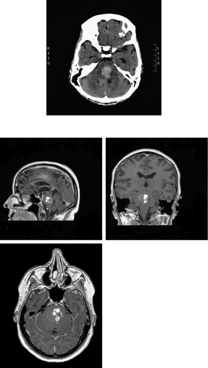
A 53-year-old Caucasian woman presented with sudden onset occipital headache with double vision, right-sided facial numbness and loss of balance, which developed over a few minutes. She was found to have a right-sided gaze palsy and a right internuclear opthalmoplegia (one-and-a-half syndrome), right-sided upper motor neuron (UMN) facial weakness and associated right-sided limb ataxia. A plain CT scan of the head showed a large right-sided pontine haemorrhage (see Figure 18.1). A brain MRI showed a 30 × 25 × 25mm right-sided pontine multicystic space-occupying lesion with surrounding haemorrhage. The ventricular system was of normal size with no signs of hydrocephalus (see Figure 18.2a–c).
The patient made a slow, but meaningful neurological recovery over a period of months. She experienced four further episodes of spontaneous bleeding over a course of 3 years. Each episode typically resulted in neurological deterioration followed by slow improvement. The patient had no previous family history of cerebral cavernous malformations.
 Learning point Brainstem gaze centres and the medial longitudinal fasiculus
A simplified model of gaze would involve three regions—the frontal gaze centres with control of saccadic eye movements, the occipital gaze centres for pursuit and accommodation, and the
brainstem gaze centres. These form a complicated internuclear connection network between the oculomotor, trochlear, and abducent nuclei, the vestibular brainstem nuclei and the mesencephalic nucleus of trigeminal nerve which provides feedback concerning head movement. The horizontal gaze centre is located in the abducent nucleus and possibly the parapontine reticular formation. The medial longitudinal fasiculus (MLF) connects the ipsilateral abducent nucleus with the contralateral oculomotor nucleus. Damage to the MLF results in internuclear opthalmoplegia (INO). Damage to the abducent nucleus, in addition, would result in ‘one-and-a-half’ syndrome, where there is complete paralysis of horizontal pursuit movement in the ipsilateral eye (the eye is fixed), and ipsilateral horizontal conjugate gaze palsy with preserved convergence.
Note that the nucleus of the facial nerve is in the proximity of the abducent nucleus.
The case was discussed at the local neurovascular Multidisciplinary team (MDT) meeting. Surgical excision of the lesion was proposed to the patient, but the patient was inclined to explore a non-operative management option. Therefore, the case was referred to the radiosurgery MDT meeting and the patient was deemed an appropriate candidate for treatment with radiosurgery. An application to the Primary Care Trust (PCT) was made to fund the treatment.
172 |
Challenging concepts in neurosurgery |
Figure 18.1 A plain CT scan of head showing hyperdensity in the pons in keeping with an acute haemorrhage.
(a) |
(b) |
(c)
Figure 18.2 (a,b,c) Sagittal, coronal, and axial MRI scans showing a multicystic lesion (cavernoma) in the pons.
Case 18 Brainstem cavernous malformation |
173 |
Discussion
Cavernous malformations or cavernomas are angiographically occult vascular malformations that affect the brain and spinal cord. Walter Dandy described the first surgical excision of a brainstem cavernous malformation in 1928 in a 31-year-old man, who presented with ‘stiffness in his right leg in spells’; Dandy stated that the patient ‘was living and well’ at follow up [1].
The true incidence of cavernous malformation became clear only following the development of MRI, as the vast majority of lesions are not detectable on angiography. Cavernomas of the CNS affect 0.4–0.9% of the population and account for 8–15% of all vascular malformations with 9–35% of cavernomas affecting the brainstem [2]. Post-mortem studies suggest that nearly 4% of the population have cavernous malformations [3], but with the ready availability of MRI in modern practice, the detection of incidental lesions (incidentalomas) has become more common, leading to an increasing number of referrals to neurosurgeons.
Cavernous malformations were thought to be purely congenital lesions; however, this has proven not to be the case as long-term follow-up especially of the familial variety showed that some lesions can develop de novo. Follow-up has also shown that those lesions are not static and can increase in size with time [3].
Cavernomas are histologically composed of sinusoidal layers of immature vascular endothelium, with evidence of chronic haemorrhage with haemosiderin-laden macrophages in the periphery of the lesion. There is no brain parenchyma within the lesion and no shunting. Gliotic changes are seen around the lesion. Occasionally, cystic formation signifying previous bleeding can be seen and some lesions have calcium deposits within them [4].
Presentation depends on the location of the lesion. Supratentorial lesions present with seizures in 50% of cases, focal neurological deficit in 30% of patients, and headache in 25% of patients. Patients can also present acutely with intracerebral haemorrhage and symptoms due to mass effect.
Infratentorial lesions very rarely present with seizures (3%) and usually present with focal neurological deficit due to haemorrhage, which can lead to mass effect and occasionally obstructive hydrocephalus. Brainstem and spinal cord cavernomas have a variety of presentations. Due to the fact that the brainstem and the spinal cord are composed of densely packed nuclei and tracts cavernomas present early with neurological deficit depending on the location and size of the lesion. There is a spectrum of deficit from gaze palsy, long tract signs to coma, and death from brainstem damage.
Bleeding risk is difficult to ascertain, especially as these lesions exhibit microhaemorrhages, explaining the presence of haemosiderin around the lesions even when there are no documented episodes of haemorrhage. The estimated risk of clinically significant haemorrhage in non-brainstem cavernomas is 0.1–1% per lesion per year. The risk of rebleeding is 4.5% if a single haemorrhage has occurred, but is much higher if there have been two or more clinically significant bleeds. Brainstem cavernomas have a higher rate of haemorrhage, this is largely due to the fact that even minor haemorrhages are symptomatic due to the tightly packed nuclei and tracts in the brainstem. The estimated risk of bleeding is 5% per year with a documented risk of rebleeding as high as 30% per year, the interval between haemorrhages is unpredictable [2].
A familial form of CCM has been described and linked to genetic mutations affecting the long arm of chromosome 7 (7q) [CCM1], the short arm of chromosome 7 (7p)

174
 Expert comment
Developmental venous anomalies (DVAs) can be useful radiological landmarks, but are not useful operatively, when one is generally trying to avoid them.
 Learning point
When cavernomas bleed, the haematoma tends to be ‘intralesional’, which leads to expansion of the lesion and
resultant mass effect in contrast to arteriovenous malformations (AVMs), which cause bleeding into the brain tissue and or
the subarachnoid space. For this reason cavernomas do not generally present with
subarachnoid haemorrhage (SAH).
 Expert comment
Cavernomas can change dramatically in their radiological appearance over time due to repeated haemorrhages. This may have the effect of making them seem to ‘grow’ in a particular direction. Having said that, this phenomenon can result in an improvement in the suitability for operative resection.
Challenging concepts in neurosurgery
[CCM2], and the long arm of chromosome 3 (3q) [CCM3] [5]. This entity is common amongst Hispanic Americans and can often present with multiple lesions. It is now believed that more than 55% of patients with cerebral cavernomas have familial tendencies [6]. Radiological follow-up of those patients shows that the lesions can change in size with time and new lesions can appear de novo [3].
 Learning point Imaging
The development of MRI played a major role in defining and understanding cavernous malformations, which were often termed angiographically occult or cryptic vascular malformations. Occasionally, a focal capillary blush can be seen on angiography. A developmental venous anomaly is a common finding adjacent to a cavernous malformation. CT imaging is often negative, except when there is an associated haematoma or calcification. The lesions have a characteristic ‘popcorn’ appearance on MRI and a signal loss is almost always seen surrounding the lesion on T2* (gradient echo) sequences representing haemosiderin deposition from chronic bleeding. The lesions can sometimes be multicystic due to previous bleeding episodes [7].
 Learning point Cavernomas and developmental venous anomalies
Developmental venous anomalies (DVA) are thought to be caused by a foetal vascular accident resulting in an anomalous vein draining the surrounding brain tissue. The lesions have a ‘caput medusa’ appearance on imaging. They are benign and do not result in bleeding. DVAs must be preserved during surgery as they drain normal brain tissue and damaging them can lead to a venous infarct. DVAs are often associated with cavernous malformations especially in the posterior fossa. Porter et al. found a 100% association with brainstem cavernomas intra-operatively. Therefore,
they hypothesized that DVAs may play a role in the formation of brainstem cavernomas and their postoperative recurrence because of this intimate association [2].
Brainstem cavernomas pose a challenging pathological entity. The commonest location is the pons in 60% followed by the midbrain, then the medulla oblongata. Due to the fact that the brainstem contains tightly-packed cranial nerve nuclei and long tract fibres, the slightest damage could lead to a catastrophic deficit. Therefore, brainstem cavernomas tend to present earlier and with much smaller haematomas than cavernomas in other locations in the CNS. They can present due to progressive neurological deficit from mass effect or direct damage to neural structures. Patients can also present acutely due to haemorrhage and/or obstructive hydrocephalus, which can lead to coma and death. Posterior fossa cavernomas do not present with seizures. Natural history studies have shown the risk of rebleeding from brainstem cavernomas to be significantly higher than cavernomas elsewhere in the CNS and can be as high as 30% per year. Cavernomas that present with haemorrhage are more likely to bleed again when compared with incidental cavernomas [2,8,9].
Management of cavernomas varies considerably with the location and size of the lesion, the symptoms associated with it, and patient specific factors, i.e. age and other comorbidities. Asymptomatic (incidental) lesions can be treated conservatively with clinical and radiological follow-up. Patients who present with epilepsy may benefit from surgical excision of the lesion with the surrounding haemosiderin ring, although surgery is not guaranteed to stop the seizures.

Case 18 Brainstem cavernous malformation |
175 |
Patients who present with mass effect or obstructive hydrocephalus from acute haemorrhage may require emergency evacuation of the haematoma, excision of the cavernoma and CSF diversion if required. In general, deep-seated lesions or lesions in eloquent locations are best treated conservatively if safe surgical excision is not possible.
The management of brainstem cavernomas and spinal cord cavernomas differs from the management of cavernomas elsewhere in the CNS due to the much higher risk of rebleeding, and the fact that very little mass effect or haemorrhage can lead to catastrophic consequences. Some authors recommend surgical excision through skull base approaches to lesions that abut the pial surface or only have a thin layer of tissue covering in symptomatic patients with two or more previous episodes of haemorrhage or one episode of haemorrhage that did not lead to a full recovery. Asymptomatic patients or patients with one previous episode of haemorrhage who go on to make a full recovery, could be treated conservatively along with patients with deep-seated brainstem lesions [2,10–13].
Another relatively modern treatment modality is stereotactic radiosurgery, i.e. Gamma knife or Cyberknife to lesions in eloquent locations when surgery is contraindicated, or is expected to result in significant morbidity or mortality. Hasegawa et al. published long-term follow-up results of eighty-two patients with high risk cavernomas treated with gamma knife in the University of Pittsburgh, Pennsylvania, between 1987 and 2000. They showed a significant reduction of the annual risk of bleeding, which was most pronounced after 2 years of treatment with minimal side effects from the treatment itself [14].
To date, there is no Class 1 evidence that stereotactic radiosurgery works, due to the difficulty in performing such a study. Advocates of surgery remain sceptical about this form of treatment [2,10].
 Clinical tip Surgical approaches to brainstem cavernomas—the two-point method
Surgical approaches to brainstem cavernomas require a highly-skilled surgical team with meticulous pre-operative planning, including specialised imaging modalities such as tractography, which may play an increasing role in the future planning of surgical approaches. It is absolutely crucial to plan the entry point to the brainstem pre-operatively and not to rely heavily on neuronavigation systems as even a minimal amount of brain shift can be misleading. This is generally not a problem when dealing with large lesions that reach the surface.
A good planning strategy is to draw a line from the centre of the lesion to the most superficial point and out through the skull. This line can be used as a guide to decide on the best surgical corridor to use in order to avoid retraction and minimize injury to neural tissue. The approaches commonly utilized are the retrosigmoid, the suboccipital, the far lateral, and the subtemporal approaches [11–13,15,16].
 Clinical tip
When operating, ensure you have the best equipment available. Do not compromise on the patient’s position, the quality of dissectors, retractors, bipolar coagulation forceps, suction, or microscope. For a brainstem cavernoma operation, everything needs to be working at its best, especially the surgeon!
A final word from the expert
Never operate on brainstem cavernomas if avoidable; many neurovascular surgeons wait for more than one symptomatic bleed. Timing is crucial—the patient must have some salvageable neurology that one would expect to be irreversibly lost if surgery was not undertaken.
176 |
Challenging concepts in neurosurgery |
References
1.Brown DL, Archer SB, Greenhalgh DG, et al. Inhalation injury severity scoring system: a quantitative method. Journal of Burn Care & Rehabilitation 1996; 17: 552–7.
2.Porter RW, Detwiler PW, Spetzler RF, et al. Cavernous malformations of the brainstem: experience with 100 patients. Journal of Neurosurgery 1999; 90: 50–8.
3.Zabramski JM, Wascher TM, Spetzler RF, et al. The natural history of familial cavernous malformations: results of an ongoing study. Journal of Neurosurgery 1994; 80: 422–32.
4.Gault J, Sarin H, Awadallah NA, et al. Pathobiology of human cerebrovascular malformations: basic mechanisms and clinical relevance. Neurosurgery 2004; 55: 1–16; discussion 16–17.
5.Mindea SA, Yang BP, Shenkar R, et al. Cerebral cavernous malformations: clinical insights from genetic studies. Neurosurgery Focus 2006; 21: e1.
6.Gunel M, Awad IA, Finberg K, et al. A founder mutation as a cause of cerebral cavernous malformation in Hispanic Americans. New England Journal of Medicine 1996; 334: 946–51.
7.Rigamonti D, Drayer BP, Johnson PC, et al. The MRI appearance of cavernous malformations (angiomas). Journal of Neurosurgery 1987; 67: 518–24.
8.Fritschi JA, Reulen HJ, Spetzler RF, et al. Cavernous malformations of the brain stem. A review of 139 cases. Acta Neurochirugia (Wien) 1994; 130: 35–46.
9.Zimmerman RS, Spetzler RF, Lee KS, et al. Cavernous malformations of the brain stem. Journal of Neurosurgery 1991; 75: 32–9.
10.Abla AA, Lekovic GP, Garrett M, et al. Cavernous malformations of the brainstem presenting in childhood: surgical experience in 40 patients. Neurosurgery 2010; 67: 1589–98; discussion 1598–9.
11.Abla AA, Turner JD, Mitha AP, et al. Surgical approaches to brainstem cavernous malformations. Neurosurgery Focus 2010; 29: E8.
12.Garrett M, Spetzler RF. Surgical treatment of brainstem cavernous malformations. Surgical Neurology 2009; 72 (Suppl. 2): S3-9; discussion S9–10.
13.Wang CC, Liu A, Zhang JT, et al. Surgical management of brain-stem cavernous malformations: report of 137 cases. Surgical Neurology 2003; 59: 444–54; discussion 454.
14.Hasegawa T, McInerney J, Kondziolka D, et al. Long-term results after stereotactic radiosurgery for patients with cavernous malformations. Neurosurgery 2002; 50: 1190–7; discussion 1197–8.
15.Brown AP, Thompson BG, Spetzler RF. The two-point method: evaluating brain stem lesions. Barrow Neurological Institute Quarterly 1996; 12: 20–4.
16.Degn J, Brennum J. Surgical treatment of trigeminal neuralgia. Results from the use of glycerol injection, microvascular decompression, and rhizotomia. Acta Neurochirugia (Wien) 2010; 152: 2125–32.