

CASE
9Bilateral vestibular schwannomas: the challenge of neurofibromatosis type 2
Patrick Grover
 Expert commentary Robert Bradford
Case history
A 17-year-old female presented to her GP with a 1-year history of left-sided hearing loss, worsening balance, generalized headaches, and numbness affecting the left side of her face and tongue. She had previously been treated for left-sided otitis media and externa, and had undergone ear syringing for wax. Her previous medical history included meningitis as a child, but she had suffered no permanent neurological deficits. She had no family or social history of note.
On examination she had multiple café au lait spots over her abdomen and nodular fleshy polyps on her elbows, knees, and shins. She had bilateral sensorineural hearing deficits worst on the left side. Light touch and pin prick sensations were reduced on the left side of her face in the V2 and V3 dermatomes. Corneal reflex was absent in the left eye. There was uvular deviation to the left and tongue deviation to the left with right-sided wasting, consistent with glossopharyngeal and hypoglossal cranial nerve deficits. There was no facial nerve dysfunction. Cerebellar examination elicited slight left-sided dysmetria.
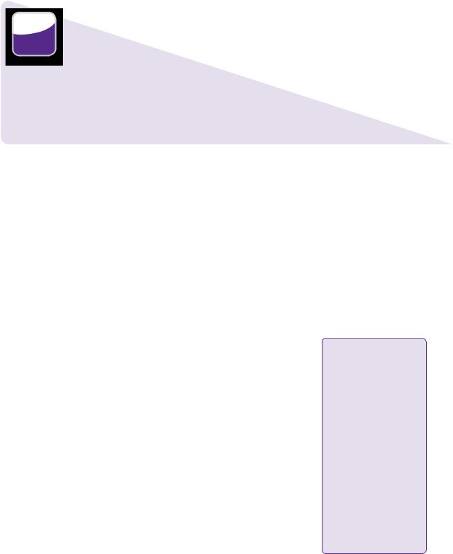
MRI demonstrated bilateral homogenously enhancing cerebellopontine angle mass lesions expanding the internal acoustic meati (Figure 9.1). The left-sided lesion measured 37 × 27 × 25mm and caused significant compression of the pons and medulla. The right-sided lesion was smaller at 23 × 14 × 11mm. Two further small extra-axial enhancing lesions were present in the right olfactory groove and abutting the right petrous ridge. There was no associated hydrocephalus. Audiometry confirmed bilateral sensorineural hearing loss particularly on the left side.
These findings were consistent with bilateral vestibular schwannoma and additional supratentorial meningiomata, with an underlying diagnosis of NF2. The patient’s symptoms and signs were predominantly localized to the larger left cerebellopontine angle lesion, which was debulked through a left retrosigmoid craniotomy and sub-occipital approach. The patient was started on dexamethasone (4mg qds po/iv). Post-operatively, the patient experienced hearing loss in her left ear, but no additional cranial nerve dysfunction. She subsequently developed hydrocephalus, for which a ventriculo-peritoneal shunt was inserted, and recovered well thereafter.
The gross pathological appearance of the resected fragments was of tan/grey pieces of tissue with a granular appearance. Histology showed multiple interlacing bundles of spindle cells with no atypical features, characteristic of a WHO Grade I schwannoma (Table 9.1).
 Learning point Diagnostic criteria for neurofibromatosis type 2
The Manchester criteria are the most widely used criteria for defining neurofibromatosis [1]. They describe neurofibromatosis type 2 (NF2) associated lesions as meningioma, glioma, schwannoma, neurofibroma, or posterior subcapsular lenticular
opacities. Diagnosis requires one of the following:
●Bilateral vestibular schwannomas
●A first-degree relative with NF2 and either a unilateral vestibular schwannoma or two NF2 associated lesions.
●Unilateral acoustic neuroma and two NF2 associated lesions.
●Multiple meningiomas and either a unilateral acoustic neuroma or two NF2 associated lesions.
84 |
Challenging concepts in neurosurgery |
(a) |
(b) |
(c) |
(d) |
(e) |
(f) |
Figure 9.1 MRI images demonstrating preand post-surgical appearance of bilateral vestibular schwannomas and additional intracranial meningiomas. (a) T1-weighted axial MRI. (b) T1-weighted with gadolinium. (c) T2-weighted. (d) Sagittal T1-weighted with gadolinium. (e) T1-weighted with gadolinium demonstrating olfactory groove meningioma. (f) T1-weighted with gadolinium after two surgical resections of the left tumour.
Her 6-month follow-up MRI unfortunately revealed progressive growth of the left-sided tumour and the patient underwent further planned subtotal excision via a translabyrinthine approach (Figure 9.1). Post-operative MRI scans at 3 and 11 months showed stable appearances of the remaining right-sided tumour and small left-sided residual. The patient has not experienced any further progression of her symptoms and has retained hearing in her right ear with moderate loss to 55 dB at 2000Hz and 30–40dB at high frequencies (Figure 9.2). She has no hearing in the left ear. The patient’s family are currently undergoing genetic testing, although no other member has clinical symptoms or signs of neurofibromatosis.
Table 9.1 Histopathological classification of central and peripheral nerve sheath tumours based on the 2007 World Health Organization (WHO) classification [30].
Subtypes |
Histological variants |
Description |
Schwannoma |
Cellular, plexiform, |
Encapsulated nerve sheath tumour composed of |
(neurilemoma, |
melanotic |
well-differentiated neoplastic Schwann cells |
neurinoma) |
|
|
Neurofibroma |
Plexiform |
Unencapsulated fusiform nerve sheath tumour |
|
|
composed of a mix of neoplastic Schwann cells, |
|
|
perineural cells, and fibroblasts in a collagen matrix |
Perineuroma |
Perineuroma, malignant |
Tumours of perineural cells, which form either |
|
perineuroma |
intraneurally encasing the nerve or within soft tissue |
Malignant |
Epitheloid, melanotic, MPNST |
Malignant and invasive peripheral nerve sheath |
peripheral nerve |
with glandular differentiation, |
tumour composed of spindle cells. Hypercellular |
sheath tumour |
MPNST with mesenchymal |
with high mitotic activity, hyperchromatic nuclei |
(MPNST) |
differentiation |
and necroses |
|
|
|
Perry A LD, Scheithauer BW, Budka H, von Diemling A. World Health Organization Classification of Tumours of the Central Nervous System, ed 4. Lyon: IARC, 2007.

Right
|
–10 |
|
125 |
|
|
||
|
0 |
|
|
|
10 |
|
|
|
20 |
|
|
(dB) |
30 |
|
|
40 |
|
|
|
level |
60 |
|
|
Heaning |
50 |
|
|
70 |
|
|
|
|
|
|
|
|
80 |
|
|
|
90 |
|
|
|
100 |
|
|
|
110 |
|
|
|
120 |
|
|
|
|
|
|
|
130 |
|
|
|
140 |
|
|
|
|
|
|
|
|
|
Case 9 Bilateral vestibular schwannomas |
85 |
||||||||
|
Frequency (Hz) |
|
|
|
Left |
|
|
Frequency (Hz) |
|
|
|
|
||||
250 |
500 |
1000 |
2000 |
4000 |
8000 |
–10 |
–10 |
125 |
250 |
500 |
1000 |
2000 |
4000 |
8000 |
–10 |
|
|
|
|
|
|
|
|
|
|
|
|
|
|
|
|||
|
|
|
|
|
|
0 |
0 |
|
|
|
|
|
|
|
0 |
|
|
|
|
|
|
|
10 |
10 |
|
|
|
|
|
|
|
10 |
|
|
|
|
|
|
|
20 |
20 |
|
|
|
|
|
|
|
20 |
|
|
|
|
|
|
|
30 |
30 |
|
|
|
|
|
|
|
30 |
|
|
|
|
|
|
|
40 |
40 |
|
|
|
|
|
|
|
40 |
|
|
|
|
|
|
|
50 |
50 |
|
|
|
|
|
|
|
50 |
|
|
|
|
|
|
|
60 |
60 |
|
|
|
|
|
|
|
60 |
|
|
|
|
|
|
|
70 |
70 |
|
|
] |
× |
|
|
|
70 |
|
|
|
|
|
|
|
80 |
80 |
|
|
|
|
|
|
80 |
|
|
|
|
|
|
|
|
|
|
|
] |
|
] |
|
|
|||
|
|
|
|
|
|
90 |
90 |
|
× |
× |
] |
|
90 |
|
||
|
|
|
|
|
|
|
|
|
× × |
|
||||||
|
|
|
|
|
|
100 |
100 |
|
|
|
|
|
× |
100 |
|
|
|
|
|
|
|
|
|
× |
|
|
|
|
|
||||
|
Right ear masked air conduction |
110 |
|
|
|
|
|
× |
110 |
|
||||||
|
|
|
|
|
|
|
|
|||||||||
|
120 |
|
|
× |
× |
× × |
× × |
× |
120 |
|
||||||
|
Right ear air conduction |
|
|
|
|
|
||||||||||
|
|
|
130 |
|
|
|
|
|
|
|
130 |
|
||||
] |
Left ear masked bone conduction |
|
|
|
|
|
|
|
|
|||||||
× |
Left ear air conduction |
|
|
|
140 |
|
|
|
|
|
|
|
140 |
|
||
Figure 9.2 Post-surgical audiometry demonstrating bilateral sensorineural hearing loss with no useful hearing on the left side.
Discussion
Vestibular schwannomas are benign tumours that are said to originate predominantly from the superior vestibular nerve. Their incidence is estimated to be between two and twenty cases per million worldwide, comprising approximately 5–10% of intracranial tumours [2]. They arise in the cerebellopontine angle accounting for approximately 90% of mass lesions in this location [3]. Speculation that their incidence may be increased by mobile phone exposure has been the subject of several studies, most of which have found no association [4]. However, the recent multi-national INTERPHONE case-control study suggested an increased incidence of ipsilateral tumours after 10 years usage [5]. They compared 678 cases of vestibular schwannoma and 3553 controls, retrospectively, interviewing participants to determine mobile phone usage. There was no overall increase in the risk of developing vestibular schwannoma (OR 0.9, 95% CI 0.7–1.1), but the risk of developing a tumour on the same side of the head as reported phone usage was statistically greater after 10 years (OR 1.8, 95% CI 1.1–3.1).
Vestibular schwannomas arise from the neurolemmal sheath of the vestibular nerve at the junction between oligodendrocytes and Schwann cells known as the Obersteiner–Redlich zone, approximately 8–12mm from the brainstem. They are characterized by spindle cells arranged in tightly compact fascicles within Antoni type A areas, and loosely compacted in Antoni type B areas. Bilateral vestibular schwannomas are pathognomonic of NF2 and are caused by mutations of the neurofibromin 2 gene found on chromosome 22q. This codes for tumour suppressor protein merlin, also known as schwannomin, which has roles in cellular proliferation and cell adherence [6]. Mutations in neurofibromin 2 are also found in a significant proportion of sporadic vestibular schwannomas [7].
Nearly all patients present with unilateral hearing loss with or without tinnitus. Balance disturbance affects around two-thirds of patients and headaches are similarly common. Facial numbness due to trigeminal nerve involvement can be a feature in up to one-third, whereas symptoms relating to the other surrounding cranial nerves are uncommon. On examination, a sensorineural hearing deficit is
 Learning point Genetics of neurofibromatosis type 2
NF2 is autosomal dominant with high penetrance in inherited cases leading to clinical manifestations by the age of 60 in almost all affected individuals [8]. However, more than 50% of cases are sporadic and, of these, approximately one-third are mosaic—only a proportion of cells carry the mutation, which
occurs after conception [8]. These patients will have a milder clinical course and a less than 50% chance of passing the mutation to their offspring.
86 |
Challenging concepts in neurosurgery |
 Expert comment The presentation of neurofibromatosis type 2
In adulthood, NF2 most commonly presents with symptoms and
signs of vestibular schwannoma, particularly unilateral hearing loss. A smaller proportion are diagnosed from alternative lesions, such as intracranial meningiomas causing seizures and headaches, ocular tumours leading to visual loss, spinal masses resulting in limb weakness and sensory disturbance, and cutaneous stigmata. More severe cases present in the paediatric population, where symptoms and signs from these alternative lesions are more prevalent [11].
almost universal. Nystagmus, facial hypoaesthesia and an abnormal corneal reflex are each elicited in approximately one-third of patients. Facial weakness and oculomotor nerve palsy may be detected infrequently. Large tumours (>3cm) can present with symptoms and signs of brainstem compression, lower cranial nerve damage, ataxia, or raised intracranial pressure due to obstruction of the fourth ventricle causing non-communicating hydrocephalus [9,10].
 Clinical tip Cranial nerve examination in vestibular schwannoma
The differential diagnosis of a patient with unilateral hearing and balance loss remains wide and a thorough cranial nerve examination is crucial. Although intimately associated with the VIIIth cranial nerve (Figure 9.3), the VIIth nerve is much less often clinically affected by compression. Symptoms and signs of trigeminal nerve involvement are slightly more common although still seen in less than 20% of cases [10]. Loss of the corneal reflex is a particularly useful sign as it often precedes facial hypoaesthesia [9].
c
b
a
Figure 9.3 Vestibular schwannoma and relations to cranial nerves. a, vestibular schwannoma; b, facial nerve (VII); c, vestibulocochlear nerve (VIII).
The investigation of choice in patients clinically suspected of vestibular schwannoma is MRI [12,13]. Our recommendation is that any patient with unilateral sensorineural hearing loss should have an MRI scan [13]. The spatial resolution of MRI is now such that the sensitivity of non-contrasted thin-slice T2and T2*-weighted imaging approaches 100% for cerebellopontine angle lesions with a specificity of between 90 and 100% [12,14]. Contrasted scans only improve sensitivity if dedicated internal acoustic meatus (IAM) views are not undertaken and are usually unnecessary.
Abnormalities of the auditory brainstem response waveform are reliably found in tumours greater than 1cm in size, but this test is not specific and is now rarely performed as MRI has become more widely available [15]. CT with contrast will miss up

Case 9 Bilateral vestibular schwannomas |
87 |
to 10% of cases, but may be useful to demonstrate bony anatomy. Audiometry is of value in serial monitoring of hearing but its use as a diagnostic tool is limited to the detection of hearing loss in advance of scanning. Speech discrimination is typically disproportionately impaired relative to pure tone threshold.
 Learning point
The American Academy of Otolaryngology-Head and Neck Surgery (AAO-HNS) hearing classification is the most widely used in vestibular schwannoma assessment (see Table 9.2) [16]
Table 9.2 The American Academy of Otolaryngology-Head and
Neck Surgery (AAO-HNS) hearing classification
Class |
Pure tone threshold (dB) |
Speech discrimination (%) |
|
|
|
A |
≤30 |
≥70 |
B |
30.1–50 |
≥50 |
C |
>50 |
≥50 |
D |
Any level |
<50 |
|
|
|
Vestibular schwannomas may be managed conservatively, surgically or with radiotherapy. Combined modality treatment has also been successfully reported, e.g. sub-total debulking followed by gamma knife therapy for large vestibular schwannomas [17]. Smouha et al. (2005) carried out a meta-analysis of the conservative management of 1345 cases in twenty-one studies, with a mean follow-up of 3.2 years [18]. Mean growth rate was 1.9mm/year with 43% of cases progressing, and 57% not growing or regressing. Fifty-one percent of the 347 patients in whom data was available lost hearing. It is important to remember this represents a small selected cohort of patients not felt suitable for immediate intervention. For example, of 432 patients referred with vestibular schwannomas to the Northwestern otolaryngology unit in Chicago, USA, fifty-three were initially managed conservatively [19]. These patients tend to be older (mean age = 62) with smaller tumours (mean size = 11.8mm) [18]. It is also important to note that these tumours may show non-linear growth, with enlargement after a period (which may be many years) with no apparent growth.
Selecting which patients are suitable for conservative management is difficult due to a lack of factors predictive of clinical progression. Twenty percent of patients in the Smouha et al. meta-analysis failed conservative management, and progressed to surgery or radiotherapy. Mean growth rate at 1 year has been shown to be predictive of the eventual need for treatment in conservatively managed patients but not routinely so [20,21].
 Expert comment Suitability for conservative management
A suitable patient is one who makes a fully-informed decision, based on balancing the risks and benefits in collaboration with their surgeon. Patients with small masses and preserved hearing may wish to forgo the risk of losing their hearing in the short-term, accepting a greater risk of hearing loss and other complications in the future. Older patients and those with multiple comorbidities have a higher risk of peri-operative complications, which might outweigh the potential benefits of surgery.
Tumours may be approached surgically via three approaches. The two most commonly used are retrosigmoid transmeatal and translabyrinthine, with the middle cranial fossa approach less prevalent. The retrosigmoid transmeatal approach
88 |
Challenging concepts in neurosurgery |
a
c
d
b
 Expert comment
Indications for intervention in vestibular schwannoma in NF2 used in our practice include tumours that are:
●Greater than 3cm.
●2–3cm if enlarging on serial imaging.
●1–2cm if growing, with poor hearing. In those with good hearing, hearing preservation surgery may be undertaken with cochlear nerve implant if required
 Expert comment
Cerebrospinal fluid leakage
CSF leakage is a common and troublesome complication of vestibular schwannoma surgery causing significant morbidity. Rates tend to be highest with the translabyrinthine approach. The incidence can be minimized with meticulous dural closure (not possible with translabyrinthine surgery), intra-operative fat and fascia grafting, use of fibrin glue, and post-operative lumbar CSF drainage.
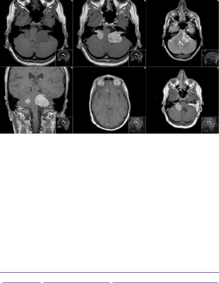
Figure 9.4 Vestibular schwannoma as seen through a retrosigmoid transmeatal approach. a, retractor; b, cerebellum; c, vestibular schwannoma; d temporal bone.
enables access to almost all tumours with the possibility of hearing preservation (Figure 9.4) [22]. However, cerebellar retraction is required and the facial nerve is encountered late, resulting in higher rates of facial nerve palsy.
The translabyrinthine approach sacrifices vestibular and cochlear function on that side, but has the advantage of being a predominently extracranial approach with early identification of the facial nerve. Preservation rates of greater than 90% for small tumours and 50% for large tumours may be expected with this technique [23]. The middle cranial fossa approach is largely extradural and enables hearing preservation also, but is limited to small intracanalicular tumours, and carries a high risk of damage to the geniculate ganglion and facial nerve palsy [24]. It is important to recognize that these rates are case series from experienced centres representing exceptional outcomes. Rates of serviceable hearing preservation after surgery, for example, are not typically as high as quoted in the literature. Furthermore, all procedures carry a risk of CSF leakage in between 10 and 20% of cases, and a small risk of mortality (<1%).
Stereotactic radiosurgery and fractionated radiotherapy are increasingly used in the management of small vestibular schwannomas. A non-randomized prospective study of eighty-two patients treated at the Mayo clinic, Minnesota, USA, compared gamma knife stereotactic radiosurgery in forty-two patients, with microsurgery carried out on thirty-six patients for tumours of similar size and clinical profile [25]. At a mean of 42 months follow-up, facial nerve function was preserved in 96% of the gamma knife group and 75% of the surgical group, while useful hearing was maintained in 63% of gamma knife patients compared with only 5% of the surgical cohort. Operated patients also had significantly worse physical functioning, and pain at 3- and 12-month follow-up.

Case 9 Bilateral vestibular schwannomas |
89 |
These data demonstrate a significantly better side effect profile for stereotactic radiosurgery, but it should be noted that evidence on long-term tumour control is lacking as the gamma knife has been in use in its current form only since 1985, with progressive dose reduction over the past 20 years. Furthermore, there is concern regarding the development of radiation-induced tumours or malignant transformation, which again may only be uncovered after a long follow-up period. At present, the risk appears to be in the region of 1:5000–1:10,000 for sporadic tumours, but it is probably much greater for NF2 tumours, which are inherently less stable. Multiple modalities of radiotherapy administration have shown efficacy in acoustic neuroma including Linear Accelerator (LINAC) radiosurgery [26] and Cyberknife® [27], with the longest follow-up for gamma knife single fraction treatment. Staged or fractionated protocols apply lower radiation doses multiple times and may result in reduced rates of damage to surrounding structures [28]. From our 12 years’ experience of fractionated radiotherapy, we quote control rates of greater than 90%, with a 3% risk of cranial nerve damage and 50% risk of loss of useful hearing.
A final word from the expert
Treatment of these tumours will increasingly depend on less invasive modalities including focused radiation techniques and molecular chemotherapeutics. Radiosurgery is significantly less effective in NF2-associated schwannomas and this represents a particular challenge
in management. Clinical trials of bevacizumab (a VEGF inhibitor), and lapatinib (a tyrosine kinase inhibitor) in NF2 are ongoing [29].
Acknowledgements
The authors wish to thank Professor Shakeel Saeed who proof-read the article and contributed to the expert commentary.
References
1.Evans DG, Baser ME, O’Reilly B, et al. Management of the patient and family with neurofibromatosis 2: a consensus conference statement. British Journal of Neurosurgery 2005; 19 (1): 5–12.
2.Tos M, Stangerup SE, Caye-Thomasen P, et al. What is the real incidence of vestibular schwannoma? Archives of Otolaryngology—Head and Neck Surgery 2004; 130 (2): 216–20.
3.Brackmann DE, Bartels LJ. Rare tumors of the cerebellopontine angle. Otolaryngology— Head and Neck Surgery 1980; 88 (5): 555–9.
4.Han YY, Kano H, Davis DL, et al. Cell phone use and acoustic neuroma: the need for standardized questionnaires and access to industry data. Surgical Neurology 2009; 72 (3): 216–22 ; discussion 222.
5.INTERPHONE Study Group. Acoustic neuroma risk in relation to mobile telephone use: results of the INTERPHONE international case-control study. Cancer Epidemiology 2011; 35 (5): 453–64.
6.Sughrue ME, Yeung AH, Rutkowski MJ, et al. Molecular biology of familial and sporadic vestibular schwannomas: implications for novel therapeutics. Journal of Neurosurgery 2011; 114 (2): 359–66.
90 |
Challenging concepts in neurosurgery |
|
|
7. |
Irving RM, Moffat DA, Hardy DG, et al. Somatic NF2 gene mutations in familial and non- |
|
|
familial vestibular schwannoma. Human Molecular Genetics 1994; 3 (2): 347–50. |
|
8. |
Evans DG, Sainio M, Baser ME. Neurofibromatosis type 2. Journal of Medical Genetics |
|
|
2000; 37 (12): 897–904. |
|
9. |
Harner SG, Laws ER, Jr. Clinical findings in patients with acoustic neurinoma. Mayo |
|
|
Clinic Proceedings 1983; 58 (11): 721–8. |
|
10. |
Matthies C, Samii M. Management of 1000 vestibular schwannomas (acoustic neuromas): |
|
|
clinical presentation. Neurosurgery 1997; 40 (1): 1–9; discussion 9–10. |
|
11. |
Evans DG, Birch JM, Ramsden RT. Paediatric presentation of type 2 neurofibromatosis. |
|
|
Archives of Diseases of Childhood 1999; 81 (6): 496–9. |
|
12. |
Fortnum H, O’Neill C, Taylor R, et al. The role of magnetic resonance imaging in the iden- |
|
|
tification of suspected acoustic neuroma: a systematic review of clinical and cost effective- |
|
|
ness and natural history. Health Technology Assessment 2009; 13 (18): iii–iv, ix–xi, 1–154. |
|
13. |
Wright A, Bradford R. Management of acoustic neuroma. British Medical Journal 1995; |
|
|
311 (7013): 1141–4. |
|
14. |
Soulie D, Cordoliani YS, Vignaud J, et al. MR imaging of acoustic neuroma with high resolu- |
|
|
tion fast spin echo T2-weighted sequence. European Journal of Radiology 1997; 24 (1): 61–5. |
|
15. |
Schmidt RJ, Sataloff RT, Newman J, et al. The sensitivity of auditory brainstem response |
|
|
testing for the diagnosis of acoustic neuromas. Archives of Otolaryngology—Head and |
|
|
Neck Surgery 2001; 127 (1): 19–22. |
|
16. |
Monsell EM. New and revised reporting guidelines from the Committee on Hearing and |
|
|
Equilibrium. American Academy of Otolaryngology-Head and Neck Surgery Foundation, |
|
|
Inc. Otolaryngology—Head and Neck Surgery 1995; 113 (3): 176–8. |
|
17. |
van de Langenberg R, Hanssens PE, Verheul JB, et al. Management of large vestibular |
|
|
schwannoma. Part II. Primary gamma knife surgery: radiological and clinical aspects. |
|
|
Journal of Neurosurgery 2011; 115 (5): 885–93. |
|
18. |
Smouha EE, Yoo M, Mohr K, et al. Conservative management of acoustic neuroma: a |
|
|
meta-analysis and proposed treatment algorithm. Laryngoscope 2005; 115 (3): 450–4. |
|
19. |
Wiet RJ, Zappia JJ, Hecht CS, et al. Conservative management of patients with small |
|
|
acoustic tumors. Laryngoscope 1995; 105 (8 Pt 1): 795–800. |
|
20. |
Deen HG, Ebersold MJ, Harner SG, et al. Conservative management of acoustic neuroma: |
|
|
an outcome study. Neurosurgery 1996; 39 (2): 260–4; discussion 264–6. |
|
21. |
Sughrue ME, Kane AJ, Kaur R, et al. A prospective study of hearing preservation in |
|
|
untreated vestibular schwannomas. Journal of Neurosurgery 2011; 114 (2): 381–5. |
|
22. |
Gormley WB, Sekhar LN, Wright DC, et al. Acoustic neuromas: results of current |
|
|
surgical management. Neurosurgery 1997; 41 (1): 50–8; discussion 58–60. |
|
23. |
Sterkers JM, Morrison GA, Sterkers O, et al. Preservation of facial, cochlear, and other |
|
|
nerve functions in acoustic neuroma treatment. Otolaryngology—Head and Neck Surgery |
|
|
1994; 110 (2): 146–55. |
|
24. |
Shelton C, Brackmann DE, House WF, et al. Middle fossa acoustic tumor surgery: results |
|
|
in 106 cases. Laryngoscope 1989; 99 (4): 405–8. |
|
25. |
Pollock BE, Driscoll CL, Foote RL, et al. Patient outcomes after vestibular schwannoma |
|
|
management: a prospective comparison of microsurgical resection and stereotactic |
|
|
radiosurgery. Neurosurgery 2006; 59 (1): 77–85. |
|
26. |
Friedman WA. Linear accelerator radiosurgery for vestibular schwannomas. Progress in |
|
|
Neurological Surgery 2008; 21: 228–37. |
|
27. |
Chang SD, Gibbs IC, Sakamoto GT, et al. Staged stereotactic irradiation for acoustic neu- |
|
|
roma. Neurosurgery 2005; 56 (6): 1254–61 ; discussion 1261–3. |
|
28. |
Williams JA. Fractionated stereotactic radiotherapy for acoustic neuromas. International |
|
|
Journal of Radiation Oncology • Biology • Physics 2002; 54 (2): 500–4. |
|
29. |
Chang, S.D., et al. Staged stereotactic irradiation for acoustic neuroma. Neurosurgery, |
|
|
2005. 56 (6): 1254–61 ; discussion 1261–3. |
|
30. |
Perry A LD, Scheithauer BW, Budka H, et al. World Health Organization Classification of |
|
|
Tumours of the Central Nervous System, 4th edn. Lyon: IARC, 2007. |