


CASE
12Deep brain stimulation for debilitating Parkinson’s disease
Jonathan A. Hyam
 Expert commentary Alexander L. Green and Tipu Z. Aziz
Case history
A 70-year-old man was referred to the functional neurosurgical service with a diagnosis of idiopathic Parkinson’s disease (PD) for 16 years. His symptoms were rigidity, bradykinesia, and dyskinesias, causing disability and limitations in his activities of daily living, such as washing himself, cutting up food, writing, and safely using appliances unsupervised. He had no disturbance of awareness, sensory-perceptual function, thought, or intellectual function.
Since the time of his diagnosis his oral medications included madopar (containing L-dopa and a levodopa (L-dopa) decarboxylase inhibitor to reduce its breakdown outside the brain), selegiline (a monoamine oxidase inhibitor), pergolide (a dopaminergic agonist), and he had an apomorphine (another dopaminergic agonist) pump in situ. However, his medical therapy had resulted in dyskinesias, 6 years after the diagnosis of PD. After adjustment to sinemet and apomorphine, he experienced a medication ‘off’ state for 75% of the day with motor symptoms breakthrough despite medication with resulting bradykinesia and rigidity; and medication ‘on’ state for 25% of the day.
 Learning point Pharmacological agents used in Parkinson’s disease
Levodopa therapy was a breakthrough in the management of PD during the 1960s [1]. Due to the eventual motor complications of its use, a variety of other drugs have been developed for PD, which act on dopaminergic and non-dopaminergic systems. In early PD, there is no single first choice drug, and therapy with L-dopa, dopaminergic agonists, or monoamine oxidase-B inhibitors, is recommended [2]. Table 12.1 describes the range of pharmacological agents currently used in PD [3].
Table 12.1 Pharmacological agents used in PD
Type |
Example |
Side effects |
Dopamine |
L-dopa |
Motor fluctuations, dyskinesias |
|
Madopar, Sinemet (L-dopa |
|
|
+ decarboxylase inhibitor) |
|
Dopaminergic agonist |
Bromocriptine, |
Hallucinations, sleepiness, |
|
apomorphine, |
impulsive behaviour, |
|
pramipexole |
e.g. gambling, hypersexuality |
Anticholinergics |
Benzhexol |
Central/peripheral autonomic disturbance |
Monoamine oxidase inhibitors |
Selegiline |
Sleep disturbance, light-headedness |
Catechol-O-methyltransferase |
Entacapone |
Augmented dopa-induced dyskinesias, |
Inhibitors |
|
|
Glutamate-antagonist; ? |
Amantadine |
Hallucinations, depression |
dopamine reuptake blocker |
|
|
|
|
|
 Expert comment
There are many treatments for Parkinson’s disease and DBS should be considered as one of many options, although not always the most suitable. As these are complex patients, they should be assessed by a multidisciplinary team including neurologist, surgeon, specialist nurse, neuropsychologist, and other relevant healthcare professionals
116 |
Challenging concepts in neurosurgery |
|
|
||
|
|
On examination, he had no arm tremor. Movements were bradykinetic. Dyskinesias, |
|||
|
particularly on the right, and bilateral cog-wheel rigidity were present. Cranial nerve |
||||
|
and peripheral neurological examinations were otherwise normal. Micrographia was |
||||
|
demonstrated. Unified Parkinson’s disease Rating Scale (UPDRS) scores were Part 1: |
||||
|
5/16; Part 2: 3/56 on, 25/56 off; Part 3: 15/104 on, 43/104 off. Formal neuropsycho- |
||||
|
logical evaluation using interview and battery tests did not reveal significant psycho- |
||||
|
logical pathology. Brain MRI was normal. |
||||
|
|
Learning point Unified Parkinson’s disease Rating Scale |
|||
|
|
UPDRS was developed to monitor the severity and progression of PD, whereby several existing scales |
|||
|
|
were incorporated into one, allowing more efficient and flexible patient assessment. It is extensively |
|||
|
|
used by neurologists across the world with 87% reporting its use in trials and 70% using it in clinical |
|||
|
|
practice [4]. The Movement Disorders Society judged the UPDRS as providing a comprehensive |
|||
|
|
assessment of the motor aspects of PD especially, more than the non-motor aspect, although some |
|||
|
|
items had low or adequate interand intra-rater reliability [4]. It is divided into four categories |
|||
|
|
evaluating: |
|
|
|
|
|
● I: mentation, behaviour, and mood. |
|
|
|
|
|
● II: activities of daily living. |
|
|
|
|
|
● III: motor examination. |
|
|
|
|
|
● IV: complications of therapy. |
|
|
|
|
|
The Modified Hoehn & Yahr Staging and Schwab & England ADL Scales were added later (Table 12.2). |
|||
|
|
Table 12.2 Unified PD Rating Scale |
|
|
|
|
|
|
|
|
|
|
|
Part |
Aspects of disease |
Factors included |
|
|
|
I |
Mentation, behaviour and mood |
Intellect, thought disorder, depression |
|
|
|
II |
Activities of daily living* |
Falls, dressing, swallowing, hygiene, utensil handling |
|
|
|
III |
Motor examination |
Tremor, posture, rigidity, speech, gait, bradykinesia |
|
|
|
IV |
Complications of therapy |
Dyskinesias, on/off fluctuations, orthostasis |
|
|
|
V |
Modified Hoehn & Yahr staging |
Disease severity, uni/bilateral, balance, independence |
|
|
|
VI |
Schwab & England ADL Scale |
Independence/dependence, showering, swallowing, |
|
|
|
|
|
bladder/bowel |
|
*Denotes patients tested in both on and off PD states. Modified from Fahn et al. [5].
When discussing the aims of surgery with the patient, amelioration of the bradykinesia, dyskinesias, and rigidity were agreed to be the most important. The patient was offered and accepted bilateral subthalamic nucleus (STN) deep brain stimulation.
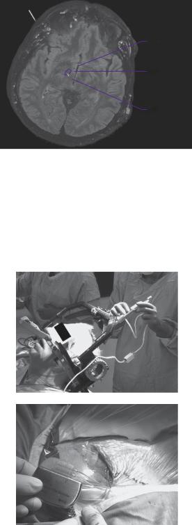
A stereotactic pre-operative MRI was performed to define the subcortical nuclei for electrode targeting (see Figure 12.1). At the beginning of the procedure, a stereotactic frame was applied to the patient’s head under local anaesthesia and a stereotactic CT performed. CT is less susceptible to spatial artefacts and is registered with the MRI. The subthalamic nucleus was identified using the patient’s imaging, and with the assistance of registration with a brain atlas to help confirm and plot the target coordinates. The patient was taken to theatre and bilateral craniostomies were fashioned under local anaesthetic. A 1.8-mm diameter radiofrequency electrode was passed to target, monitoring impedance to detect transgression of the ventricle, which can lead to electrode misplacement. The DBS electrode was then inserted in its place (Figure 12.2a). A neurologist objectively assessed the contralateral limb rigidity during test stimulation to ensure electrode position produced clinical benefit. The
Case 12 Deep brain stimulation for debilitating Parkinson’s disease |
117 |
Third ventricle
Subthalamic nucleus traversed by electrode
Red nucleus
Figure 12.1 Reconstructed three-dimensional brain MRI in axial section with deep brain electrode trajectory to subthalamic nucleus (circled) planned on neuronavigation workstation.
procedure was repeated on the contralateral side. On placement of the contralateral radiofrequency electrode to target, there was an improvement in rigidity, independent of stimulation, a phenomenon known as ‘stun’, whereby the mechanical interruption or microlesioning of the target nucleus produces a temporary therapeutic effect. Although it confirms that the target produces clinical efficacy, it also prevents the fine-tuning of electrode placement based on further clinical examination during the procedure.
(a)
(b)
Figure 12.2 (a) Deep brain electrode implanted using stereotactic frame attached to patient. (b)
Implanted pulse generator to be internalised within subclavicular pocket
118 Challenging concepts in neurosurgery
A post-operative stereotactic CT head was performed with head frame and localizer still attached, which verified the electrode contacts’ position in the STNs. On returning to theatre, under general anaesthesia, extension leads were connected to the electrodes and tunnelled behind the ear, into a subclavian pocket that had been fashioned. The implanted pulse generator was connected to the extension leads and placed in the pocket, which was then closed (Figure 12.2b). The patient was woken in recovery and had suffered no neurological deterioration. The stimulator was not initially activated. There was a unilateral improvement in rigidity and dyskinesia as a result of the intra-operative stun effect. Stimulation was activated 2 weeks post-operatively at 1.5V, 90 microseconds and 130Hz, allowing the acute changes of surgery including stun to settle down so that stimulation titration was performed without such a confounding factor.
Post-operative UPDRS scores at 6 months were Part 1: 1/16; Part 2: 8/56 on, 24/56 off; Part 3: 8/104 on, 34/104 off.
There was a marked improvement in Part III of the UPDRS with improvements in rigidity, dyskinesias, and bradykinesia on examination. There was no deterioration in mood or cognition detected.
Discussion
PD is a neurodegenerative disorder caused, in part, by the loss of dopaminergic neurones in the substantia nigra (pars compacta). This results in disruption of the normal oscillatory and synchronous neuronal activity between the cortex, globus pallidus interna (GPi) and STN, The three cardinal clinical manifestations of PD are bradykinesia, tremor, and rigidity. Gait and postural instability is often also seen [6]. The place of surgery in the management of PD has been cyclical. It was once the mainstay of treatment, in the form of ablative surgeries, such as pedunculotomy, and was then made largely redundant by the advent of dopaminergic drugs. However, it was found that dopaminergic drugs caused side effects including dyskinesias, which could be severely incapacitating. Once again, surgery (commonly taking the form of DBS) became an important modality in the management of PD, not only to treat the cardinal symptoms of the disease itself, but also to treat the dyskinetic side effects of medical therapy.
Patient selection
The commonest reasons for poor outcomes after DBS are: poor patient selection, poor operative electrode placement, and inadequate stimulation programming [7]. In DBS for PD, the ideal patient characteristics are a patient with idiopathic PD with an excellent response to L-dopa, particularly the medication motor ‘on’ state. Broadly, DBS surgery is offered to those who suffer from intractable tremor, debilitating side effects of medical therapy, such as dyskinesia, are of a younger age, and have a psychological and physical health sufficient to tolerate surgery and ongoing stimulation management as an outpatient. Patients with psychiatric diagnoses of major depression, acute psychosis, and dementia are excluded [7]. Therefore, a movement disorder neurologist and clinical psychologist/psychiatrist should form part of the DBS team, as well as the surgeon. A multidisciplinary approach is key to the management of these patients. Furthermore, identification of the most debilitating symptoms for the individual patient are critical as this determines whether non-surgical

Case 12 Deep brain stimulation for debilitating Parkinson’s disease
therapies have been exhausted for a given symptom profile, the location of the deepbrain stimulator placement, and allows for an estimation of the chances that DBS will be beneficial.
Location, location, location
Depending on the site targeted by DBS, a variety of symptoms can be ameliorated (see ‘Learning point: Parkinson’s disease symptoms and relevant deep-brain stimulation target nuclei’). It is therefore critical to tailor the targeting to the individual patient. DBS for PD is supported by the NICE Guidelines of 2006 with particular reference to STN, GPi and thalamic stimulation [2].
Subthalamic nucleus
The cardinal symptoms of PD, namely bradykinesia, rigidity, and tremor, as well as dyskinesias resulting from medication, can all be ameliorated by STN DBS to varying degrees. The STN was identified as a target in PD as a direct result of primate models [8,9]. As STN DBS diminishes these symptoms, the pharmacological therapy can be reduced together with their resulting side effects, particularly the incidence and severity of dyskinesias. Krack et al. demonstrated that STN DBS improved the motor symptoms of PD, improved activities of daily living, and reduced medication requirements [10]. The multicentre PD SURG Trial randomly assigned 366 patients with advanced PD to immediate surgery with best medical therapy or best medical therapy alone [11]. This was effectively a study of STN DBS surgery as only four patients received stimulation of a different nucleus, i.e. the GPi. The PD SURG trial found that DBS produced clear advantages compared with maximal medical therapy in clinical assessments and patient-assessed quality of life at 1 year follow-up. DBS conferred improvements in mobility and the activities of daily living domains of the PDQ-39 questionnaire, the total UPDRS, particularly part IV including time and severity of dyskinesias and ‘off’ periods, and a fall in daily dopaminergic drug requirement by a third. PD SURG found an adverse surgery-related event in 19% of patients. There was one procedure-related death, but no suicides [11].
Adverse effects of STN DBS attributable to the subthalamic location itself include psychiatric and cognitive disturbances, reflecting the STN’s role in association and limbic circuits [12]. PD SURG found no decline in cognition as rated by the dementia rating scale (DRS-II), although its sensitivity to cognitive decline has been questioned [13]. Speech decline after surgery was detected on detailed neuropsychological testing, with a reduced verbal fluency and vocabulary [11]. Decline in cognition and mood has been inconsistently reported by other series, but appears to affect 1–2% of patients [14].
Globus pallidus interna
The GPi has been an important target in PD for the amelioration of dyskinesias. Its impact on bradykinesia and rigidity is becoming increasingly recognized. In a multicentre RCT, the cooperative studies programme (CSP) 468 Study Group demonstrated that STN and GPi DBS were more efficacious than medical therapy alone, in terms of increased time in the PD on state without dyskinesias, increased motor function, and a variety of quality of life measures [15]. After randomization between STN and GPi stimulation, they later demonstrated that both targets produced equivalent efficacy in motor function improvement measured by the UPDRS
119
 Expert comment
Patient selection for DBS is one of the most important aspects. A good candidate is generally one who has a good response to
L-dopa, but either the side effects of the treatment are too severe (dyskinesia) or motor on–off fluctuations predominate. Tremor can also be treated successfully. Approximately 10% of patients with PD are suitable.

120 Challenging concepts in neurosurgery
Part III at 24 months [16]. Although dopaminergic drug requirement was lowered to a greater degree by STN stimulation, it also led to a decline in mood and visuomotor processing speed compared with GPi stimulation. Given that cognitive and mood disturbance was also found less after GPi compared with STN stimulation in other studies [17, 18, 19], pallidal stimulation is an important option for treating bradykinesia, rigidity, and dyskinesias in PD, and is a valid target in the case presented here.
Thalamus
Tremor amelioration is one of the oldest indications for functional neurosurgery after Irving Cooper’s serendipitous observations in the 1950s [20,21]. Tremor can be treated by DBS of the motor thalamus or the STN. Thalamic DBS should be reserved for cases in which tremor is the predominant debilitating symptom and where the other cardinal symptoms of PD or drug side effects have not and are not expected to manifest [7]. Within the motor thalamus, the ventralis intermedius nucleus (VIM) is the commonest target, but the ventralis oralis nucleus (VOP), intimately related to it, is an alternative [22]. As this patient was not troubled by tremor, thalamic stimulation would not be an appropriate choice for him.
Pedunculopontine nucleus
Postural instability and gait freezing have historically not responded well to DBS nor L-dopa therapy. However, a novel target, the pedunculopontine nucleus (PPN), was identified in primate studies [23, 24, 25], as a reticular nucleus located at the junction of the mesencephalon and pons [Jenkinson et al. 2006; 26]. In humans with advanced PD, PPN stimulation results in improvements in measurements of gait, posture, and balance [27, 28, 29]. As these were not prominent symptoms in this gentleman’s PD, PPN stimulation would not be an appropriate choice for him.
 Clinical tip Indications and patient selection for DBS in PD
Patient selection is critical and only a minority of PD sufferers are appropriate for DBS. The factors recommended to confer good outcome from DBS can be divided into three broad categories relating to the PD itself and response to L-dopa, psychiatric and psychological factors, and general surgical factors.
Parkinson’s disease features
●Idiopathic.
●Excellent response to L-dopa.
●Dyskinesias.
●Intractable tremor.
Psychiatric
●No dementia.
●No major depression.
●No acute psychosis.
●Cognition status good.
General
●Younger age.
●Fitness for neurosurgery.

Case 12 Deep brain stimulation for debilitating Parkinson’s disease |
121 |
 Learning point Parkinson’s disease symptoms and relevant deep-brain stimulation target nuclei
Depending on the electrode target, DBS can confer benefit on a range of symptoms in PD. Establishing the symptoms most deleterious to the individual patient is therefore crucial to planning DBS in order to provide as much benefit as possible (Table 12.3). Some targets benefit a greater range of symptoms than others [2,16,27].
Table 12.3 PD symptoms and relevant deep-brain stimulation target nuclei
Symptom |
Target |
Tremor |
Thalamus, STN |
Bradykinesia, rigidity |
STN, GPi |
Dyskinesia |
STN, GPi |
Postural instability, gait freezing |
PPN |
|
|
 Clinical tip Accurately implanting deep brain stimulation electrodes
Several measures to optimize the accuracy of deep brain electrode implantation are undertaken. Their utilization varies depending on the case and the unit.
Neuroimaging
MRI provides definition of the subcortical structures for targeting. CT provides greater spatial accuracy as it is less subject to artefacts than MRI. Fusion of the two modalities provides the advantages of both.
Intra-operative neurological assessment
Test stimulation and clinical assessment while the patient is awake provides rapid feedback on the clinical effect of stimulation and adverse effects, and allows optimization of electrode depth. The anaesthetist’s role is therefore crucial. This is not suitable for patients who would not tolerate surgery while awake, such as those in whom their movement disorder is so severe.
Microelectrode recording
Localization of cell groups within the target nucleus by depth recordings from multiple fine microelectrodes provides neurophysiological targeting feedback. Disadvantages include longer operative time and a concern of increase risk of intracranial haemorrhage due to multiple electrode passes [30].
Lesional surgery
Creating a lesion, rather than chronically implanting an electrode is an important alternative for clinicians and patients to consider. Historically, deep brain ablational surgery preceded DBS, which is not suitable for all patients. Lesions of the GPi (pallidotomy) or motor thalamus (thalamotomy) can confer similar efficacy to DBS [31,32] and benefits from subthalamotomy have also been reported [17,33], and are therefore useful to consider in PD patients. DBS is an expensive therapy on account of the hardware costs of the electrode and pulse generator and also the subsequent need for follow-up and battery replacement surgeries. The advantage of a lesion is that it is a one-off therapy and does not require continued follow-up nor is there any hardware to manage. Therefore, determining factors include the patient’s tolerance and compliance with intensive follow-up, and their agreement to undergo further battery change procedures to maintain stimulation, their cognitive level, expectations and level of neurological risk they deem acceptable, bilateral symptoms (bilateral thalamotomy has an unacceptably high risk of speech and swallowing

122 Challenging concepts in neurosurgery
disturbance [34,35], hardware and infection fears, and local economic factors. The lesion, however, is an irreversible and unmodifiable therapy. DBS electrodes have the advantage that they can be switched-off or removed if causing adverse effects and the stimulation parameters can be titrated to the patient’s needs in addition to allowing adjustment over time as their tolerance or disease state changes.
A final word from the expert
There are likely to be two main future developments and these are equivalent to a ‘space race’ between improving technology and other biological treatments. For example, electrode design is advancing rapidly with improvements in electric field shaping and other modalities, such as optogenetics. On the other hand, there have been huge recent developments
in stem cell research, viral vectors, and growth factor infusions with the aim of restoring ‘normal’ brain.
References
1.Cotzias GC, Papavasiliou PS, Gellene R. L-dopa in Parkinson’s syndrome. New England Journal Medicine 1969; 281(5): 272.
2.National Institute for Health and Clinical Excellence (NICE). Parkinson’s diseases: diagnosis and management in primary and secondary care, NICE Clinical Guideline 35. London: NICE, 2006. Available at: http://www.nice.org.uk/nicemedia/ live/10984/30088/30088.pdf.
3.Kalinderi K, Fidani L, Castor Z, et al. Pharmacological treatment and the prospect of pharmacogenetics in Parkinson’s disease. International Journal of Clinical Practice 2011; 65(12): 1289–94.
4.Movement Disorder Society Task Force on Rating Scales for Parkinson’s Disease. The Unified Parkinson’s Disease Rating Scale (UPDRS): status and recommendations. Movement Disorders 2003; 18(7): 738–50.
5.Fahn S, Elton RL, Members of the UPDRS Development Committee. Unified Parkinson’s Disease Rating Scale. In: S Fahn, CD Marsden, DB Calne, et al. (eds), Recent developments in Parkinson’s disease vol. 2 (pp. 153–64). Florham Park, NJ: Macmillan Health Care Information 1987.
6.Williams D, Tijssen M, van Bruggen G, et al. Dopamine-dependent changes in the functional connectivity between basal ganglia and cerebral cortex in humans. Brain 2002; 125: 1558–69.
7.Volkmann J. Selecting appropriate Parkinson’s patients for deep brain stimulation. In:P Bain, T Aziz, X Liu, et al. (eds), Deep brain stimulation (pp. 75–83). Oxford: Oxford University Press, 2009.
8.Aziz TZ, Peggs D, Sambrook MA, et al. Lesion of the subthalamic nucleus for the alleviation of 1-methyl-4-phenyl-1,2,3,6-tetrahydropyridine (MPTP)-induced parkinsonism in the primate. Movement Disorders 1991; 6: 288–92.
9.Bergman H, Wichmann T, Delong MR. Reversal of experimental parkinsonism by lesion of the subthalamic nucleus. Science 1990; 249: 1436–8.
10.Krack P, Batir A, Van Blercom N, et al. Five year follow-up of bilateral stimulation of the subthalamic nucleus in advanced Parkinson’s disease. New England Journal of Medicine 2003; 349: 1925–34.
11.Williams A, Gill S, Varma T, et al., on behalf of the Parkinson’s disease Surgical Collaborative Group. Deep brain stimulation plus best medical therapy versus medical
Case 12 Deep brain stimulation for debilitating Parkinson’s disease |
123 |
therapy alone for advanced Parkinson’s disease (PD SURG trial): a randomized, openlabel trial. Lancet Neurology 2010; 9 (6): 581–91.
12.Hamani C, Saint-Cyr SA, Fraser J, et al. The subthalamic nucleus in the context of movement disorders. Brain 2004; 127: 4–20.
13.Rodriguez-Oroz MC. Deep brain stimulation for advanced Parkinson’s disease. Lancet Neurology 2010; 9(6): 558–9.
14.Woods SP, Fields JA, Troster AI. Neuropsychological sequelae of subthalamic nucleus deep brain stimulation in Parkinson’s disease: a critical review. Neuropsychology Reviews 2002; 12: 111–26.
15.Weaver FM, Follett K, Stern M, et al. Bilateral deep brain stimulation vs best medical therapy for patients with advanced Parkinson disease: a randomized controlled trial. Journal of the American Medical Association 2009; 301(1): 63–73.
16.Follett KA, Weaver FM, Stern M, et al. Pallidal versus subthalamic deep-brain stimulation for Parkinson’s disease. New England Journal of Medicine 2010; 362(22): 2077–91.
17.Walter BL, Vitek JL. Surgical treatment for Parkinson’s disease. Lancet Neurology 2004; 3: 719–28.
18.Volkmann J, Alert N, Voges J, et al. Safety and efficacy of pallidal or subthalamic nucleus stimulation in advance PD. Neurology 2001; 56: 548–51.
19.Rodriguez-Oroz MC, Obeso JA, Lang AE, et al. Bilateral deep brain stimulation in Parkinson’s disease: a multicentre study with 4 years follow-up. Brain 2005; 128: 2240–9.
20.Cooper IS. Effect of anterior choroidal artery ligation on involuntary movements and rigidity. Transactions of the American Neurological Association 1953; 3(78th meeting): 6–7.
21.Das K, Benzil DL, Rovit RL, et al. Irving S. Cooper (1922–1985): a pioneer in functional neurosurgery. Journal of Neurosurgery 1998; 89(5): 865–73.
22.Hyam J, Owen SLF, Kringelbach ML, et al. Contrasting connectivity of the ventralis intermedius and ventralis oralis posterior nuclei of the motor thalamus demonstrated by probabilistic tractography. Neurosurgery 2012; 70(1): 162–9.
23.Jenkinson N, Nandi D, Miall RC, et al. Pedunculopontine nucleus stimulation improves akinesia in a Parkinsonian monkey. NeuroReport 2004; 15: 2621–4.
24.Jenkinson N, Nandi D, Oram R, et al. Pedunculopontine nucleus electric stimulation alleviates akinesia independently of dopa-minergic mechanisms. NeuroReport 2006; 17: 639–41.
25.Nandi D, Aziz TZ, Giladi N, et al. Reversal of akinesia in experimental parkinsonism by GABA antagonist microinjections in the pedunculopontine nucleus. Brain 2002; 125(11): 2418–30.
26.Zrinzo L, Zrinzo LV, Tisch S, et al. Stereotactic localization of the human pedunculopontine nucleus: atlas-based coordinates and validation of a magnetic resonance imaging protocol for direct localization. Brain 2008; 131(6): 1588–98.
27.Plaha P, Gill SS. Bilateral deep brain stimulation of the pedunculopontine nucleus for Parkinson’s disease. NeuroReport 2005; 16: 1883–7.
28.Moro E, Hamani C, Poon YY, et al. Unilateral pedunculopontine stimulation improves falls in Parkinson’s disease. Brain 2010; 133(1): 215–24.
29.Thevathasan W, Coyne TJ, Hyam JA, et al. Pedunculopontine nucleus stimulation improves gait freezing in Parkinson’s disease. Neurosurgery 2011; 69: 1248–54.
30.Zrinzo L, Foltynie T, Limousine P, et al. Reducing hemorrhagic complications in functional neurosurgery: a large case series and systematic literature review. Journal of Neurosurgery 2012; 116(1): 84–94.
31.Bittar RG, Hyam J, Nandi D, et al. Thalamotomy versus thalamic stimulation for multiple sclerosis tremor. Journal of Clinical Neuroscience 2005; 12(6): 638–42.
32.Gross RE. What happened to posteroventral pallidotomy for Parkinson’s disease and dystonia? Neurotherapeutics 2008; 5: 281–93.
124 |
Challenging concepts in neurosurgery |
|
|
33. |
Alvarez L, Macias R, Lopez G, et al. Bilateral dorsal subthalamotomy in Parkinson’s |
|
|
disease (PD): initial response and evolution after 2 years. Movement Disorders 2002; |
|
|
17(Suppl. 5): S95. |
|
34. |
Alusi SH, Aziz TZ, Glickman S, et al. Stereotactic lesional surgery for the treatment of |
|
|
tremor in multiple sclerosis: a prospective case-controlled study. Brain 2001; 124(8): |
|
|
1576–89. |
|
35. |
Samra K, Waltz JM, Riklan M, et al. Relief of intention tremor by thalamic surgery. |
|
|
Journal of Neurology Neurosurgery and Psychiatry 1970; 33(1): 7–15. |

CASE
13Endoscopic resection of a growth hormone-secreting pituitary macroadenoma
Alessandro Paluzzi
 Expert commentary Paul Gardner
Case history
A 61-year-old male presented with a 2-year history of fatigue, erectile dysfunction, and increasing hand and shoe sizes (size 9 to size 11). He also complained of visual problems affecting his driving. His wife had reported that he had started snoring at night, and had noticed that his nose and jaw had grown to change his facial features significantly compared with photographs of him several years before.
His previous medical history was unremarkable except for hypertension.
 Learning point The signs and symptoms of acromegaly
The acral changes (from Gr akron = extremity) are the most common clinical signs that lead to the diagnosis. Hands and feet are broadened, and the fingers and toes are thickened and stubby. The nose is widened, and the cheekbones and forehead become prominent, sometimes with frontal bossing. Prognathism, maxillary widening, dental diastasis, and macroglossia are also common.
In addition to the typical dysmorphic facial and body features, acromegaly is associated with a number of systemic complications, including hypertension, caradiomyopathy, diabetes mellitus, sleep apnoea syndrome, and colon cancer. These account for the associated mortality risk in acromegalic patients compared with the normal population [1]. Treatment of each specific co-morbidity greatly improves the general prognosis of the patients [2]. Furthermore, the systemic comorbidities, together with the presence of macroglossia and jaw malocclusion, need to be taken into account pre-operatively before removal of a pituitary adenoma, since they increase the anaesthetic risk of these patients.
On examination, the features of acromegaly were noted. His blood pressure was 170/102 on lisinopril/hydrocholorthyazide and random blood glucose was 8.3mmol/L (normal range 3.9–5.5mmol/L). Visual field assessment demonstrated gross bitemporal hemianopia, and this was confirmed on Humphrey visual field automated testing (Figure 13.2e).
Endocrine tests showed a random growth hormone (GH) level of 58ng/ mL (normal range 0–5 ng/mL) and IGF-1 level of 667ng/mL (reference range: 71–290ng/mL). Other endocrine tests revealed hypothyroidism with decreased free T4 at 0.48ng/dL (normal range 0.8–1.8ng/dL) and normal TSH at 0.520μIU/mL
126 Challenging concepts in neurosurgery
(normal range 0.300–5000μIU/mL). He also displayed hypogonadotropic hypogonadism with decreased LH at 0.3mIU/mL (normal range 1–5.6mIU/mL) and FSH at 1.5mIU/mL (normal range 1.5–14.3mIU/mL) and undetectable testosterone <1ng/dL (normal range 250–1100ng/dL). His AM cortisol was also low at 1μg/dL (normal AM range 7–25μg/dL) with an ACTH of 15pg/mL (normal range 9–46pg/mL).
T1-weighted MRI with contrast revealed a large sellar lesion with suprasellar extension consistent with pituitary macroadenoma measuring 3.3 × 2.6 × 3.7cm (Figure 13.1). The tumour extended laterally beyond the lateral wall of the cavernous internal carotid artery, suggesting a high probability of cavernous sinus invasion (Knosp grade III) (Figure 13.1 a, c, e).
(a) |
(b) |
(c) |
(d) |
Naso-septal flap
(e) (f)
Figure 13.1 Preand post-operative (12 months) gadolinium-enhanced T1 MR imaging of the macroadenoma. (a, b) Axial views. The arrow points to the portion of the adenoma invading
the left cavernous sinus. (c, d) Coronal views. On the pre-operative scan (c) the most lateral border of the adenoma on the left side extends beyond the lateral edge of the carotid artery indicating, according to the Knosp classification, high probability of cavernous sinus invasion. (e, f) sagittal views. In the post-operative scan (f) the enhancing tissue at the level of the planum sphenoidale corresponds to the muco-perichondrial naso-septal flap used to repair the intra-operative
dural opening.

Case 13 Endoscopic resection of a macroadenoma |
127 |
 Learning point Knosp classification
In 1991, Engelbert Knosp proposed a radiological classification to predict the likelihood of cavernous sinus invasion from a pituitary adenoma. He studied the pre-operative MRI scans of 25 pituitary adenomas that were confirmed surgically to have invaded the cavernous sinus space. Five ‘Knosp grades’ were defined by the relationship of the adenoma’s lateral edge with the internal carotid artery, as shown on the most representative coronal post-contrast T1 slice. Grade 0 represents the normal condition, and Grade 4 corresponds to the total encasement of the intracavernous carotid artery. According to this classification, surgically proven invasion of the cavernous sinus space was present in all Grade 4 and 3 cases and in all but one of the Grade 2 cases; no invasion was present in Grade 0 and Grade 1 cases.
In view of the recent history of visual deterioration and the diagnosis of acromegaly from a GH-secreting adenoma the patient was advised to undergo surgical intervention. He consented to an expanded endonasal approach (EEA) for resection of the pituitary macradenoma.
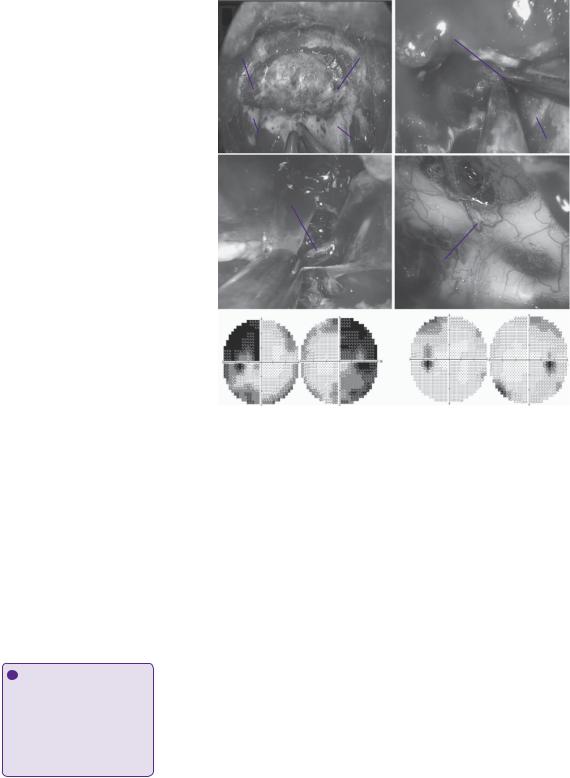
During the operation, marked expansion of the sella was noticed. After initial bony exposure of the sella and both cavernous sinuses (Figure 13.2a), the tumour was debulked using a ‘2-sucker technique’ (Figure 13.2b). The adenoma was found to have invaded the medial wall of the left cavernous sinus and to extend into the medial compartment of the cavernous sinus. Complete resection of this component of the tumour was achieved with the help of a 45-degree angled endoscope. The inferior hypophyseal artery was identified and coagulated (Figure 13.2c). To avoid herniation of arachnoid through the enlarged diafragma sellae during the initial steps of the tumour debulking, the suprasellar portion of the tumour was addressed only at the end, using again a 45-degree angled endoscope (Figure 13.2d). Both superior hypophyseal arteries were visualized and preserved. Gross total resection of the tumour was achieved. The repair of the dural defect was carried out using a pedicled muco-perichondrial naso-septal flap (Figure 13.1f).
The patient made a satisfactory post-operative recovery. His vision subjectively improved immediately post-operatively and formal visual field assessment 2 weeks and 6 months later demonstrated an objective substantial decrease in the visual field defects bilaterally (Figure 13.2f). Post-operative MRI scans at 3, 6, and 12 months (Figure 13.1b, d, f) demonstrated gross total resection without any evidence of residual or recurrent tumour.
His GH on the first post-operative day was down to 0.74ng/mL (normal range 0–5 ng/mL), while the IGF-1 was still abnormal at 412ng/mL (reference range: 71–290ng/mL). Two weeks later, both levels were normal, with a random GH of 0.40ng/mL and IGF-1 of 113ng/mL and the MRI scan at 1 month showed no evidence of residual adenoma. Both endocrinological and radiographic results were taken with caution at this stage, since it is well known that during the first 3 months post-operatively they can be misleading. During subsequent follow-up, the clinical features of acromegaly gradually improved and biochemical cure was maintained at 7 months and at his last follow-up 1 year post-operatively.
The patient was also medically treated with oral hydrocortisone 10mg bd, transdermal testosterone 5g/day, and levothyroxine 100μg/day for panhypopituitarism that was present preoperatively.
128 |
Challenging concepts in neurosurgery |
R-CS |
L-CS |
(a) |
R-ICA |
L-ICA |
|
|
|
|
Inf. hypophyseal A. |
|
(c)
(e)
L-CS
(b) |
L-ICA |
|
Optic chiasm
(d)
(f)
Figure 13.2 (a) Intra-operative endoscopic picture of the initial bony exposure from cavernous sinus to cavernous sinus. (b) Intra-operative picture with a 45-degree angled endoscope of the left medial cavernous sinus opened; two angled suction tubes are used to remove the tumour from this compartment. The left cavernous internal carotid artery is exposed. (c) During dissection of the medial cavernous sinus, the left inferior hypophyseal artery is identified. Failure to recognize it can lead to its avulsion from the internal carotid artery and arterial bleeding difficult to control. (d) Intra-operative
picture with a 45-degree angled endoscope after the suprasellar component of the adenoma has been removed. It is possible to appreciate the decompressed optic chiasm. (e) Pre-operative Humphrey’s visual field test displaying a bi-temporal hemianopsia. (f) Post-operative examination 6 months after the operation showing a dramatic improvement.
R-CS = right cavernous sinus; L-CS = left cavernous sinus; R-ICA = right internal carotid artery; L-ICA = left internal carotid artery; Inf. Hypophyseal A. = inferior hypophyseal artery.
 Learning point Incidence of pituitary adenomas
Pituitary adenomas represent the third most common primary brain tumour after gliomas and meningiomas, with a frequency in the population of 14.4% from autopsy studies.
Discussion
Pituitary tumours represent the third most common primary brain tumour after gliomas and meningiomas [3]. Autopsy studies confirmed a frequency in the population of 14.4% [4]. The prevalence of acromegaly is in the order of 40–125 cases per million and its annual incidence is 3 or 4 cases per million [5,6] although recent studies suggest that it might as high as 1034 cases per million [7,8].
Apart from the classic dysmorphic features of acromegaly or gigantism (if the over-secretion takes place before the growth plates have closed), patients develop systemic complications over time including hypertension, cardiomyopathy, diabetes,

Case 13 Endoscopic resection of a macroadenoma
sleep apnoea, arthritis, carpal tunnel syndrome, and colon cancer. These account for the associated 2–2.5 times increased mortality risk in acromegalic patients compared to the normal population [1].
Mass effect from macroadenomas can lead to visual field defects, hypopituitarism, headache, and oculomotor nerve deficit. Due to the insidious development of these signs and symptoms, it is estimated that the interval between their onset and the diagnosis of acromegaly is about 7–8 years [9], although a more recent study reports that this delay has now reduced to 2–3 years [10]. The delay in the diagnosis might explain why the majority of GH-secreting pituitary adenomas are macroadenomas (>1cm in diameter).
MRI represents the gold-standard imaging for the diagnosis of a pituitary adenoma. The typical appearance is of a well-circumscribed lesion isointense to grey matter on non-contrasted sequence. On T1-weighted sequence, the normal posterior lobe of the pituitary gland looks brighter, possibly due to the presence of myelin. The heterogenous appearance in macroadenomas often reflects the presence of haemorrhage, necrosis, or proteinaceous material within cystic degeneration. After contrast injection, pituitary adenomas classically enhance to a lesser extent than the surrounding normal gland. The ability to discriminate between normal gland and adenoma, however, is limited by a reduced size of tumour, often being challenging in the case of small microadenomas.
 Evidence base Natural history of pituitary adenomas [11]
The only evidence about the natural history of untreated pituitary adenomas comes from studies on pituitary incidentalomas (PIs) or non-functioning-pituitary adenomas (NFPAs) managed conservatively. A recent systematic review and meta-analysis pooled together patients from eleven such studies, all of them being non-comparative cohort studies. The median follow-up was 3.9 years (range 1–15). The differentiation between PIs and NFPAs was not feasible and the quality of these studies was judged to be suboptimal and with several methodological limitations. The conclusions
were that the incidence of tumour growth in PIs/NFPAs is higher in macroadenomas and solid lesions in comparison with microadenomas and cystic lesions. There was a trend that did not reach statistical significance for greater incidence of pituitary apoplexy and new endocrine dysfunction worsening in macroadenomas compared with microadenomas.
The treatment of GH secreting pituitary adenomas aims to:
●Achieve biochemical cure: the normalization of GH and/or IGF-1 levels has been shown to reduce the mortality risk to the one of the normal population [12]. The most recent criteria for biochemical cure are a normal IGF-1 and GH < 0.4ng/mL or a random GH < 1.0ng/mL. 11 Most of the papers in the literature, however, still reports the old criteria set in previous guidelines of
normal IGF-1 and GH < 1ng/mL after OGTT or random GH < 2.5ng/mL at least 3 months post-operatively [13]
●Control the signs and symptoms of acromegaly: improve physical appearance, voice, mouth occlusion, etc.
●Reduce mass effect from the tumour: improve visual fields and pituitary function
129
 Expert comment Mortality in acromegaly
Patients with acromegaly due to a GH-secreting pituitary adenoma have a risk of mortality that is 2–2.5 times that of the normal population [1].
 Expert comment
The clinical suspicion of acromegaly needs to be confirmed biochemically. An increased serum GH level that is not suppressed
by oral glucose tolerance testing (OGTT) to less than 1mg/L (3mIU/L) and increased IGF-1 above the age-adjusted normal range are diagnostic. For a woman aged between 40 and 60, the normal range is 237–246ng/mL, for a man it is 211–251ng/mL. More recent guidelines suggest that the cut-off of GH nadir during OGTT should be decreased to 0.4mg/L (1mIU/L), in view of the fact that modern assays have become extremely sensitive [10].
 Learning point Remission criteria for acromegaly
The 2011 guidelines by the American Association of Clinical Endocrinologists (AACE) recommend that in order to
achieve surgical cure, the following criteria need to be met at least 3 months post-operatively:
●IGF-I value within normal range for age and gender.
●GH value less than 0.4ng/mL after glucose load or a random GH value less than 1.0ng/mL.

130 Challenging concepts in neurosurgery
The AACE guidelines recommend surgery as the primary mode of therapy in all GH-secreting microadenomas and all macroadenomas with mass effect. Surgical debulking to improve the response of subsequent medical therapy is also advocated in patients with macroadenomas without mass effect and with low likelihood of surgical cure (e.g. cavernous sinus invasion) [10]
The trans-sphenoidal (sublabial and transeptal microscopic or endonasal endoscopic) route is widely accepted as the standard approach for the majority of pituitary adenomas, while the role of craniotomy, even for large suprasellar tumours, is gradually being replaced by the EEA.
In the UK, the NICE recommends the use of endoscopic trans-sphenoidal removal of pituitary adenomas, since it results in comparable surgical outcomes to conventional surgery, may shorten operation time, and the complication rate is less than that of conventional surgery [14]. These guidelines were issued based on the evidence available in 2003. Since then, further larger studies, all retrospective reviews, have confirmed the efficacy and safety of the procedure with comparable or better results than microscopic surgery in terms of outcome [15–22].
 Clinical tip Follow-up tests
The ideal time to assess whether the resection has been complete and the patient is in biochemical remission is about 3 months post-operatively. Before then the radiological, as well as
endocrinological investigations can be misleading [26]
 Expert comment
In patients who have failed surgical resection, or without radiological evidence of residual tumour, but persistently elevated biochemical markers, the use of somatostatin analogues (SSAs) and/or GH antagonists can be recommended as adjuvant therapy. In selected cases, these drugs have also a role as primary medical therapy, namely in those patients in whom surgery is contraindicated or is unlikely to achieve satisfactory results [10]
 Expert comment
Compared with traditional microscopic trans-sphenoidal approaches, the endoscopic approach offers technical advantages in particular situations:
●Extension to or invasion of the cavernous sinus (CS): by removing the bone covering at least the medial part of both cavernous sinuses, it becomes possible to retract them laterally to reach the areas normally difficult to visualize with the microscope. This is facilitated by the wide angle view and proximity of the endoscope, and can be augmented by the used of angled endoscopes (usually 45 degrees). In the case of secreting tumours with CS invasion it is possible to excise the medial cavernous wall and aim for complete resection of the adenoma, provided this does not extend to the lateral CS wall, where the oculomotor nerves are located. The use of intra-operative neurophysiological monitoring electromyelography (EMG) plays an important role in avoiding injury to these nerves.
●Suprasellar extension: for adenomas with suprasellar extension that do not descend into the sella (fibrous, dumb-bell shaped, previous radiation or pharmacological therapy), the endoscopic
trans-tuberculum approach offers improved access to the suprasellar cistern and its contents. This is particularly important for visualizing the superior hypophyseal arteries, hence preserving the blood supply to the stalk and the optic chiasm.
●Pars intermedia/posterior lobe lesions: in less common situations where a functioning microadenoma is located in the pars intermedia or in the posterior lobe, an ‘infrasellar’ approach with a 45-degree endoscope allows access to these lesions from a caudo-rostral direction without the need to damage or split an intact anterior lobe
To assess whether biochemical cure has been achieved it is advisable to wait 3–6 months before conducting the above mentioned biochemical tests [23,24]. For reasons that are unclear, it takes months for the IGF-1 levels to normalize even after complete resection of the adenoma [25]. A similar rule applies to post-operative MRI. Early MRI is often difficult to interpret due to the presence of blood products, absorbable gelatin materials or fat, all of which take several weeks to disappear. For this reason the first post-operative scan should be performed about 3 months after surgery [26].

Case 13 Endoscopic resection of a macroadenoma |
131 |
 Learning point Somatostatin analogues and growth hormone (GH) receptor antagonists [10]
Octreotide and lanreotide are the two most common SSAs used in acromegaly, they have similar efficacy profiles and newer formulations for im or deep sc injections enable them to be administered once a month. SSAs are effective in normalizing IGF-I and GH levels in approximately 55% of patients. The clinical and biochemical responses to SSAs are inversely related to tumour size and degree of GH hypersecretion. SSAs reduce pituitary tumour size modestly in approximately 25–70% of patients, therefore, they should not be relied on for decompression of local structures in the presence of mass effects. Their side effects include
gastrointestinal upset, malabsorption, constipation, gallbladder disease, hair loss, and bradycardia. They can cross the placenta and, although a sc injection of octreotide can cause an acute decrease in uterine artery blood flow, longer use of octreotide does not appear to cause adverse effects
on the course of pregnancy, on delivery, or on foetal development. There have been a number of cases in which octreotide was used in pregnant patients and most of these pregnancies were uneventful. In a few cases of pregnant patients given SSA therapy, the resultant infants were small for gestational age, although the causality was not clear.
Pegvisomant is a GH receptor antagonist that seems to be effective regardless of baseline tumour size or degree of GH hypersecretion. It normalizes IGF-I values in more than 90% of the patients, including patients who are partially or completely resistant to other medical therapies, and it is effective at improving glucose homeostasis in patients with associated diabetes mellitus. Side effects of pegvisomant, include flu-like illness, allergic reactions, and increase in liver enzymes. Tumour enlargement has been infrequently associated with use of pegvisomant, therefore, serial monitoring with pituitary MRI scans is suggested. Specific recommendations for the use of pergvisomant during pregnancy cannot be made since the experience is limited to a single case in which pegvisomant administration was well tolerated, the patient’s condition was well controlled, and the infant was normal in size and health.
Compared with conventional adjuvant radiotherapy for residual disease, stereotactic radiosurgery has the theoretical advantage of potentially stabilizing the disease and reducing tumour size, while sparing the optic apparatus and the pituitary function, provided the adenoma is more than 5mm away from them [27]. Another advantage of stereotactic radiosurgery over conventional radiotherapy seems to be the shorter mean time to achieve biochemical remission (2 years) [28] The biochemical remission rates reported in the literature vary between 17 and 50%, although the follow-up is limited to a period between 2 and 5 years [29–34]. In terms of tumour control, up to 75% of the patients treated with radiosurgery achieve a decrease in tumour size, a result similar to conventional radiotherapy [30–33].
A final word from the expert
There is not enough evidence to favour surgery versus conservative management in asymptomatic patients with non-functioning adenomas, since their life-time risk of developing visual field defects, hypopituitarism, or pituitary apoplexy is not known.
However, these studies seem to suggest that patients with macroadenomas are at a higher risk than patients with microadenoamas to develop such events.
132 |
Challenging concepts in neurosurgery |
References
1.Terada T, Kovacs K, Stefaneanu L, et al. Incidence, pathology, and recurrence of pituitary adenomas: study of 647 unselected surgical cases. Endocrine Pathology 1995; 6: 301–10.
2.Ezzat S, Asa SL, Couldwell WT, et al. The prevalence of pituitary adenomas: a systematic review. Cancer 2004; 101: 613–19.
3.Holdaway IM, Rajasoorya C. Epidemiology of acromegaly. Pituitary 1999; 2: 29–41.
4.Alexander L, Appleton D, Hall R, et al. Epidemiology of acromegaly in the Newcastle region. Clinical Endocrinology (Oxford) 1980; 12: 71–79.
5.Daly AF, Rixhon M, Adam C., et al. High prevalence of pituitary adenomas: a crosssectional study in the province of Liege, Belgium. Journal of Clinical Endocrinology and Metabolism 2006; 91: 4769–75.
6.Schneider HJ, Sievers C, Saller B., et al. High prevalence of biochemical acromegaly in primary care patients with elevated IGF-1 levels. Clinical Endocrinology 2008; 69: 432–435.
7.Swearingen B, Barker FG II, Katznelson L, et al. Long-term mortality after transsphenoidal surgery and adjunctive therapy for acromegaly. Journal of Clinical Endocrinology and Metabolism 1998; 83: 3419–26.
8.Rajasoorya C, Holdaway IM, Wrightson P, et al. Determinants of clinical outcome and survival in acromegaly. Clinical Endocrinology (Oxford) 1994; 41: 95–102.
9.Nachtigall L, Delgado A, Swearingen B, et al. Changing patterns in diagnosis and therapy of acromegaly over two decades. Journal of Clinical Endocrinology and Metabolism 2008; 93: 2035–41.
10.Katznelson L, Atkinson JL, Cook DM, et al. American Association of Clinical Endocrinologists Medical Guidelines for Clinical Practice for the Diagnosis and Treatment of Acromegaly—2011 update: executive summary. Endocrine Practice 2011; 17 (4): 636–46.
11.Karavitaki N, Collison K, Halliday J, et al. What is the natural history of nonoperated nonfunctioning pituitary adenomas? Clinical Endocrinology (Oxford) 2007; 67 (6): 938–43.
12.Holdaway IM, Bolland MJ, Gamble GD. A meta-analysis of the effect of lowering serum levels of GH and IGF-I on mortality in acromegaly. European Journal of Endocrinology 2008; 159: 89–95.
13.Cook DM, Ezzat S, Katznelson L, et al. AACE medical guidelines for clinical practice for the diagnosis and treatment of acromegaly. Endocrine Practice 2004; 10: 213–25. [Published corrections appear in Endocrine Practice 2005; 11: 144; and 2008; 14: 802–3.]
14.National Institute of Health and Clinical Excellence. Endoscopic transsphenoidal pituitary adenoma resection, NICE interventional procedures guidance IPG32. Available at: http://guidance.nice.org.uk/IPG32 (accessed on 24 January 2012).
15.Messerer M, De Battista JC, Raverot G, et al. Evidence of improved surgical outcome following endoscopy for nonfunctioning pituitary adenoma removal. Neurosurgical Focus 2011; 30 (4): E11.
16.Dorward NL. Endocrine outcomes in endoscopic pituitary surgery: a literature review. Acta Neurochirugia (Wien) 2010; 152 (8): 1275–9.
17.Gondim JA, Schops M, de Almeida JP, et al. Endoscopic endonasal transsphenoidal surgery: surgical results of 228 pituitary adenomas treated in a pituitary center. Pituitary 2010; 13 (1): 68–77.
18.D’Haens J, Van Rompaey K, Stadnik T, et al. Fully endoscopic transsphenoidal surgery for functioning pituitary adenomas: a retrospective comparison with traditional transsphenoidal microsurgery in the same institution. Surgical Neurology 2009; 72 (4): 336–40.
19.Tabaee A, Anand VK, Barrón Y, et al. Endoscopic pituitary surgery: a systematic review and meta-analysis. Journal of Neurosurgery 2009; 111 (3): 545–54.
20.Dehdashti AR, Ganna A, Karabatsou K, et al. Pure endoscopic endonasal approach for pituitary adenomas: early surgical results in 200 patients and comparison with previous microsurgical series. Neurosurgery 2008; 62 (5): 1006–15.
Case 13 Endoscopic resection of a macroadenoma |
133 |
21.Frank G, Pasquini E, Farneti G, et al. The endoscopic versus the traditional approach in pituitary surgery. Neuroendocrinology 2006; 83 (3-4): 240–8.
22.Kabil MS, Eby JB, Shahinian HK. Fully endoscopic endonasal vs. transseptal transsphenoidal pituitary surgery. Minimally Invasive Neurosurgery 2005; 48 (6): 348–54
23.Carmichael JD, Bonert VS, Mirocha JM, et al. The utility of oral glucose tolerance testing for diagnosis and assessment of treatment outcomes in 166 patients with acromegaly. Journal of Clinical Endocrinology and Metabolism 2009; 94: 523–7.
24.Melmed S, Colao A, Barkan A, et al. Guidelines for acromegaly management: an update. Journal of Clinical Endocrinology and Metabolism 2009; 94: 1509–17.
25.Espinosa-de-Los-Monteros AL, Sosa E, Cheng S, et al. Biochemical evaluation of disease activity after pituitary surgery in acromegaly: a critical analysis of patients who spontaneously change disease status. Clinical Endocrinology (Oxford) 2006; 64: 245–9.
26.Dina TS, Feaster SH, Laws ER Jr, et al. MR of the pituitary gland postsurgery: serial MR studies following transsphenoidal resection. American Journal of Neurological Research: American Journal of Neuroradiology 1993; 14: 763–9.
27.Minniti G, Gilbert DC, Brada M. Modern techniques for pituitary radiotherapy. Reviews in Endocrine and Metabolic Disorders 2009; 10: 135–44.
28.Pollock BE, Jacob JT, Brown PD, et al. Radiosurgery of growth hormone-producing pituitary adenomas: factors associated with biochemical remission. Journal of Neurosurgery 2007; 106: 833–8.
29.Castinetti F, Taieb D, Kuhn JM, et al. Outcome of gamma knife radiosurgery in 82 patients with acromegaly: correlation with initial hypersecretion. Reviews in Endocrine and Metabolic Disorders 2005; 90: 4483–8.
30.Attanasio R, Epaminonda P, Motti E, et al. Gamma-knife radiosurgery in acromegaly: a 4-year follow-up study. Reviews in Endocrine and Metabolic Disorders 2003; 88: 3105–12.
31.Jezková J, Marek J, Hána V, et al. Gamma knife radiosurgery for acromegaly—long-term experience. Clinical Endocrinology (Oxford) 2006; 64: 588–95.
32.Pollock BE, Jacob JT, Brown PD, et al. Radiosurgery of growth hormone-producing pituitary adenomas: factors associated with biochemical remission. Journal of Neurosurgery 2007; 106: 833–8.
33.Vik-Mo EO, Oksnes M, Pedersen PH, et al. Gamma knife stereotactic radiosurgery for acromegaly. European Journal of Endocrinology 2007; 157: 255–63.
34.Zhang N, Pan L, Wang EM, et al. Radiosurgery for growth hormone-producing pituitary adenomas. Journal of Neurosurgery 2000; 93 (Suppl. 3): 6–9.