


CASE
19 Peripheral nerve injury
Sophie J. Camp
 Expert Commentary Rolfe Birch
Case history
Whilst serving abroad, a 23-year-old, right-handed, male soldier sustained life-threat- ening injuries from an improvised explosive device (IED) blast. These included a closed head injury (petechial haemorrhage in the basal ganglia), a right mandibular fracture, a comminuted left olecranon fracture, soft tissue damage to the left forearm, bilateral hand injuries (soft tissue damage to all fingers of the right hand, right ring proximal phalanx fracture, soft tissue damage to all fingers of the left hand), abdominal wound to the left iliac fossa, perineal injury, an open pelvic fracture, a right anterior acetabular fracture, and severe lower limb injuries, requiring bilateral below knee amputations.
The patient had no previous past medical history and was not taking any regular medication. He had no family history of note and lived with his parents. He smoked 20 cigarettes per day, and regularly drank large amounts of alcohol.
The patient underwent multiple surgical procedures of relevance to the upper limbs: open reduction and internal fixation (ORIF) to the left olecranon with debridement of the distal left humerus, debridement of the left upper limb with local fasciocutaneous flap to cover the instrumentation at the left elbow, left ulnar nerve graft at the elbow (7cm defect in the nerve) using the right common peroneal nerve taken from the amputated limb, right-hand debridement with closure of wounds on the index, middle, ring, and little fingers, and closed reduction of a proximal phalanx fracture of the right ring finger with K wire and rotation flap, split skin graft to the left forearm, and removal of plate from proximal left ulna due to infection.
The patient had a prolonged stay in the Intensive Therapy Unit. He reported that at no stage did he feel pain in his upper limb. There was a history of quadriplegia initially. However, the patient noted sensation and strong movement of the lower limbs within 7 days. With regard to the upper limbs, he was unable to move or feel both of these for 6–7 weeks. Recovery of sensation started in the right upper limb at 7 weeks from injury and followed thereafter in the left upper limb. This proceeded from distal to proximal in both upper limbs. Movement had commenced in the right hand by 9 weeks, and in the left 11–12 weeks.
 Clinical tip Symptoms to note
When taking the history it is imperative to note the nature and distribution of pain, abnormal sensation, alteration or loss of sensibility, weakness, motor paralysis, and impairment of function. There may be no pain or it may be delayed in onset. It may be episodic or continuous. The presence of severe crushing or burning pain, typically in the forearm, and hand or leg and foot, and shooting pain in the distribution of the nerve(s) indicates neuropathic pain. Associated abnormal sensations suggest continued action of the noxious agent [1].

178 |
Challenging concepts in neurosurgery |
The patient was first reviewed in the War Nerve Injuries Clinic 40 weeks post-injury, and then subsequently at monthly intervals. Appearance of the proximal right upper limb was unremarkable. Clawing of the right metacarpophalangeal (MCP) joints was noted, with deformity of the right ring finger (proximal phalanx fracture), and bilateral scarring of the fingers (soft tissue injuries). The left forearm sustained massive, circumferential, soft tissue destruction, with extensive muscle loss (split skin graft noted). However, pulses were preserved throughout the left upper limb. The left elbow was ankylosed. No Horner’s sign was detected or clinical evidence of phrenic nerve injury.
Neurological examination of the right upper limb revealed intact sensation to light touch and pin prick. Temperature sense and joint position sense were preserved in the right upper limb. Sensation to light touch and pin prick were present in the left upper limb, except in the left forearm containing the split skin graft and in the distribution of the left ulnar nerve, where it was abnormal to light touch and absent to pin prick. Temperature sense was impaired in the left upper limb. Joint position sense (at the thumb) was preserved. These findings did not change during the subsequent clinic reviews (over an 8-week period).
With regard to the motor examination, tone was normal in the upper limbs. Power gradings at first clinic review and at a later time point are detailed in Table 19.1.
Table 19.1 Medical Research Council (MRC) power grading of selected muscles in the upper limbs at two time points
Muscle: innervation |
Right |
|
|
Left |
|
|
T0 |
T1 |
|
T0 |
T1 |
|
|
||||
Trapezius: CN XI |
4 |
5 |
5 |
5 |
|
Serratus anterior: C5,6,7 |
5 |
5 |
5 |
5 |
|
Supraspinatus: C5,(6) |
4 |
4 |
3 |
4 |
|
Infraspinatus: C5,(6) |
4 |
4 |
4 |
4 |
|
Latissimus dorsi: C7 |
5 |
5 |
4 |
4 |
|
Deltoid (all heads): C(5),6 |
2 |
3 |
1 |
1 |
|
Clavicular head of pectoralis major: C6,(7) |
5 |
5 |
5 |
5 |
|
Sternal head of pectoralis major: C7,(8) |
4 |
4 |
0 |
0 |
|
Triceps (all heads): C6,7,8 |
3 |
4 |
0 |
0 |
|
Biceps: C6 |
5 |
5 |
|
Present, but difficult to |
|
Brachioradialis: C6 |
5 |
5 |
|
examine due to ankylosis |
|
Extensor carpi radialis longus and brevis |
4 |
4 |
|
Active, but complete loss of |
|
(ECRL & B): C6,(7) |
|
|
|
continuity of extensor muscles |
|
Abductor pollicis longus (APL): C8 |
5 |
5 |
0 |
0 |
|
Extensor pollicis longus (EPL): C8 |
5 |
5 |
0 |
0 |
|
Flexi carpi radialis (FCR): C(6),7 |
5 |
5 |
0 |
0 |
|
Flexi carpi ulnaris (FCU): C(7),8 |
4 |
4 |
0 |
0 |
|
Flexor digitorum superficialis (FDS): C(7),8 index |
4 |
5 |
3 |
4 |
|
middle |
4 |
5 |
3 |
3 |
|
ring |
0 |
1 |
2 |
2 |
|
little |
0 |
2 |
0 |
0 |
|
Flexor digitorum profundus (FDP): C8,T1 index |
4 |
4 |
3 |
3 |
|
middle |
3 |
3 |
2 |
2 |
|
ring |
0 |
0 |
1 |
1 |
|
little |
0 |
0 |
0 |
0 |
|
Flexor pollicis longus (FPL): C8 index |
3 |
4 |
3 |
4 |
|
Flexor pollicis brevis (FPB): T1 |
3 |
4 |
3 |
3 |
|
Interossei: T1 |
0 |
0 |
0 |
0 |
|
|
|
|
|
|
|
T0: time zero (first clinic review); T1: 8 weeks later; CNXI: cranial nerve XI (spinal accessory nerve)

Case 19 Peripheral nerve injury |
179 |
Reflexes were present in the upper limbs. Tinel’s sign was negative over the supraclavicular and infraclavicular brachial plexus bilaterally. A positive Tinel’s sign was noted at the proximal repair line of the left ulnar nerve and, again, present 15cm distal to the medial epicondyle. Initially, the proximal Tinel’s sign was stronger. Eight weeks later, the distal Tinel’s sign had progressed distally and was stronger. Sudomotor function was normal in the right upper limb, but absent on the left.
Examination of the shoulders on first clinic review revealed fixed deformity bilaterally. The passive inferior scapulohumeral angle (a measure of elevation at the glenohumeral joint) was 70 degrees on the right, 40 degrees on the left. Eight weeks later, following intensive physiotherapy, this had improved to 130 degrees on the right and 110 degrees on the left. The active values at the later review were 130 and 90 degrees on the right and left, respectively.
 Clinical tip Detailed examination
When undertaking the clinical examination it should be possible to ascertain the level and depth of the lesion. A sound grasp of the level of the branches of the trunk nerve and of the contribution to that nerve coming from the individual spinal nerves is a prerequisite [1]. To this end, the latest
(4th) edition of Aids to the Examination of the Peripheral Nervous System by Michael O’Brien (2010) is invaluable [2]. The DVD companion ‘Examination of the Limb Muscles’ by Birch [1] Surgical Disorders of the Peripheral Nerves is also useful.
 Learning point Types of brachial plexus injury
Brachial plexus injuries can be divided into preganglionic and postganglionic lesions. Preganglionic injuries have been further classified by Bonney [3]:
●Type A: roots torn central to transition zone; true avulsion.
●Type B: roots torn distal to transition zone.
●Dura torn within spinal canal; dorsal root ganglion (DRG) displaced into neck.
●Dura torn at mouth of foramen; DRG more or less displaced.
●Dura not torn; DRG not displaced.
●Dura not torn; DRG not displaced; either ventral or dorsal root intact.
Post-ganglionic lesions, as for all peripheral nerves, can be subdivided using Seddon’s classification [4]:
●Neurapraxia: due to blunt trauma; myelin injury or ischemia; complete recovery without degeneration.
●Axonotmesis: axonal loss; complete peripheral degeneration occurs, but recovery follows due to preservation of axon sheaths and the internal architecture.
●Neurotmesis: epineurium disrupted; loss of anatomical and functional continuity.
 Expert comment Tinel’s sign [5]
Tinel’s sign, elicited by percussion along the course of a nerve from distal to proximal, is signified by pins and needles, or abnormal sensations, which may be painful, in the distribution of the nerve. It is an important aid to diagnosis. In the conscious patient, it may be strongly positive on the day of injury, indicating the torn or ruptured axons. Absence of a Tinel’s sign over a spinal nerve that is not working suggests either conduction block or avulsion. Over time, Tinel’s sign continues to provide much information, including:
●In axonotmesis, or after a repair that is going to be successful, the centrifugally moving Tinel’s sign is persistently stronger than that at the suture line.
●After a repair that is going to fail, the Tinel’s sign at the suture line remains stronger that that at the growing point.
●Failure of distal progression of the Tinel’s sign in a closed lesion indicates rupture or other injury not susceptible of recovery by natural process.
●A positive Tinel’s sign means the lesion is degenerative, not a conduction block, for at least a significant number of axons within the nerve [1].
180 |
Challenging concepts in neurosurgery |
|
|
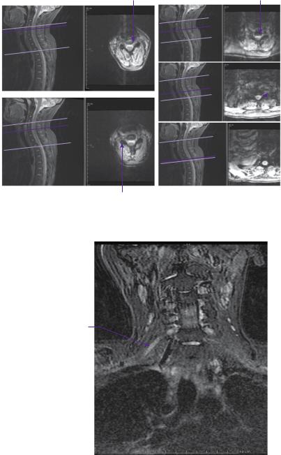
MRI of the cervical spine and brachial plexus bilaterally (Figures 19.1 and 19.2) |
|
|
showed oedema along the C5 and C6 nerve roots bilaterally, worse on the left than |
|
|
the right, extending into the supraclavicular fossae. However, there was no pseudo- |
|
|
meningocele to indicate nerve root avulsion. |
|
|
C5 nerve root |
C7 nerve root |
|
(c) |
||
(a) |
|
|
C8 |
|
|
|
nerve |
|
|
|
|
|
|
|
root |
|
(d) |
||
T1  nerve
nerve
root
(b) |
(e) |
C6 nerve root
Figure 19.1 T2-weighted STIR MRI axial images showing the spinal nerves as they emerge from the cervical cord (a:C5; b:C6; c:C7; d:C8; e:T1).
Hyperintense
C6 nerve trunk
Figure 19.2 Coronal T2-weighted STIR MRI of the brachial plexus, illustrating the oedema along the upper brachial plexus nerve roots.

Case 19 Peripheral nerve injury |
181 |
 Learning point Imaging of the brachial plexus
●MRI of the brachial plexus has been used to discriminate preand post-ganglionic lesions. Features of preganglionic injury include: spinal cord oedema, lateral displacement of the spinal cord, a syrinx, absence of root in the canal or intervertebral foramina, traumatic meningocele, haemorrhage/scarring in the spinal canal, and denervation of erector spinae muscles (shown by wasting and fatty infiltration over a timescale of ≥15 days). Spinal cord oedema is thought to
signify root avulsion central to the transition zone. Post-ganglionic injuries can also be identified, appearing as swelling of the nerve trunks on T1-weighted imaging and increased signal intensity on T2-weighted imaging [6,7].
●CT myelography allows demonstration of the intradural nerve roots [8]. Root avulsion can be identified by the absence of continuity of the root with the cord. Traumatic meningoceles are also visible. However, the C8 and T1 roots can be difficult to visualize in continuity due to their oblique path, and interference from the shoulders.
●High resolution ultrasound has the potential to allow in depth visualization of the brachial plexus and peripheral nerves. Preliminary studies of peripheral nerves are encouraging [9,10] and suggest that in the initial period following injury, prior to haematoma transformation into scar tissue, high resolution ultrasound may permit early detection of ruptures and other injuries to the nerves.
The patient underwent detailed electrophysiological examination 10 months post-injury, and Quantitative Sensory Testing (QST) almost 12 months from injury. The results are shown in Tables 19.2 and 19.3. They are of limited use in the left upper limb, as they reflect the second, more distal injury of the ulnar nerve, with the median and radial nerves affected by the severe soft tissue damage to the left forearm. The late F wave response was detectable in the right upper limb.
 Learning point Electrophysiological studies
Neurophysiological investigations have a role in determining conduction across a lesion from the outset. Although they are difficult to interpret in the context of nerve injury at multiple levels.
●SSEPs establish the presence or absence of a lesion proximal to the sensory dorsal root ganglion.
●Standard nerve conduction studies assess large, fast-conducting, alpha and beta nerve fibres, which mediate fine touch (velocity 40–60m/s), and deep afferent pathways.
●Small, myelinated (velocity 3–10m/s; A delta) fibres and non-myelinated (C) fibres are involved in pain and temperature sensation, and require the specialized techniques of QST.
●Electromyography records and analyses the spontaneous, insertional, and volitional electrical activity of the muscle.
●Neurapraxia is demonstrated by conduction block or slowing at the level of the lesion, and normal conduction distal to the lesion.
●Axonotmesis leads to fibrillations, compound sensory action potential (CSAP) and compound motor action potential (CMAP) reduction in proportion to the axonal loss. The latter manifest at least 7 days after injury, once Wallerian degeneration has occurred.
●Neurotmesis is demonstrated by fibrillation potentials and absent CSAPs and CMAPs
Intra-operative neurophysiological testing is invaluable. It can guide dissection, identify the region of nerve injury, protect against iatrogenic damage, and monitor function in both sensory and motor nerves [1].

182 |
Challenging concepts in neurosurgery |
|
|
|
Table 19.2 Neurophysiological results |
|
|
|
|
|
|
|
Motor conduction studies |
Right |
Left |
|
Median (to abductor pollicis brevis |
Latency from wrist:4.1ms |
Absent |
|
(APB)) |
amplitude:0.6mV |
|
|
|
latency from elbow: variable |
|
|
Ulnar (to abductor digiti minimi) |
Latency from wrist: 3.2ms |
Absent |
|
|
Amplitude: 2mV |
|
|
|
Latency below elbow: 8.1ms |
|
|
|
Amplitude: 2mV |
|
|
|
Latency above elbow: 9.7ms |
|
|
|
Amplitude: 2mV |
|
|
|
MCV (wrist to elbow): 47m/ms |
|
|
|
MCV (across elbow): 52m/ms |
|
|
Sensory conduction studies |
|
|
|
Median (terminal phalanx of |
Latency: 2.3ms |
Absent |
|
middle finger to palm and wrist) |
Amplitude: 5μV |
|
|
|
SCV: 53m/ms |
|
|
Median (index proximal phalanx |
Latency: 2.3ms |
Absent |
|
to wrist) |
Amplitude: 1.2μV |
|
|
|
SCV: 54m/ms |
|
|
Ulnar (little finger to wrist) |
Latency: 1.9ms |
Absent |
|
|
Amplitude: 2μV |
|
|
|
SCV:56m/ms |
|
|
Radial (first metacarpal to forearm) |
Latency: 1.7ms |
Absent |
|
|
Amplitude: 8μV |
|
|
|
SCV: 54m/ms |
|
|
Lateral antebrachial |
Latency: 1.6ms |
absent |
|
|
Amplitude: 5μV |
|
|
|
SCV: 50m/ms |
|
|
Medial antebrachial |
Latency: 2.0ms |
Absent |
|
|
Amplitude: 3μV |
|
|
|
SCV: 66m/ms |
|
|
Electromyography (EMG) |
|
|
|
Deltoid |
Few fibrillations and positive sharp waves; |
Polyphasics, |
|
|
excess polyphasics, moderately reduced, 2mV |
moderate, 3–4mV |
|
Infraspinatus |
Polyphasics, moderate, 3–4mV |
Not assessed |
|
Biceps |
Polyphasics, moderate, 3–4mV |
Polyphasics, |
|
|
|
moderate/ |
|
|
|
reduced, 2–3mV |
|
Triceps |
Polyphasics, moderate, 3–4mV |
Polyphasics, |
|
|
|
moderate, 2–3mV |
|
Brachioradialis |
Polyphasics, moderate, 3–4mV |
Not assessed |
|
FDS |
Polyphasics, moderate, 3–4mV |
Not assessed |
|
FDP |
Polyphasics, moderate, 3–4mV |
Not assessed |
|
Interossei (first dorsal) |
Few units only |
No units |
|
APB |
Few units only |
Few units only |
|
|
|
|
ms: millisecond; mV: millivolts; μV: microvolts; MCV: motor conduction velocity; SCV: sensory conduction velocity in metre/microsecond

|
|
Case 19 Peripheral nerve injury |
183 |
|
Table 19.3 Quantitative Sensory Testing |
|
|
|
|
|
|
|
|
|
QST |
Right |
Left |
|
|
Thermal thresholds |
Normal |
Normal, but elevated in |
|
|
|
|
hypothenar region |
|
|
Vibration thresholds |
Mod. elevated right index |
Elevated in index (marginally), |
|
|
|
finger and thumb |
ring and little fingers |
|
|
Joint position sense |
Preserved |
Preserved aside from little finger |
|
|
Pin prick |
Normal |
Reduced in little finger and |
|
|
|
|
ulnar border of forearm; |
|
|
|
|
hypersensitivity C5 |
|
|
Monofilaments |
Normal |
Mod. elevated in arm and hand |
|
|
|
|
at No.10 |
|
|
Sweating |
Preserved in palm |
Preserved in palm |
|
|
|
|
|
|
|
mod.: moderately.
The patient continues to undergo intensive inpatient neuro-rehabilitation, currently targeting the range of movement throughout the upper limbs, together with strengthening exercises, and intermittent splinting of the left upper limb.
Discussion
This patient had many potential reasons for his sensory and motor deficits. He was initially quadraplegic, which suggested a cervical cord injury. With the episodes of hypovolaemic and subsequent septic shock, and the preferential recovery of the lower limbs, poor perfusion of the cervical cord and, hence, the possibility of a central cord syndrome was considered. Anterior cord injury was another potential diagnosis, while a partial Brown–Séquard syndrome may be postulated, as multiple brachial plexus nerve root avulsions carries the inevitably degree of CNS involvement. However, the patient’s tone always remained normal. Other potential diagnoses included ITU polyneuropathy, hypoperfusion of the brachial plexus bilaterally, and blast injury to the brachial plexus bilaterally. The polyneuropathy of critical illness has been postulated to be a neurological manifestation of the systemic inflammatory response syndrome [11]. Corticosteroids and neuromuscular blocking agents, poor glycaemic control, and immobility may also be contributory factors [12,13], as may a low albumin. Critical care polyneuropathy may be induced by a diffuse compartment syndrome, with increased pressure in the endoneurial, perineurial, and epineurial compartments.
Evaluation of the left upper limb in this case was complicated by the extensive destruction of muscle and skin, the anklyosis of the left elbow, and the presence of an ulnar nerve lesion, i.e. a second level peripheral nerve lesion, which was grafted soon after injury. The neurophysiological studies, at 10 months post-injury, indicate moderate function of the proximal muscles of the left upper limb. The left median nerve and superficial radial nerve are in continuity, as shown by the preservation of sensation, and the activity of muscles supplied by the median nerve. The absence of conduction on the formal neurophysiological testing, at 10 months post-injury, is predictable, due to the forearm soft tissue destruction and ulnar nerve lesion. The Tinel’s sign over the left ulnar nerve indicates axonal severance with regeneration
 Expert comment Preganglionic brachial plexus lesions
Root avulsion can be detected by the electrodiagnostic triad of:
●Normal CSAP and a preserved histamine response.
●Denervation of paraspinal muscles.
●Loss of the SSEP.
This triad, together with evidence of fibrillations and absence of voluntary motor unit activity in peripheral muscles supplied by the root, distinguish a proximal lesion from more distal pathology [1].
 Expert comment
In blast injury, the patient is exposed to a shock wave at close range, without any fracture, or signs of significant soft tissue injury. The underlying mechanism is, as yet, not fully understood. Large fibre conduction recovers, but the small fibres repair more slowly, and sometimes imperfectly [14].

184
 Expert comment
It is generally advocated that patients with traumatic peripheral nerve injuries, including closed brachial plexus traction lesions, should undergo surgery as early as possible, as this has been shown to optimize recovery.
In this case, however, bilateral exploration of the brachial plexus was not advised. This was due to the patient’s poor systemic state and multiple co-existing injuries. Furthermore, in the acute stage, there was no definitive evidence of brachial plexus nerve damage amenable to surgical intervention.
Challenging concepts in neurosurgery
through the graft. Indeed, subsequent electrophysiological studies show early recovery, although the graft of the left ulnar nerve.
A diagnosis of bilateral brachial plexus conduction block at the root level was reached in this patient due to the notable absence of pain, lack of Horner’s sign, intact phrenic nerves, and absence of supraclavicular or infraclavicular Tinel’s sign. These features together with the neurophysiological results indicate a preganglionic conduction block, rather than an avulsion or rupture. There is evidence of physical continuity of motor axons to the small muscles of the right hand. The EMGs at 10 months post-injury showed only a few motor axons under volition, but clear evidence of continuity. These features are consistent with considerable axonopathy. On the right, the F wave indicated at least partial preservation or continuity of the large afferent and efferent myelinated pathways. To date, the right C6, C7, and C8 roots have shown better recovery than C5 and T1. However, the prognosis is favourable, especially as sympathetic function throughout the right upper limb is present. The QSTs show all modalities recovering, even the finer fibres.
 Evidence base Outcome following brachial plexus injury
●Kato et al. [15] studied 137 patients who underwent brachial plexus repair. Of the sixty who underwent early surgery (within 1 month), 56.7% had a good result, i.e. the patient regained at least one function. Of those in whom surgery was delayed by more than 6 months, only 13.6% showed a good result.
●Berman et al. [16] showed that repair of the plexus offers the patient the possibility of pain relief. Of 116 patients with proven root avulsions who underwent nerve transfer and/or grafting, 88% experienced severe pain pre-operatively. This was reduced to 34% at 3 years post-operatively. These findings are supported by later work [15,17]. Hence, even when repair fails to restore worthwhile
function, reinnervation by some means alleviates pain. It seems that reinnervation of muscle is more important than that of the skin.
 Clinical tip Surgical intervention for traumatic brachial plexus lesions
●The avulsed ventral root may be repaired by:
●Transfer to an adjacent ruptured stump.
●Transfer to a normal nerve (for example, the spinal accessory nerve).
●Rarely, by direct repair to a stump in the spinal canal.
●By re-implantation.
●Preganglionic avulsion lesions of the brachial plexus may be considered for re-implantation, if within 1 month of injury. Contraindications include severe head, chest, or visceral injuries, rupture or occlusion of the subclavian or vertebral artery, cervical or upper thoracic vertebral fractures, and spinal cord injury. These exclusion criteria do eliminate approximately 25% of individuals who
present with avulsion injury. The pure lateral approach, as detailed in [18], allows access to the intraand extraspinal regions of the brachial plexus. It involves exposure of the anterior and posterior tubercles, and the transverse processes at the relevant spinal levels. The posterior tubercle and part of the lateral mass are then resected to allow direct access to the spinal cord. The dura is opened longitudinally and the grafts fed through the intervertebral canals and implanted via small slits in the pia as close to the ventral root exit zone as possible. Published results of long-term functional recovery are encouraging, although sample size is small [19, 20, 21].
●The approach to post-ganglionic lesions is dependent on the target region of the brachial plexus [1] describes a medial extension of the transverse supraclavicular approach, which he and Bonney adopted. It allows access to the first part of the subclavian and vertebral arteries and the whole of the brachial plexus. Fiolle and Delmas [22] added a vertical limb to the transverse supraclavicular wound, with the potential to display the supra-, retro-, and infraclavicular plexus, the second part of the subclavian artery to the terminal axillary artery, and the subclavian and axillary veins deep to the clavicle. The infraclavicular part of the brachial plexus can be displayed by full opening of the deltopectoral interval, and division of the pectoralis minor tendon. Finally, the posterior subscapular route provides access to the most proximal parts of the nerves [23].

Case 19 Peripheral nerve injury |
185 |
 Expert comment Peripheral nerve surgery
●External neurolysis involves dissection outside of the epineurium, i.e. freeing the nerve from a constricting or distorting agent.
●Internal or interfascicular neurolysis requires exposure of the bundles by epineurotomy and their separation.
●Nerve repair entails the restoration of healthy conducting elements without tension. This may be by direct suture or grafting. The nerve must be adequately prepared, that is, the ends cut back progressively, until healthy bundles are exposed. This may be 1–2mm in an urgent case, typically 5–10mm, but up to 4cm in delayed cases or where infection has occurred. Intra-operative neurophysiological assessment is used in conjunction with visualization and gentle palpation
of the nerve. If the gap after resection is small and the repaired nerve lies without tension, little mobilization of the nerve is required and end-to-end suture is appropriate. This involves
matching bundle (fascicle) to bundle, with additional epineurial sutures. This is rarely appropriate in supraclavicular brachial plexus injuries. Grafting is undertaken using cutaneous nerves from the damaged limb if feasible. In brachial plexus surgery, this is typically the medial cutaneous nerve of the forearm (MCNF), taken through a straight incision. The sural nerve is also often required,
which is taken through a midline incision running laterally in the distal aspect of the leg, to a point mid-way between the posterior aspect of the lateral malleolus and the lateral margin of the Achilles tendon. In cases where C5 and/or C6 are ruptured or avulsed, the superficial radial nerve makes an excellent graft, and the lateral cutaneous nerve of the forearm may also prove useful. The grafts are cut to the appropriate length and sewn into place, the suture uniting the epineurium of the graft to the perineurium of the stump bundle. The repaired nerve must lie in healthy tissue, with full thickness skin overlying.
●Nerve transfer is appropriate in cases of brachial plexus root avulsion not amenable to reimplantation. Nerve transfer has the optimum chance of success if it is transferred to a nerve and muscle of roughly equivalent size, without an interposed nerve graft [24].
●Muscle transfers may be used once the neurological prognosis is known.
The patient continues to work with an extensive multi-disciplinary neurorehabilitation team. To date, his management has rested on accurate diagnosis and prognostic information, together with intensive neuro-rehabilitation. Stiffness in the shoulders continues to be addressed, as does that in the small joints of the hands. Surgery is now indicated to overcome the clawing of the right MCP joints. Although small muscle activity continues to recover, their weakness rules out useful function in his dominant hand. Extensor carpi radialis longis (ECRL) transfer via Brandt’s method is planned, followed by resurfacing of the left forearm together with an assessment of the continuity of musculotendinous units. This would provide the left median and ulnar nerves with the best possible conditions for recovery.
 Learning point Rehabilitation
Neuro-rehabilitation is a mainstay of treatment for patients with peripheral nerve lesions. Active and close involvement of relevant surgical specialties is also essential. Further operative procedures may form an integral part of the rehabilitation process, to manage neuropathic pain, decompress nerves, or provide full thickness skin cover to aid pain management and enhance nerve regeneration [14].
Neuro-rehabilitation involves a MDT whose purpose is to:
●Objectively assess disability and accurately measure the outcome of treatment.
●Reduce the degree of disability by physical and other therapies.
●Return the patient to his/her original work, a modified role, or to alternative employment.
●Restore the patient’s ability to live in his/her own home, to enjoy recreation and social interaction, and to be independently mobile.
Neuro-rehabilitation often involves functional splinting, which may prevent deformity, enhance use by placing joints in a useful position, and strengthen selective muscles [1].
186 |
Challenging concepts in neurosurgery |
A final word from the expert
Peripheral nerve injuries offer many challenges, especially those that are proximal, involving the nerve plexus, and/or are complicated by other injuries. Secondary distal damage
to a peripheral nerve can further confuse the clinical picture. A thorough history and examination, together with appropriate imaging and neurophysiological investigations, allow accurate diagnosis and prognosis. The management of such severe blast injury to peripheral nerves is a new phenomenon requiring further research. In this case, a diagnosis of conduction block at the root level was made using all available clinical details and investigations. Despite the patient’s quadriplegic presentation, a positive prognosis was delivered. Further neuro-rehabilitation, in its broadest sense, remains the long course ahead for this patient.
References
1.Birch R. Surgical disorders of the peripheral nerves, 2nd edn. London: SpringerVerlag, 2011.
2.O’Brien M. Aids to the examination of the peripheral nervous system, 5th edn. Kidlington: Saunders/Elsevier, 2010.
3.Bonney G. Clinical Neurophysiology in Peripheral Nerve Injuries. In: R Birch, G Bonney, CB Wynn Parry (eds), Surgical disorders of the peripheral nerves (pp. 196–8). Edinburgh: Churchill Livingstone, 1998.
4.Seddon HJ. Three types of nerve injury. Brain 1943; 66: 237–88.
5.Tinel J. Nerve wounds. London: Balliere Tindall and Co., 1917. [Authorised translation by F Rotherwell, revised and edited by CA Joll.]
6.Hems TEJ, Birch R, Carlstedt T. The role of magnetic resonance imaging in the management of traction injuries to the adult brachial plexus. Journal of Hand Surgery 1999; 24b: 550–5.
7.Tavakkolizadeh A, Saifuddin A, Birch R. Imaging of adult brachial plexus traction injuries. Journal of Hand Surgery 2001; 26B: 183–91.
8.Carvalho GA, Nikkhah G, Matthies G, et al. Diagnoses of root avulsions in traumatic brachial plexus injuries: value of computerised tomography myelography and magnetic resonance imaging. Journal of Neurosurgery 1997; 86: 69–76.
9.Cokluk C, Aydin K. Ultrasound examination in the surgical treatment for upper extremity peripheral nerve injuries. Part I: Turkish Neurosurgery 2007a 17: 197–201.
10.Cokluk C, Aydin K. Ultrasound examination in the surgical treatment for upper extremity peripheral nerve injuries. Part II: Turkish Neurosurgery 2007b 17: 277–82.
11.Visser LH. (2006). Critical illness polyneuropathy and myopathy: clinical features, risk factors and prognosis. European Journal of Neurology 2006; 13: 1203–12.
12.Schweickert WD, Hall J. ICU-acquired weakness. Chest 2007; 11: 1541–9.
13.Latronico N, Bolton CF. Critical illness polyneuropathy and myopathy: a major cause of muscle weakness and paralysis. Lancet Neurology 2011; 10: 931–41.
14.Birch R, Eardley WGP, Ramasamy A, et al. Nerve injuries sustained during warfare: Part II: Outcomes. Journal of Bone and Joint Surgery (British) 2012; 94B: 529–35.
15.Kato N, Htut M, Taggart M, et al. The effects of operative delay on the relief of neuropathic pain after injury to the brachial plexus. Journal of Bone and Joint Surgery 2006; 88B: 756–9.
16.Berman J, Taggart M, Anand P, (1995) The effects of surgical repair on pain relief after brachial plexus injuries. In: Association of British Neurologists Proceedings, University of Liverpool, April 1995, JNNP; 44: 5–7.
Case 19 Peripheral nerve injury |
187 |
17.Taggart M. (1998) Relief of pain with operation. In:Birch R, Bonney G, WynnParry CB. (eds) Surgical disorders of the peripheral nerves. Edinburgh: Churchill Livingstone, 373–405.
18.Camp SJ, Carlstedt T, Casey ATH. Technical note: pure lateral approach to intraspinal re-implantation of the brachial plexus. Journal of Bone and Joint Surgery (British) 2010; 92B: 975–9.
19.Carlstedt T. (2007) Central nerve plexus injury. In:T Carlstedt,(ed.), Central nerve plexus injury. London: Imperial College Press.
20.Carlstedt T, Anand P, Hallin R, et al. Spinal nerve root repair and reimplantation of avulsed ventral roots into the spinal cord after brachial plexus injury. Journal of Neurosurgery 2000; 93: 237–47.
21.Carlstedt T, Grane P, Hallin RG, et al. Return of function after spinal cord implantation of avulsed spinal nerve roots. Lancet 1995; 346: 1323–5.
22.Fiolle J.Delmas J. In: CG Cumston (transl. ed.), The surgical exposure of the deep seated blood vessels (pp. 61–7). London: Heinemann, 1921.
23.Kline DG, Kott J, Barnes G, et al. Exploration of selected brachial plexus lesions by the posterior subscapular approach. Journal of Neurosurgery 1978; 49: 872–9.
24.Addas BMJ.Midha R. Nerve transfers for severe nerve injury. Neurosurgery Clinics 20: Peripheral Nerve Injury 2009; 20(1): 27–38.