

CASE
2 Glioblastoma multiforme
Mohammed Awad
 Expert commentary Kevin O’Neill
Case history
A 59-year-old, right-handed male, with a background of hypertension and previous transient ischaemic attack (TIA), presented to his general practitioner with a 3-week history of intermittent, but progressively worsening right arm numbness. This was followed by speech disturbance a week later, and an outpatient computed tomography (CT) head scan was ordered. On examination, he was found to have a normal conscious level with a mild expressive dysphasia. He was also found to have a mild pyramidal weakness (MRC (Medical Research Council) 4/5) and sensory disturbance involving the right arm.
 Learning point Dysphasia
There are several subtypes of dysphasia, however, they broadly fall into one of three syndromes— expressive dysphasia, receptive dysphasia, or global dysphasia. Expressive dysphasia, also known as motor dysphasia, is a conscious difficulty in the expression of speech, including speech initiation, proper grammatical sequencing, and proper word forming and articulation. Patients can fully understand what is told to them and can fully follow commands, but speech is slow and ‘forced’, and
features short phrases. Receptive dysphasia is essentially the reverse of expressive in that the patient’s speech may appear quite fluent and articulate, although it may not necessarily make sense and these patients are unaware of their mistakes. They find it difficult to comprehend spoken language and or word–object relations and therefore show some difficulty in following spoken commands. Wernicke’s dysphasia is the most common of the receptive dysphasias. Conduction dysphasia, also known as associative dysphasia, is relatively uncommon and only amounts to 10% of the presenting dysphasias. It is caused by damage to the arcuate fasciculus, essentially disconnecting Broca’s from Wernicke’s, and results in difficulty with repetition. Patients may also suffer the inability to describe people or objects in the proper terms. Global aphasia results from damage to all three regions—Broca’s, the arcuate fasciculus, and Wernicke’s areas, which results in total language disturbance.
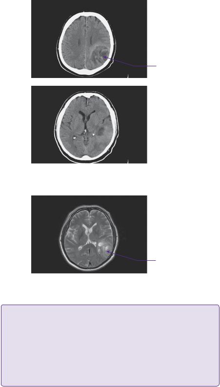
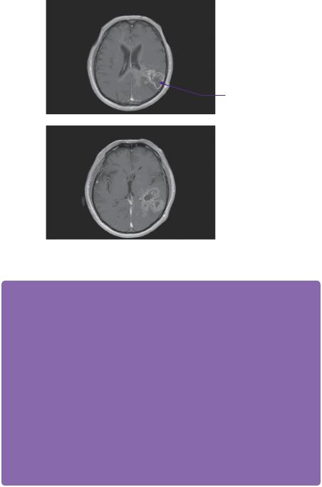
The CT head scan demonstrated areas of low attenuation within the left temporoparietal region that enhanced heterogeneously with surrounding mass effect and oedema (Figure 2.1). An enhanced MRI scan confirmed a left temporoparietal intrinsic space-occupying lesion. The lesion was of mixed intensity on the T2-weighted image, but predominantly isoto hyperintense (Figure 2.2). The flair demonstrated the vasogenic oedema spreading predominantly in the white matter directed around and away from the lesion. On a T1 post-contrasted scan, the lesion showed peripheral rim enhancement with a presumed necrotic, non-enhancing centre (Figure 2.3).
In view of the perilesional oedema and mass effect, dexamethasone was administered with a proton pump inhibitor for gastric protection.
 Learning point Hemisphere dominance and language
Language functions, such as vocabulary, grammar, and literal meaning are areas that reside in the dominant hemisphere of an individual. In right-handed individuals, this is the left hemisphere in approximately
90–95% of people. In left-handed people, the dominant hemisphere is still left-sided in 63–71%. The main areas of language are Broca’s in the posterior inferior frontal gyrus and Wernicke’s in the superior temporal gyrus. They are connected by a white matter tract, known as the arcuate fasciculus.
12 |
Challenging concepts in neurosurgery |
Area of spaceoccupying low density in parietal region
Figure 2.1 CT—axial scan images revealing a low density space-occupying lesion in the posterior temporal and parietal lobes with heterogenous contrast uptake.
Mixed attenuation area with posterior ventricular horn effacement
Figure 2.2 T2-weighted axial MRI showing the iso-hyperintense lesion with evidence of encroachment of mass effect on surrounding eloquent areas.
 Learning point Vasogenic oedema and dexamethasone
Vasogenic oedema occurs around tumours and inflammation as a result of a breakdown of the blood–brain barrier. This causes an influx of proteins into the extracellular space from the intravascular compartment and water follows by the process of osmosis. In normal circumstances, these proteins would not pass through tight junctions, but these are disturbed by the presence of the tumour. The exact mechanism of action of corticosteroids around tumours is unknown, but it is thought that dexamethasone works to reduce the inflammatory response around the tumour and, therefore, restore some element
of normality to the blood–brain barrier. It is also thought to decrease oedema by the effect on bulk flow away from the tumour, although corticosteroids decrease capillary permeability in the tumour itself [1]. VEGF inhibitors, such as bevacizmab, as an alternative to steroids, have also been reported to reduce vascular permeability effectively and thereby brain tumour oedema in the clinical setting [2].
Case 2 Glioblastoma multiforme |
13 |
Heterogenous contrast uptake favouring the periphery of the tumour
Figure 2.3 T1-weighted images with gadolinium showing peripheral contrast uptake of the heterogeneous intrinsic lesion.
 Expert comment The function of the multidisciplinary team
The neuro-oncology multidisciplinary team (MDT) brings together all the necessary clinical expertise to optimize a brain tumour patient’s care. This framework guidance was implemented by the National Institute for Health and Clinical Excellence (NICE) through its Improving Outcome Guidance (IOG) guidelines. NICE is a special health authority of the English National Health Service (NHS). NICE publishes guidelines in three areas—the use of health technologies within the NHS (such as the use of new and existing medicines, treatments, and procedures), clinical practice (guidance on the appropriate treatment and care of people with specific diseases and conditions), and guidance for public sector
workers on health promotion and ill-health avoidance. The MDT’s members will include neuro-oncologists (neurosurgeons, clinical oncologists), neuroradiologists, neuropathologists, psychiatrist/ psychologists, clinical nurse specialists, physiotherapists, occupational therapists, and clinical trials co-ordinators.
The team should handle all neuro-oncology referrals by making recommendations about further management based on diagnosis. This will be based on current best evidence-based practice, including NICE guidance and should also provide the opportunity for eligible patients to be entered into clinical trials. The MDT may make recommendations for further investigation or clinical assessment if there
are uncertainties about the case or the evidence for treatment benefit is unclear. It is now considered a core element in the patient pathway, is usually established in a designated neuro-oncology centre, and involves both inpatient care and outpatient follow-up. The overall goal of the MDT is to expedite and improve patient care, ultimately leading to better outcomes and survival. It is now mandatory to have an MDT-centric pathway to manage neuro-oncology patients in the UK.
The patient’s symptoms resolved after 3 days on dexamethasone. His case was discussed in the neuro-oncology MDT meeting. The patient scored highly on the Karnofsky performance scale (90/100) and, after discussion with the patient about the management options (risks as well as benefits of surgery with adjuvant therapies versus no treatment), the decision was taken to proceed to craniotomy and debulking, with subsequent adjuvant therapy dictated by the pathology results.
14 |
Challenging concepts in neurosurgery |
 Learning point Karnofsky score
The Karnofsky performance status score is an attempt to quantify ‘well-being’. It is used to determine whether patients are fit enough to withstand and benefit from standard treatments, or whether they should be exposed to less radical therapy instead. It is also used as a measure of quality of life. The Karnofsky score runs from 100 to 0, where 100 is ‘perfect’ health and 0 is death. This scoring system is named after Dr David A. Karnofsky, who described the scale with Dr Joseph H. Burchenal in 1949, and it is still used today.
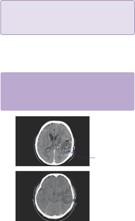
The patient underwent craniotomy and debulking of the tumour assisted by magnetic resonance (MR)-directed image guidance with 3D intra-operative ultrasound (SonowandTM). Post-operatively he was mildly dysphasic again, but this resolved within 1 week of surgery. His post-operative CT scan showed a good clearance of the bulk of the tumour (see Figure 2.4).
 Clinical tip Combined intra-operative MR-directed image guidance and ultrasound
SonowandTM is a combined navigation console and intra-operative ultrasound. Pre-operative DICOM magnetic resonance imaging (MRI) or CT images are uploaded and when matched with the patient can be used for flap planning, as standard with all navigation consoles. However, the SonowandTM has the advantage of being able to update the ‘road-map’ being used, by providing a 3D ultrasound image as the surgery progresses, which provides real-time up-to-date images allowing for brainshift and aids with visualization of nearby vessels. The authors believe it is a great visualization surgical aid that aids with extensive and safer resections.
Post-operative tumour cavity with low density air bubbles under craniotomy flap
Figure 2.4 Post-operative CT axial scan showing good surgical resection of the tumour.

Case 2 Glioblastoma multiforme |
15 |
Enhancement pattern suggesting tumour recurrence with extension to craniotomy flap
Figure 2.5 T1-weighted MRI with gadolinium at 1 year post-resection, showing tumour recurrence at the site of the previous resection.
He later received standard dose conformal external beam radiotherapy (2Gy fractions daily to a total of 60Gy over 6 weeks). This is a typical regimen and each 2Gy fraction takes approximately 3 minutes. He was also given six monthly cycles of temozolomide chemotherapy. This is currently first line chemotherapy for glioblastoma multiforme (GBM) in the UK and is given for 6–12 months, depending on tumour grading, Karnofsky grade of the patient, response while undergoing treatment, and genetic marker studies. In this case, a 6-month MRI scan showed no progression, but a 12-month follow-up scan revealed significant recurrence (Figure 2.5).
Discussion
GBM is the most common form of malignant primary brain tumour. In the UK, the incidence is approximately 2–3 cases per 100,000 people per year and it accounts for approximately 20% of all primary intracranial tumours. GBM is derived from glial cells, and is thought to arise either de novo as primary GBM or secondary to malignant progression from a low-grade astrocytoma (Chapter 21). There is a greater male predilection for reasons unknown[3].
16
 Learning point Presentation times of glioblastoma
Astrocytic tumour cells may diffusely infiltrate cortex without initially affecting neuronal function. However, eventually neighbouring neurons are damaged or isolated, and patients develop neurological symptoms. In this way, GBM may be very large before patients become symptomatic, although growth
in eloquent areas will manifest relatively early.
Challenging concepts in neurosurgery
 Learning point The WHO classification of astrocytomas—I–IV
Grade I lesions are those with low proliferative potential and may be cured following surgical resection alone. Grade II lesions are generally infiltrative and despite showing low proliferative activity, they usually recur after surgery. Most grade II tumours transform to higher grades over time. WHO defines diffusely infiltrative astrocytic tumours with cytological atypia alone as Grade II (diffuse astrocytoma). WHO Grade III lesions (anaplastic astrocytoma) show histological evidence of malignancy, including nuclear atypia and brisk mitotic activity. Finally, WHO Grade IV lesions are
cytologically malignant, mitotically active, necrosis-prone neoplasms with endovascular proliferation typically associated with rapid disease progression and a dismal prognosis. Grade IV tumours tend to show widespread infiltration of surrounding tissue and some may demonstrate craniospinal dissemination.
GBM commonly presents with neurological deficit, seizures, and signs of raised intracranial pressure. Approximately 30–50% will present with non-specific headache, which may become characteristic of raised intracranial pressure (ICP). 30–60% will present with seizures and, depending on the location, these may be simple, focal, or generalized. Focal neurological deficit is a presenting feature in 40–60% and 20–40% present with mental status changes. The location of the tumour usually delineates the neurological deficit and the time to presentation, as tumours in more eloquent areas tend to present sooner.
Intrinsic high-grade tumours may show a variety of appearances on CT and MRI. This is dependent on the presence or absence of low grade areas around the tumour, possible calcification, the rate of growth, the degree of necrosis, and the presence of any haemorrhage.
CT scans will usually demonstrate an area of low density with surrounding mass effect and after contrast administration, usually a ragged ring of enhancement, surrounded by oedema. The mass effect may be minimal and local, but may be severe enough to cause midline shift and compression of the ventricles, and may even cause hydrocephalus.
On MRI, high-grade intrinsic lesions show as low density areas on T1-weighted imaging that ring enhance, also usually in a ragged fashion, with contrast administration. On T2-weighted images they show as heterogeneous masses, usually with marked extensive surrounding oedema, predominantly in the white matter. The fluid-attenuated inversion recovery (FLAIR) sequence shows the oedema even more extensively. It is difficult, however, to distinguish between oedema and tumour infiltration, as both are of high intensity on the FLAIR sequence. The differential diagnosis of ring-enhancing lesions includes metastases, abscess, and parasitic infections. Other image sequences, such as apparent diffusion coefficient (ADC) map and gradient echo, may be helpful, with the clinical history, to differentiate between these. MR spectroscopy may also be useful.
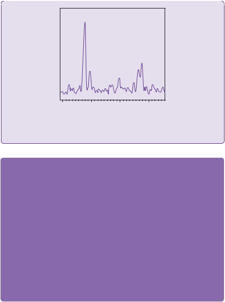
 Learning point Magnetic resonance spectroscopy of glioblastoma multiformes
MR spectroscopy gives additional information to aid with diagnosis. Glioblastomas typically demonstrate high levels of choline, lactate, and lipid, and low levels of N-acetylaspartate (NAA) and creatine. Generally, the more malignant the lesion, the higher the choline-to-creatine peak ratio, with an increased lactate peak, and decreased NAA peak ratio. A typical graph of a sampled glioblastoma is shown in Figure 2.6.
(continued)
Case 2 Glioblastoma multiforme |
17 |
|
|
|
|
CHO
LAC
CR
NAA
4 |
3 |
2 |
1 |
|
|
ppm |
|
Figure 2.6 A typical MR spectroscopy graph of sampled glioblastoma tumour tissue.
CHO: choline; CR: creatine; NAA: N-acetylaspartate; LAC: lactate; ppm: parts per million.
 Expert comment Imaging glioblastoma multiformes
When imaging all gliomas, including GBM, I will always require a structural MRI, which will include a T1-weighted series of images, with and without contrast to determine the enhancing bulk of
the tumour. I will also want, as standard, a T2 sequence with a T2 FLAIR to get an estimate of the extent of tumour and or any oedema within the surrounding parenchyma. In essence, one has to assess the location and extent of the tumour. From this I can assess its contribution to local mass effect and estimate the degree of diffuse invasion. Ultimately, we know that radical resection can improve the patient’s outlook and response to adjuvant treatments, but this has to be balanced against inflicting morbidity or deficit that could worsen their prognosis. In certain cases, where tumours look resectable, but are close to eloquent areas or tracts, I will request functional MRI and/or tractography. Most tumours, particularly the lower grade gliomas, undergo physiological scanning with spectroscopy and cerebral blood volume maps to build a database of MRI biomarkers, particularly, if we are following tumours for any length of time. Again, all gliomas that require surgery will have a neuronavigation thin slice acquisition scan to be able to use this now standard technology. I incorporate as much imaging information into that system as I can for the purposes of accurate navigation. In addition, my preferred mode of intra-operative imaging to update the pre-operative image data is 3D ultrasound, which provides similar, but complimentary information to the MRI,
and will take into account resection and brain shift. In addition to intra-operative imaging, awake craniotomy and cortical mapping are useful adjuncts to avoid neurological deficit during surgery near eloquent cortex.
Gliomas are composed of a heterogeneous mix of poorly differentiated neoplastic astrocytes. Glioblastomas are distinguished from WHO grade III astrocytomas by the presence of necrosis and endothelial hyperplasia. Both usually form in the cerebral white matter. In adults, this is usually in the cerebral hemispheres supratentorially, but in children it is not unusual for the primary location to be the brainstem. Approximately, half of the supratentorial tumours occupy more than one lobe or are bilateral. The classic ‘butterfly appearance’ (Figure 2.7) develops as a result of growth across the corpus callosum. Grade III and IV tumours most commonly can develop de novo or can be the result of a transformation from a lower grade astrocytoma (less than 10%). These secondary GBMs are more common in a younger age

18 |
Challenging concepts in neurosurgery |
 Learning point Risk factors for glioblastoma multiformes
●Male.
●Older age: over 50 years old.
●Caucasians and Asians.
●Low-grade astrocytoma.
●Having one of the following genetic disorders is associated with an increased incidence of gliomas: neurofibromatosis, tuberous sclerosis, von Hippel–Lindau (VHL) disease, Li–Fraumeni syndrome, Turcot syndrome.
Enhancement pattern of a ‘butterfly glioma’ crossing the midline through the body and rostrum of the corpus callosum
Figure 2.7 T1-weighted MRI with gadolinium. Axial and coronal sections showing the classic appearance of a butterfly glioma.
group (average age 45) versus primary GBMs (average age 62) [4]. Rarely, high-grade gliomas may seed through the CSF to distant cranial or spinal sites. They can cause meningeal gliomatosis and, indeed, may be found in CSF, resulting in high protein content.
High-grade gliomas are invariably difficult to treat due to the degree of diffuse infiltration in the surrounding brain making surgical resection incomplete, the lack of efficacy of standard radiotherapy, and a lack of effective chemotherapy. As a result they are currently considered incurable. Treatment is ultimately palliative and aimed at increasing the length of survival and quality of life.
The options are:
●Purely conservative and supportive (palliative care).
●Radiotherapy with or without chemotherapy.
●Surgery with or without radio and/or chemotherapy.
Surgery may be used simply to achieve a histological diagnosis through a biopsy or may reduce the tumour bulk, either to reduce mass effect, or to allow adjuvant therapy its best chance by reducing the tumour cell load.
Clinical research is emerging that shows that radically extensive volume resections have a greater impact on length of survival and, therefore, in the UK the trend is moving towards radical debulking of high-grade lesions with subsequent adjuvant radioor chemotherapy if the patient is fit enough.

Case 2 Glioblastoma multiforme |
19 |
 Learning point Extent of resection
A landmark paper that assessed the length of survival of patients with glioblastomas according to the extent of resection was a multivariate analysis of 416 patients [5]. The conclusion was that a significant survival advantage was associated with resection of 98% or more of the tumour volume (median survival 13 months), compared with 8.8 months for resections of less than 98%. Many other studies have reached similar conclusions. Stummer et al. [6] looked at the extent of glioblastoma resections with the aid of 5-ALA fluorescence. This was a randomized study of 270 patients. Half underwent surgical resection guided by fluorescence achieved with 5-ALA and the remainder under white light. They found that 65% of the 5-ALA patients had complete
resections of the pre-operative MRI-enhancing areas versus only 36% under white light. They also found that 41% of the 5-ALA resected patients had progression-free survival at 6 months compared with 21% for the control group. Vuorinen et al. [7] found a survival advantage of >2 months for craniotomy and surgical resection versus biopsy for GBM patients. They also concluded that craniotomy and debulking offered a modest survival advantage over biopsy in elderly patients with a poor Karnofsky score, unsuitable for other adjuvant therapies. In a study of 500 patients with newly-diagnosed glioblastoma operated between 1997 and 2009. Evidence has emerged from studies showing that the extent of resection at repeat craniotomy for recurrent glioblastoma predicted overall survival. They concluded that even if initial resection had not been optimal, the repeat craniotomy should attempt to achieve macroscopic complete resection if at all possible, as this significantly improved survival benefit
 Clinical tip Maximizing resection, while minimizing neurological deficit
Following the advice of Lacroix et al. [5], the evidence suggests that attempting a radical (>98%) resection will significantly increase life expectancy. However, this should not be at the expense of quality of life. In order to avoid or minimize damage to surrounding functioning brain and en-passant vessels, one should always use the adjuncts of image guidance, Moreso, the use of intra-operative ultrasound, or MRI will allow for a larger resection, while keeping within the tumorous tissue and not beyond in eloquent areas. The use of the ‘angio mode’ on intra-operative ultrasound (SonowandTM) will also allow for the visualization of en-passant vessels and help keep them intact, thereby potentially reducing the incidence of post-operative deficit. Stummer et al. [8] also demonstrated that operating on high-grade tumours with the aid of 5-ALA microscopy greatly aided the surgeon
in visualizing abnormal tissue, and therefore in obtaining an extensive resection. The crucial point with surgery for glioblastomas is not to attempt a complete resection if there is a risk of leaving permanent damage, given that these are currently incurable tumours. Many surgeons will now obtain intra-operative histology (smear, frozen section), in order to confirm the diagnosis and decide how aggressive they will be with their resections.
 Expert comment Intracavity chemotherapy
Gliadel® wafers represent an alternative approach to the delivery of chemotherapy in malignant glioma. Gliadel® wafers contain Carmustine® and are designed to release this agent over a 2–3-week period. Gliadel® wafers are placed on the surface of the resected tumour. For recurrent malignant glioma one randomized control trial (RCT) compared the efficacy of Gliadel® with that of placebo in patients with recurrent glioma. No significant survival advantage was seen in the primary analysis, however, a survival advantage for Gliadel® was observed in patients with GBM after adjustment for prognostic factors. This suggests that Gliadel® may increase overall survival in some patients with
recurrent resectable malignant glioma. As such patients generally have a poor outlook, any treatment that has the potential for prolonging life without significant adverse events should be considered
an option. However, given that no subgroups had been identified beforehand, the results of the
subgroup analysis of GBM patients in that trial should be interpreted with caution.
(continued)

20 |
Challenging concepts in neurosurgery |
|
|
|
|
The strongest evidence for their use involves trials in newly-diagnosed malignant glioma. Two RCTs compared the efficacy of Gliadel® with placebo in patients with newly-diagnosed gliomas. In the largest RCT to date, patients who received Gliadel® for newly-diagnosed malignant glioma were reported to have experienced a 2-month improvement in median survival compared with patients who received placebo (p = 0.017). In addition, analysis of the survival curves revealed a significant 27% reduction in risk of mortality for patients who received Gliadel® (p = 0.018). A survival advantage with Gliadel® in patients with GBM was not detected, but the trial was not designed to make comparisons between histological subgroups. Because the researchers in another randomized trial were unable to obtain sufficient Gliadel®, that trial included only 32 patients newly diagnosed with malignant glioma, instead of the anticipated 100. Although a survival benefit was reported for Gliadel® in the overall patient population and in patients with GBM, no conclusions could be reached, based on the small number of patients enrolled. Both studies reported similar adverse events in the treatment and control arms. The most common adverse events associated with Gliadel® were hemiplegia, convulsions, confusion, and brain oedema. The most commonly reported adverse events among patients who received placebo were convulsions, confusion, brain oedema, and aphasia. A significantly higher number of patients experienced intracranial hypertension in the Gliadel® arm of the Westphal trial. Because neither trial included a comparison with systemic therapy, the possible contrast between
the adverse event rates associated with interstitial chemotherapy wafers and the rates expected with systemic chemotherapy is unclear. Given that the largest trial demonstrated a survival advantage
in the Gliadel® treatment arm, Gliadel® may be considered an option in the subgroup of patients with newly-diagnosed resectable malignant gliomas. However, the exact patient population (based on age, histology, performance status, and so on) that may benefit from Gliadel® is unclear; further investigation is needed. In addition, no comparison has been performed between the efficacy of interstitial and systemic chemotherapy; clinicians should therefore review the latest evidence for the benefit of systemic chemotherapy in patients with newly-diagnosed malignant glioma.
As a result of these studies Carmustine® wafers (Gliadel®) are now recommended for use by NICE for selected patients, provided the following criteria are satisfied:
●Pre-operative MRI suggestive of newly-diagnosed high-grade glioma (HGG).
●Discussion before surgery in a neuro-oncology MDT.
●Surgical resection by a specialist neurosurgical oncologist.
●Surgical resection of more than 90% of the tumour.
●Intra-operative pathological confirmation of HGG.
●The ventricle is not widely opened.
A recent national audit yet to be published suggested that brain tumour surgeons are not considering the use of wafers in many patients that may be eligible. I believe that most surgeons are concerned about potential complications, such as brain oedema, wound healing and infection rates, and perhaps also cost in a period of reducing spending cuts in healthcare.
The prognosis for these high-grade tumours is very poor, although over the last 10 years, it has improved somewhat due to improved surgery and better chemotherapy agents. The average survival without any treatment is 3–6 months, but with aggressive treatment in the form of surgery, radiotherapy and chemotherapy, this can commonly be 1–2 years [9]. Older patients and patients with neurological deficit at presentation carry a worse prognosis. Conversely, younger patients, under 50 years, with a good initial Karnofsky Performance score of >70, and those with surgical resections >98% carry a better prognosis [5].
Death from GBM is usually secondary to raised ICP or severe neurological deterioration allowing opportunistic infections or thromboembolic events to supercede.
There are several molecular markers associated with outcome prediction in glial tumours. The 1p/19q codeletion strongly predicts response to treatment and survival in oligodendroglial tumours. The Methylguanine-methyltransferase promoter methylation, which is thought to render the cells more vulnerable to alkylating chemotherapy

Case 2 Glioblastoma multiforme |
21 |
agents is also associated with a better outcome. Hegi et al. [10] tested the relationship between MGMT silencing in the tumour and the survival of patients enrolled in a randomized trial comparing radiotherapy alone with radiotherapy and adjuvant temozolomide. They concluded that patients with glioblastoma containing a methylated MGMT promoter benefitted from temozolomide, whereas those who did not have a methylated MGMT promoter did not have such a benefit. Their work has led to significant changes in oncological practice. Finally, and more recently, mutations of the IDH1 gene have been found in 40% of gliomas and are inversely correlated to grade. IDH1 mutation is a strong and independent predictor of survival [11]. These are all DNA characteristics intrinsic to the patient and currently cannot be altered externally.
Long-term disease-free survival is possible, but these tumours usually reappear, often within 3cm of the original site, and 10–20% may develop new lesions at distant sites (termed multifocal GBM). Further radical surgery, perhaps supplemented with adjuvant radiosurgery and/or suitable chemotherapy may lead to additional prolongation of life, but the benefits of such treatment need to be assessed realistically and quality of life must also be taken into account.
A final word from the expert
Clearly, the prognosis for patients with malignant glioma remains poor, with median survival in the region of 12–14 months. This has only changed marginally over the last few decades with the use of systemic chemotherapy alongside surgical resection and radiotherapy. There are outliers who do better or worse than the median, and 3-year survival percentages do seem to be increasing with current treatment regimes. The results tell us that these tumours are very heterogenous in their genetics and response to therapy. Laboratory research has not only identified abnormal genes, but also abnormal epigenetic control of normal gene expression, which may be even more important. More and more pathways are being discovered that relate to biological behaviour and prognosis. The problem is that the cellular targets differ from tumour to tumour. That is why I believe the future of GBM management will become much more tailored to the individual patient. This will mirror the trend in surgery with improved tumour identification techniques already seen with fluorescent markers and intra-operative imaging technology. As research is broadening there will also be an increasing use of physical treatments, such as particle beam and other forms of electromagnetic energy, as well as nanotechnology to deliver targeted therapy. The therapy will need to be effective against all cell types, rather than selecting out resistant populations. There is currently a resurgence of interest in the immunology and metabolism of these tumours in the search for magic bullets. The future holds many challenges, but much potential for improvement.
References
1.Molnar P, Lapin GD, Groothuis DR. The effects of dexamethasone on experimental brain tumors: I. Transcapillary transport and blood flow in RG-2 rat gliomas. Neuro Oncol 1995; 25(1): 19–28.
2.Gerstner ER, Duda DG, di Tomaso E, et al. VEGF inhibitors in the treatment of cerebral edema in patients with brain cancer. Nat Rev Clin Oncol 2009; 6(4): 229–36.
22 |
Challenging concepts in neurosurgery |
|
|
3. |
Ohgaki H, Kleihues P. Population-based studies on incidence, survival rates, and genetic |
|
|
alterations in astrocytic and oligodendroglial gliomas. J Neuropath Exp Neurol 2005; |
|
|
64(6): 479–89. |
|
4. |
Ohgaki H, Kleihues P. Genetic alterations and signaling pathways in the evolution of |
|
|
gliomas. Cancer Sci 2009; 100(12): 2235–41. |
|
5. |
Lacroix M, Abi-Said D, Fourney DR, et al. A multivariate analysis of 416 patients with |
|
|
glioblastoma multiforme: prognosis, extent of resection, and survival. J Neurosurg 2001; |
|
|
95(2): 190–8. |
|
6. |
Stummer W, Pichlmeier U, Meinel T, et al. Fluorescence-guided surgery with |
|
|
5-aminolevulinic acid for resection of malignant glioma: a randomised controlled |
|
|
multicentre phase III trial. Lancet Oncol 2006; 7(5): 392–401. |
|
7. |
Vuorinen V, Hinkka S, Färkkilä M, et al. Debulking or biopsy of malignant glioma in |
|
|
elderly people—a randomised study. Acta Neurochir (Wien) 2003; 145: 5–10. |
|
8. |
Stummer W, Pichlmeier U, Meinel T, Wiestler OD, Zanella F, Reulen HJ, ALA-Glioma |
|
|
Study Group, Fluorescence-guided surgery with 5-aminolevulinic acid for resection of |
|
|
malignant glioma: a randomised controlled multicentre phase III trial. Lancet Oncol. |
|
|
2006; 7(5): 392–401. |
|
9. |
Krex D, Klink B, Hartmann C et al. Long-term survival with glioblastoma multiforme. |
|
|
Brain 2007; 130(Pt 10): 2596–606. |
|
10. |
Hegi ME, Diserens AC, Gorlia T, et al. MGMT gene silencing and benefit from temozolo- |
|
|
mide in glioblastoma. N Engl J Med 2005; 352: 997–1003. |
|
11. |
Ducray F, del Rio MS, Carpentier C, et al. Up-front temozolomide in elderly patients with |
|
|
anaplastic oligodendroglioma and oligoastrocytoma. Journal of Neuro-oncology. 2011; |
|
|
101(3): 457–462. |
|
12. |
Westphal M, Hilt DC, Bortey E, et al. A phase 3 trial of local chemotherapy with biode- |
|
|
gradable carmustine (BCNU) wafers (Gliadel wafers) in patients with primary malignant |
|
|
glioma. Neuro Oncol 2003; 5: 79–88. |
|
13. |
Westphal M, Ram Z, Riddle V, et al. Gliadel wafer in initial surgery for malignant glioma: |
|
|
long-term follow-up of a multicenter controlled trial. Acta Neurochir (Wien) 2006; 148: |
|
|
269–75. |