

Wilkinson. Essential Neurology 2005
.pdf70
The speed and extent of this process vary considerably between individuals. Some patients become disabled by impaired movement and then dementia within a few years. Others have a barely progressive tremor that may require little or no treatment for a decade or more. Most patients fall between these extremes, with symptoms that become slightly more troublesome (and require increasingly complex treatment) with each passing year. With treatment, people with Parkinson’s disease now have a normal life expectancy, unless they go on to develop dementia. This is much harder to treat and still shortens life.
Features of Parkinson’s disease
There is often a delay in diagnosing Parkinson’s disease. The early symptoms of Parkinson’s disease can be vague (see box). The signs are usually asymmetrical, leading to confusion with strokes and tumours.
The core features of Parkinson’s disease are bradykinesia and rigidity. Bradykinesia is a lack of spontaneous movements (often most noticeable as reduced blinking, lack of facial expression and reduced arm swing when walking), and a slowing of movements, especially fine repetitive ones. Rigidity is an increase in tone throughout the full range of movement, unlike spasticity which builds up and then gives way. Rigidity may have a constant resistance (like bending a lead pipe) or a juddering feel (like turning a cogwheel against a ratchet).
The rest tremor of Parkinson’s disease (in the limbs, the jaw or the lightly closed eyes) is highly characteristic, but 50% of patients do not have it at presentation and 20% never get it. Gait disturbance is usually mild in the first few years. Patients may then develop difficulty in starting to walk or in stopping (festination) or may abruptly freeze in doorways or crowds. Eventually most patients have falls. The falling is partly a consequence of slow, stiff muscles or freezing, but partly also due to a failure of more complex postural righting reflexes or orthostatic hypotension. It has a very bad effect on quality of life.
Non-motor symptoms are often equally distressing, especially depression, dementia and disturbed sleep. Depression has a prevalence of 30% and should be actively sought and treated. Dementia is unusual before 70 years but affects many patients thereafter. Losing the thread of sentences and memory loss are followed by periods of confusion, often accompanied by visual hallucinations (see p. 230). Sleep may be disrupted for several reasons. The most worrying is REM sleep behavioural disturbance, in which patients act out their vivid dreams.
CHAPTER 5
Early symptoms of
Parkinson’s disease
Commonly non-specific at first:
•aches and pains
•disturbed sleep
•anxiety and depression
•slower dressing
•slower walking
Later more specific:
•tremor
•difficulty turning in bed
•stooping or shuffling
•softer speech
•spidery handwriting
Main signs of
Parkinson’s disease
•Rigidity
•Bradykinesia
•Tremor
•Gait disturbance
•Stooped posture
Causes of reduced facial expression
•Parkinsonism
•Depression
•Severe facial weakness
•Severe hypothyroidism

PARKINSONISM, INVOLUNTARY MOVEMENTS AND ATAXIA |
71 |
Management of patients with Parkinson’s disease
The management of patients with Parkinson’s disease requires patience and persistence. The patient’s concerns and expectations may be very different from yours. It may take time to reach a shared understanding and objectives. Specialist nurses and patient groups like the Parkinson’s Disease Society are a huge help with this. Non-drug treatments should be sought where possible, and advice from speech therapists, physiotherapists, occupational therapists and dieticians is often required.
Drug schedules should be altered gradually. Side-effects from the drugs are common, making regular consultations important. As the illness evolves, drug schedules can become quite complicated, needing clear explanation and written confirmation.
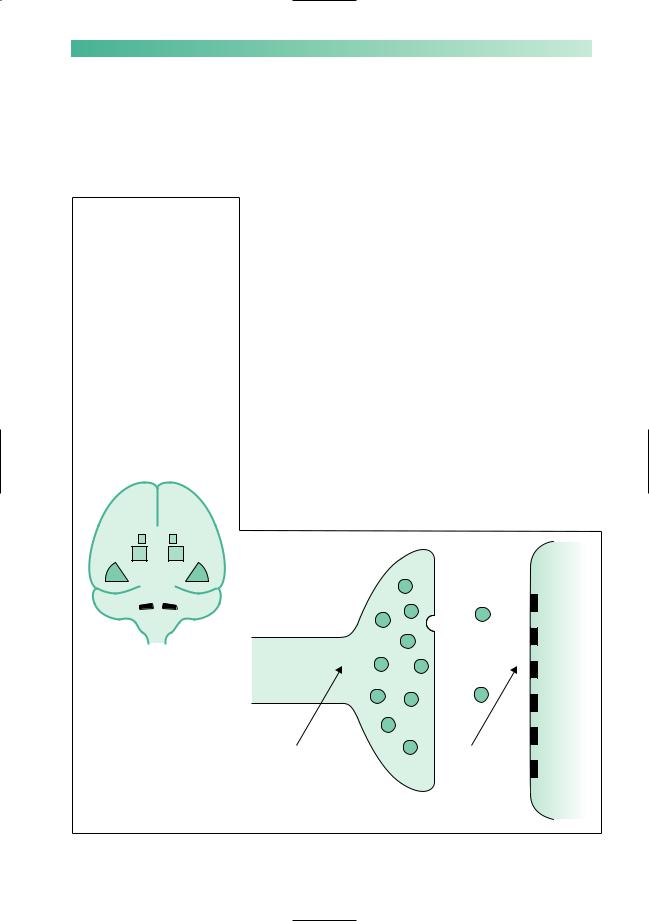
The mainstay of drug treatment is to boost dopaminergic activity in the nigrostriatal pathway, either by giving levodopa which can be turned into dopamine within the remaining neurones in the substantia nigra or by giving dopamine agonists which mimic the effect of dopamine in the striatum (Fig. 5.3). Less potent benefits can be obtained from drugs which inhibit the metabolism of dopamine by monoamine oxidase type B and catechol-O-methyl transferase, and from drugs that modify other neurotransmitters in the striatum such as amantadine and anticholinergics.
Levodopa treatment was developed in the 1960s and remains the most powerful treatment for Parkinson’s disease. Levodopa
Other drugs benefit Parkinson's disease by inhibiting the unopposed action of other neurotransmitters in the
corpus striatum, e.g. anticholinergics, glutamate antagonists
L-dopa is metabolized to dopamine and thereby elevates brain dopamine levels, without similar elevation elsewhere when given with peripheral inhibitors (such as dopa decarboxylase and COMT inhibitors)
NIGROSTRIATAL
PATHWAY
Substantia nigra
Corpus striatum
Dopamine
Dopamine agonists simulate dopamine action directly in the brain
MAO type B inhibitors inhibit
the metabolism of released dopamine thereby prolonging its action
Clearly drugs which either interfere with the biosynthesis of dopamine (tetrabenazine) or which block
dopamine receptors (phenothiazines, metoclopramide) can induce parkinsonism or aggravate it
Fig. 5.3 Diagram to show the substantia nigra in the midbrain, and the nigrostriatal pathway.

72
is absorbed from the gut, crosses the blood–brain barrier and is converted into dopamine by the enzyme dopa-decarboxylase. It is given with a dopa-decarboxylase inhibitor which does not cross into the brain. This prevents the levodopa from being converted into dopamine in the body (where it would cause nausea and vomiting) without interfering with the production of dopamine in the brain.
Most patients initially respond brilliantly to levodopa therapy. There are three subsequent problems:
•Wearing off: the response becomes shorter and less marked.
•Dyskinesia: each dose produces involuntary chorea movements.
•On–off effect: the transition between lack of response (off) and response (on) becomes rapid.
The oral dopamine agonists are not as potent as levodopa in
alleviating parkinsonism, but are less prone to cause dyskinesia and fluctuation. Young and mildly affected patients are sometimes started therefore on a dopamine agonist initially, in the hope of postponing levodopa therapy and its complications for a while. Alternatively the agonist drugs can be introduced later, to reduce the patient’s reliance on levodopa after these complications have begun. It is not clear whether one strategy is better than the other. Dopamine agonists often cause their own adverse effects in older patients, especially hallucinations.
Selegiline and entacapone prolong the duration of action of levodopa, and have a limited role in smoothing fluctuations. Amantadine can be very helpful in suppressing dyskinesia.
There are two strategies for very refractory fluctuation or dyskinesia:
•Apomorphine infusion: this parenteral dopamine agonist can be infused subcutaneously to achieve stable control. This is tricky and requires specialist medical and nursing input.
•Surgical treatments: these are aimed at inhibiting overactive parts of the basal ganglia circuitry, either with a stereotactic lesion or by implanting an electrode with an impulse generator. Targets for these operations include the internal globus pallidus within the striatum (to reduce dyskinesia) and the tiny subthalamic nucleus (to reduce the parkinsonism itself). Procedures to graft the striatum with fetal substantia nigra
cells or stem cells remain experimental.
Anticholinergic drugs are useful for treating tremor, but have much less effect on the other aspects of parkinsonism, and often seriously aggravate memory problems and hallucinations. Conversely, memory problems and hallucinations often respond to cholinergic therapy with drugs like donepezil and rivastigmine. These inhibit the cholinesterase enzyme that beaks down acetylcholine. They do not aggravate the parkinsonism. An alternative way of suppressing hallucinations (but not
CHAPTER 5
Drugs for Parkinson’s disease
Levodopa with dopa decarboxylase inhibitor:
•cobeneldopa (Madopar)
•cocareldopa (Sinemet)
Dopamine agonists:
•cabergoline
•pergolide
•ropinirole
•premipexole
•bromocriptine
•apomorphine
MAO-B inhibitor:
• selegiline
COMT inhibitor:
• entacapone
Glutamate antagonist:
• amantadine
Anticholinergics
Cholinergics:
•rivastigmine
•donepezil
Atypical neuroleptics:
•quetiapine
•clozapine
Antidepressants
The main side-effects or overdose phenomena when enhancing the failing nigrostriatal pathway
≠ Dopamine in the gut:
•nausea
•vomiting
≠Dopamine in the striatum:
• dyskinesia
≠Dopamine elsewhere in the brain:
• postural hypotension
• confusion/hallucinations
PARKINSONISM, INVOLUNTARY MOVEMENTS AND ATAXIA |
73 |
The substantia nigra, nigrostriatal pathway and corpus striatum may be involved in diffuse cerebral pathology:  viral encephalitis
 cerebrovascular disease
  severe head injury
Parkinsonian signs may feature amongst other neurological deficits subsequently
There are rarer patterns of neuronal loss from the CNS which involve nigral and striatal neurones:
 progressive supranuclear palsy (Steele–Richardson syndrome)
  multisystem CNS degeneration with autonomic failure (Shy–Drager syndrome)
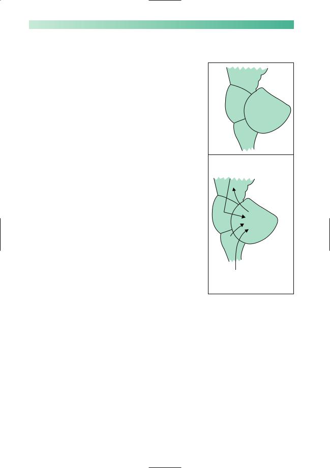
Drugs which interfere with dopamine synthesis and release by the pre-synaptic terminal of the nigrostriatal axon, A, or which block dopamine uptake by the receptors on the surface of the corpus striatal neurone, B, may induce parkinsonism
memory problems) is to use atypical neuroleptic drugs such as quetiapine or clozapine. Conventional neuroleptics (such as haloperidol or chlorpromazine) cannot be used in Parkinson’s disease because they exacerbate parkinsonism, occasionally to a fatal degree.
Other causes of parkinsonism
Parkinsonism can result from other problems in the nigrostriatal pathway (Fig. 5.4). The commonest is drug-induced parkinsonism, due to drugs which block striatal dopamine receptors, such as antipsychotics or antiemetics. Vascular parkinsonism is due to multiple small infarcts in the basal ganglia and often causes more trouble with walking than the upper limbs. Other neurodegenerative diseases can cause parkinsonism together with additional features. These include multiple system atrophy where there may be any combination of parkinsonism, cerebellar ataxia and autonomic failure, and progressive supranuclear palsy where there is usually a severe disturbance of vertical eye movements. Clinical pointers to these other causes include:
•symmetrical bradykinesia and rigidity, without tremor;
•lack of response to levodopa therapy;
•poor balance and falls in the first 2 years of the illness.
Biosynthetic pathway to form dopamine
A |
B |
e.g. |
e.g. |
reserpine |
chlorpromazine |
tetrabenazine |
haloperidol |
|
metoclopramide |
Fig. 5.4 Parkinsonism other than idiopathic Parkinson’s disease.

74 |
CHAPTER 5 |
Involuntary movements
The main categories of involuntary movements are tremor, chorea and dystonia, tics, and myoclonus.
Chorea and dystonia
Chorea refers to fidgety momentary movements which flit randomly around the body. They resemble fragments of purposeful acts or gestures. If you find yourself responding to the movements then you are likely to be dealing with chorea. Dystonia refers to slower involuntary movements that give rise to repetitive twisting or fixed abnormal postures.
There is an overlap between the two movement disorders, in both their clinical manifestations and their causes. Both are due to dysfunction in the basal ganglia. Many patients will have mainly chorea with a little dystonia, or vice versa. The term athetosis refers to slow writhing movements that are halfway between chorea and dystonia, but is now largely reserved for a form of cerebral palsy where these movements predominate.
Hemichorea and hemiballismus
If chorea affects just one side of the body it is termed hemichorea, or hemiballismus if the movements are wild and large scale. This is usually due to a small infarct in the subthalamic nucleus on the opposite side of the brain. Although the movements usually subside within weeks, they can be exhausting and can be suppressed with dopamine-blocking drugs (like haloperidol).
Huntington’s disease
The most serious cause of chorea is Huntington’s disease. This is inherited as an autosomal dominant disorder, and is one of several neurodegenerative diseases that are due to an expansion in a run of repeated CAG nucleotide triplets within a gene. It typically starts with chorea, often accompanied by impulsive or erratic behaviour, and progresses relentlessly to cause dystonia and parkinsonism together with dementia. Depression and suicide are common, but most patients ultimately succumb to the complications of immobility. People who are at risk of developing Huntington’s disease because of their family history can, if they wish, undergo predictive genetic testing. This process is surrounded by emotional issues and requires very careful genetic counselling.
Causes of chorea
Drugs:
•levodopa in Parkinson’s patients
•oral contraceptive pill
•many psychiatric drugs
Vascular disease of the basal ganglia:
•atheroma
•systemic lupus erythematosus
Degenerative diseases:
• Huntington’s disease
Post-infectious:
• Sydenham’s chorea
Other causes:
• thyrotoxicosis

PARKINSONISM, INVOLUNTARY MOVEMENTS AND ATAXIA |
75 |
|
Trinucleotide repeat diseases |
Sydenham’s chorea |
|
|
|
|
Many genes have stretches of |
This used to be a common sequel to streptococcal infection |
|
in young people, along with rheumatic fever. It is now rare in |
|
|
DNAwhere the same three |
|
|
developed countries but remains common elsewhere. Other |
|
|
nucleotides are repeated over |
|
|
post-streptococcal movement disorders are described in |
|
|
and over again (e.g. |
|
|
CAGCAGCAG etc.). Several |
Chapter 15 (p. 253). |
|
neurological diseases are due |
|
|
to expansions of these |
Drug-induced movement disorders |
|
sequences, with an excessive |
|
|
|
|
|
number of repeats. Examples |
We have seen already how tremor and parkinsonism can be in- |
|
include: |
duced by drugs. Drugs can also cause all the other movement |
|
• Huntington’s disease |
|
|
disorders, especially chorea and dystonia. Drugs which block |
|
|
• several dominantly |
dopamine receptors are the main culprit. They can cause acute |
|
inherited cerebellar ataxias |
dystonia (for example, when intravenous metoclopramide is |
|
• Friedreich’s ataxia |
given to a young person for nausea), with spasms of the face, |
|
• one form of motor neurone |
neck and arms and a phenomenon called an oculogyric crisis |
|
where the eyes roll up involuntarily. Long-term use of these |
|
|
disease |
|
|
• myotonic dystrophy |
drugs (for example, when neuroleptic drugs are used for de- |
|
pression or schizophrenia) can cause complex involuntary |
|
|
• fragile X mental retardation |
|
|
chewing and grimacing movements in the face called tardive |
|
|
|
|
|
These expansions have |
dyskinesia, which persist after the causative drug is |
|
unusual features. The |
withdrawn. |
|
expansions are unstable and |
|
|
can increase in size from one |
Focal dystonia |
|
generation to the next, leading |
|
|
to an earlier age of onset |
Dystonia can affect one localized area of the body. Common |
|
(termed anticipation). This |
|
|
forms of focal dystonia include blepharospasm, where there are |
|
|
tendency may exclusively |
|
|
repetitive spasms of eye closure that can seriously interfere with |
|
|
affect expansions transmitted |
|
|
by mothers or fathers. The |
vision; torticollis, where the head pulls painfully to one side and |
|
ways in which the extra |
may also shake; and writer’s cramp, where the forearm cramps |
|
nucleotides cause disease is a |
up and causes the hand to take on a painful twisted posture |
|
matter of intense current |
when writing is attempted. These conditions can be treated by |
|
research, but may involve |
weakening the overactive muscles with regular botulinum |
|
changes in the expression of |
toxin injections. |
|
the gene and the folding of the |
|
|
|
|
|
protein product. |
|
|
|
|
|
|
|

76
Other forms of dystonia
Dystonia can affect larger areas of the body, such as the neck and one arm, segmental dystonia, the trunk muscles, axial dystonia, or the whole of the body, generalized dystonia. Generalized dystonia usually begins in childhood and often has a genetic basis. It can be treated to a very limited extent with drugs. There is increasing interest in treatment with brain stimulator operations.
Wilson’s disease
This is a very rare metabolic disorder characterized by the accumulation of copper in various organs of the body, especially the brain, liver and cornea. It is inherited in an autosomal recessive fashion, and is due to mutations in a gene for ATP-dependent copper-transporting protein.
It is a disease of children and young adults. In the brain, it chiefly affects basal ganglia function, giving rise to all sorts of movement disorders including tremor, chorea, dystonia and parkinsonism. It can also cause behavioural disturbance, psychosis or dementia. In the liver it may cause cirrhosis and failure. In the cornea it is visible (with a slit lamp) peripherally as a brownish Kayser–Fleischer ring. Looking for this and a low serum caeruloplasmin level are ways of screening for the disease.
The importance of Wilson’s disease is that it can be treated with copper-chelating drugs (like penicillamine) if diagnosed early, when brain and liver changes are reversible.
Tics
Tics are stereotyped movements that can be momentary or more complex and prolonged. They differ from chorea in that they can be suppressed for a while by an effort of will. Simple tics, like blinking or grimacing or shrugging repeatedly, are very common in children, especially boys aged 7–10 years. In a small minority these persist into adult life. A wider range of tics, producing noises as well as movements, is suggestive of Gilles de la Tourette syndrome.
CHAPTER 5
Gilles de la Tourette syndrome
Georges Gilles de la Tourette was a nineteenth-century French neurologist who came across the disorder while attempting to classify chorea. He went on to have a distinguished career, survived being shot by the husband of a patient, and died of neurosyphilis. Gilles de la Tourette syndrome begins in children and teenagers with:
•multiple motor tics, with a gradually evolving repertoire of movements;
•phonic tics, commonly sniffs and grunts, rarely repetitive speech (echolalia) or swearing (coprolalia);
•obsessive–compulsive disorder, such as repeated checking or complex rituals.
Mild forms are common in the general population, and very common in people with learning disability. The tics respond to dopamineblocking drugs. The obsessive–compulsive disorder (which is often more disabling) may improve with selective serotonin reuptake inhibitors (like fluoxetine) or behavioural therapy. It is possible that some cases may be caused or exacerbated by autoimmune responses to streptococcal infection analogous to Sydenham’s chorea.

PARKINSONISM, INVOLUNTARY MOVEMENTS AND ATAXIA |
77 |
Myoclonus
Myoclonus produces sudden, shock-like jerks. It is a normal phenomenon in most children and many adults as they are falling off to sleep. It also occurs in a wide range of disease states, and can be due to dysfunction in the cerebral cortex, basal ganglia, brainstem or spinal cord.
Myoclonus as part of general medicine:
•hepatic encephalopathy (‘liver flap’);
•renal failure;
•carbon dioxide retention.
Myoclonus as part of degenerations of the cerebral cortex:
•Alzheimer’s disease;
•Lewy body dementia;
•Creutzfeldt–Jakob disease.
Myoclonus as part of epilepsy:
•juvenile myoclonic epilepsy (where there are jerks in the morning: ‘messy breakfast syndrome’);
•severe infantile epilepsies.
Myoclonus due to basal ganglia disease:
•jerking on attempted movement (‘action myoclonus’) after anoxia due to cardiorespiratory arrest or carbon monoxide poisoning.
Myoclonus due to brainstem disease:
•exaggerated jerks in response to sudden noise (‘startle myoclonus’) in rare metabolic and degenerative disorders.
78 |
CHAPTER 5 |
Cerebellar ataxia
Figure 5.5 is a grossly oversimplified representation of the cerebellum. The function of the cerebellum is to coordinate agonist, antagonist and synergist muscle activity in the performance of learned movements, and to maintain body equilibrium whilst such movements are being executed. Using a massive amount of input from proprioceptors throughout the body, from the inner ear and from the cerebral hemispheres, a complex subconscious computation occurs within the cerebellum. The product of this process largely re-enters the CNS through the superior peduncle and ensures a smooth and orderly sequence of muscular contraction, characteristic of voluntary skilled movement.
In man, the function of the cerebellum is seen at its best in athletes, sportsmen, gymnasts and ballet dancers, and at its worst during states of alcoholic intoxication when all the features of cerebellar malfunction appear. A concern of patients with organic cerebellar disease is that people will think they are drunk.
Localization of lesions
From Fig. 5.5 it is clear that patients may show defective cerebellar function if they have lesions in the cerebellum itself, in the cerebellar peduncles, or in the midbrain, pons or medulla. The rest of the CNS will lack the benefit of correct cerebellar function whether the pathology is in the cerebellum itself, or in its incoming and outflowing connections. Localization of the lesion may be possible on the basis of the clinical signs.
•Midline cerebellar lesions predominantly interfere with the maintenance of body equilibrium, producing gait and stance ataxia, without too much ataxia of limb movement.
•Lesions in the superior cerebellar peduncle, along the course of one of the chief outflow tracts from the dentate nucleus in the cerebellum to the red nucleus in the midbrain, classically produce a very marked kinetic tremor, as mentioned at the beginning of this chapter.
•Lesions in the midbrain, pons and medulla, which are causing cerebellar deficits by interfering with inflow or outflow pathways to or from the cerebellum, may also cause other brainstem signs, e.g. cranial nerve palsies, and/or long tract signs (upper motor neurone or sensory) in the limbs.
Midbrain
Pons
Cerebellum
Medulla
Cerebro-cerebellar input, via pons and middle cerebellar peduncle
Outflow from the cerebellum, via superior
cerebellar peduncle
Vestibulo-cerebellar input, via inferior cerebellar peduncle
Spino-cerebellar input (proprioception), via inferior cerebellar peduncle
Fig. 5.5 Highly simplified diagrammatic representation of the brainstem and cerebellum as viewed from the left.
PARKINSONISM, INVOLUNTARY MOVEMENTS AND ATAXIA |
79 |
Clinical signs of cerebellar dysfunction
The common, important clinical signs of cerebellar dysfunction are listed below.
• Nystagmus.
• Dysarthria: the muscles of voice production and speech lack coordination so that sudden irregular changes in volume and timing occur, i.e. scanning or staccato speech.
•Upper limbs: ataxia and intention tremor, best seen in movement directed towards a restricted target, e.g. the finger–nose test; dysdiadochokinesia, i.e. slow, inaccurate, rapid alternating movements.
•Lower limbs: ataxia, best seen in the heel–knee–shin test.
•Gait and stance ataxia, especially if the patient is asked to walk heel to toe, or to stand still on one leg.
• Hypotonia, though a feature of cerebellar lesions, is not very useful in clinical practice.
Cerebellar representation is ipsilateral, so a left cerebellar hemisphere lesion will produce nystagmus which is of greater amplitude when the patient looks to the left, ataxia which is more evident in the left limbs, and a tendency to deviate or fall to the left when standing or walking.
To date, it has not been possible to improve defective cerebellar function pharmacologically.
Causes of cerebellar malfunction
The common causes of cerebellar malfunction are:
•cerebrovascular disease;
•multiple sclerosis;
•drugs, especially anticonvulsant intoxication;
•alcohol, acute intoxication.
Rarer cerebellar lesions include:
•posterior fossa tumours;
•cerebellar abscess, usually secondary to otitis media;
•cerebellar degeneration, either hereditary (e.g. Friedreich’s ataxia and autosomal dominant cerebellar ataxia), alcohol induced, or paraneoplastic;
•Arnold–Chiari malformation (the cerebellum and medulla are unusually low in relation to the foramen magnum);
•hypothyroidism.