

Wilkinson. Essential Neurology 2005
.pdf160 |
CHAPTER 10 |
Skin, joints, etc.
Spinal cord
Muscle
|
Sensory |
Motor |
Reflex |
Symptoms |
|
|
|
Upper limbs |
Glove distribution of |
Weakness of grip |
|
|
tingling, pins and needles |
and fingers |
|
|
and numbness |
|
|
|
Difficulty in manipulating |
|
|
|
small objects in the fingers |
|
|
|
because of loss of |
|
|
|
sensation |
|
|
Lower limbs |
Stocking distribution of |
Foot drop |
|
|
tingling, pins and needles |
|
|
|
and numbness |
|
|
|
Unsteadiness of stance |
Loss of spring at |
|
|
and gait, especially in |
the ankles for |
|
|
the dark or when eyes |
running and climbing |
|
|
closed |
stairs |
|
Signs |
|
|
|
Upper limbs |
Glove distribution of |
Distal lower motor |
Loss of distal |
|
sensory loss, affecting |
neurone signs |
reflexes, e.g. |
|
any sensory modality |
in hands |
supinator jerks |
|
Sensory ataxia in |
|
|
|
fingers and hands |
|
|
Lower limbs |
Stocking distribution |
Distal lower motor |
Loss of distal |
|
of sensory loss, affecting |
neurone signs in |
reflexes, especially |
|
any sensory modality |
legs and feet |
ankle jerks |
|
Sensory ataxia in legs |
|
|
|
and gait |
|
|
|
Rombergism (i.e. |
|
|
|
dependence on eyes |
|
|
|
for balance) |
|
|
|
|
|
|
Fig. 10.6 Symptoms and signs of peripheral neuropathy.

PERIPHERAL NEUROMUSCULAR DISORDERS |
161 |
Common causes of peripheral neuropathy
Fig. 10.7 Common causes of peripheral neuropathy in the UK.
In developed countries the commonest identifiable causes of peripheral neuropathy are alcohol and diabetes. In other parts of the world, vitamin deficiency and leprosy cause more disease, although this is gradually changing. In both settings, many cases of peripheral neuropathy remain unexplained. Figure 10.7 lists some of the most important causes of peripheral neuropathy.
Alcoholic neuropathy
Alcoholic neuropathy is common and usually more sensory than motor. How much it is caused by the direct toxic effect of alcohol on the peripheral nerves, and how much it is due to coexistent vitamin B1 deficiency, is not completely known.
Vitamin B12 deficiency
Vitamin B12 deficiency is not a common cause of neuropathy, but is an important one to recognize because of its reversibility. Every effort should be made to reach the diagnosis before the irreversible changes of subacute combined degeneration of the spinal cord become established.
Deficiency |
Vitamin B1 in alcoholics |
|
Vitamin B6 in patients taking isoniazid |
|
Vitamin B12 in patients with pernicious |
|
anaemia and bowel disease |
Toxic |
Alcohol |
|
Drugs, e.g. isoniazid, vincristine, |
|
aminodarone |
Metabolic |
Diabetes mellitus |
|
Chronic renal failure |
Inflammatory |
Guillain–Barré syndrome |
|
Chronic inflammatory |
|
demyelinating polyneuropathy |
Paraneoplastic |
Bronchial carcinoma and other |
|
malignancies |
Connective tissue disease |
Rheumatoid arthritis |
|
Systemic lupus erythematosus |
|
Polyarteritis nodosa |
Hereditary |
Hereditary motor and sensory |
|
neuropathy (HMSN) (also known as |
|
Charcot–Marie–Tooth disease) |
Haematological |
Paraproteinaemia |
Idiopathic |
Perhaps accounting for 50% of cases |
|
|

162
Diabetes mellitus
Diabetes mellitus is probably the commonest cause of peripheral neuropathy in the Western world. It occurs in both juvenile-onset insulin-requiring diabetes and maturity-onset diabetes. It may be the first clinical suggestion of the presence of diabetes. Excellent diabetic control has been shown to prevent neuropathy, but does not reverse it once it has developed.
The commonest form of neuropathy in diabetes is a predominantly sensory one. The combination of neuropathy and atherosclerosis affecting the nerves and arteries in the lower limbs very strongly predisposes the feet of diabetic patients to trophic lesions, which are slow to heal.
There are a few unusual forms of neuropathy that may occur in patients with diabetes:
•painful weakness and wasting of one proximal lower limb, so-called diabetic lumbosacral radiculo-plexopathy or diabetic amyotrophy;
•involvement of the autonomic nervous system giving rise to abnormal pupils, postural hypotension, impaired cardioacceleration on changing from the supine to the standing position, impaired bladder, bowel and sexual function, and loss of normal sweating;
•a tendency for individual nerves to stop working quite abruptly, with subsequent gradual recovery. Common nerves to be involved are the 3rd and 6th cranial nerves and the common peroneal nerve in the leg. Involvement of several individual nerves in this way constitutes the clinical syndrome of multifocal neuropathy.
Hereditary motor and sensory neuropathy
(HMSN, also known as Charcot–Marie–Tooth disease)
There are several forms of this, with a complex genetic classification. One of the more common, HMSN type I, is due to a duplication in the gene for peripheral myelin protein 22; this and some other forms can be diagnosed with a genetic test. The illness is usually evident in teenage life and very slowly worsens over many years. Motor involvement predominates, with lower motor neurone signs appearing in the feet and legs (especially in the anterolateral muscle compartments of the calves), and in the small muscles of the hands. Pes cavus and clawing of the toes are very common consequences. Sometimes, the pathology primarily involves the axons, but more often there is demyelination and remyelination to be found in the peripheral nerves.
CHAPTER 10
Key features of HMSN
•Pes cavus
•Distal wasting (‘champagne bottle legs’)
•Distal weakness
•Absent reflexes
•Mild distal sensory loss

PERIPHERAL NEUROMUSCULAR DISORDERS |
163 |
|
|
Guillain–Barré syndrome |
|
Causes of death |
|
|
Guillain–Barré syndrome is rather different from the other |
|
|
|
|
|
Guillain–Barré syndrome can |
forms of peripheral neuropathy. This is because of its rapid |
|
be fatal, but most of the causes |
evolution over several days, because it can produce a life- |
|
are avoidable: |
threatening degree of weakness, and because the underlying |
|
• aspiration pneumonia |
pathology clearly affects the nerve roots as well as the peri- |
|
• DVT and pulmonary |
pheral nerves. |
|
embolism |
The syndrome commonly occurs a week or two after an infec- |
|
• cardiac arrhythmia |
tion, such as Campylobacter enteritis, which is thought to trigger |
|
So monitor bulbar function, |
an autoimmune response. |
|
The patient notices limb weakness and sensory symptoms |
|
|
vital capacity and the heart, |
|
|
which worsen day by day for 1–2 weeks (occasionally the |
|
|
and anticoagulate |
|
|
|
progression may continue for as long as 4 weeks). Often, the |
|
|
illness stops advancing after a few days and does not produce |
|
|
a disability that is too major. Not uncommonly, however, it |
|
|
progresses to cause very serious paralysis in the limbs, trunk |
|
|
and chest muscles, and in the muscles supplied by the |
|
|
cranial nerves. Involvement of the autonomic nerves may cause |
|
|
erratic rises and falls in heart rate and blood pressure and pro- |
|
|
found constipation. |
|
|
Patients with Guillain–Barré syndrome need to be hospi- |
|
|
talized until it is certain that deterioration has come to an end, |
|
|
because chest and bulbar muscle weakness may make ventila- |
|
|
tion and nasogastric tube nutrition essential. Daily, or twice |
|
|
daily, estimations of the patient’s vital capacity during the early |
|
|
phase of the disease can be a very valuable way of assessing the |
|
|
likelihood of the need for ventilatory support. Prompt adminis- |
|
|
tration of intravenous immunoglobulin or plasma exchange |
|
|
can prevent deterioration and the need for ventilation in many |
|
|
cases. Steroids have not been shown to be of proven benefit. |
|
|
Patients with Guillain–Barré syndrome become very |
|
|
alarmed by the progressive loss of function at the start of |
|
|
their illness. They often need a good deal of psychological and |
|
|
physical support when the disability is severe and prolonged. |
|
|
The ultimate prognosis is usually very good, however. Incom- |
|
|
plete recovery and recurrence are both well described, but by far |
|
|
the most frequent outcome of this condition is complete |
|
|
recovery over a few weeks or months, and no further similar |
|
|
trouble thereafter. |
|
|
The pathology is predominantly in the myelin rather than in |
|
|
the axons of the peripheral nerves and nerve roots, i.e. a de- |
|
|
myelinating polyneuropathy and polyradiculopathy. Recovery |
|
|
is due to the capability of Schwann cells to reconstitute the |
|
|
myelin sheaths after the initial demyelination. The involvement |
|
|
of the nerve roots gives rise to one of the diagnostic features of |
|
|
the condition, a raised CSF protein. |
164 |
CHAPTER 10 |
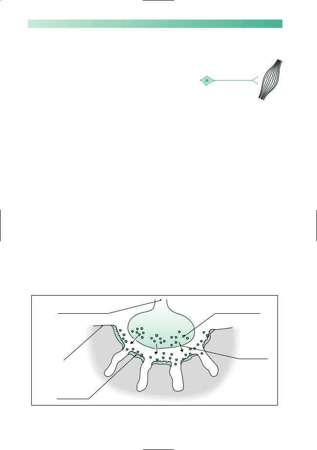
Myasthenia gravis
Myasthenia gravis is the rare clinical disease that results from impaired neuromuscular transmission at the synapse between the termination of the axon of the lower motor neurone and the muscle, at the motor end plate. Figure 10.8 is a diagram of a motor end plate. Neuromuscular transmission depends on normal synthesis and release of acetylcholine into the gap substance of the synapse, and its uptake by healthy receptors on the muscle membrane. The main pathological abnormality in myasthenia gravis at the neuromuscular junction is the presence of auto-antibody attached to receptor sites on the post-synaptic membrane. This auto-antibody both degrades and blocks acetylcholine receptor sites, thus impairing neurotransmission across the synapse.
Myasthenia gravis is an autoimmune disease in which the auto-antibody appears clearly involved in the pathogenesis of the muscle weakness.
Myasthenia gravis is rather more common in women than men. In women, it tends to occur in young adult life, and in men it more commonly presents over the age of 50 years. Various subtypes of myasthenia gravis have been distinguished according to age and sex prevalence, HLAtype associations, incidence of auto-antibodies, and other characteristics.
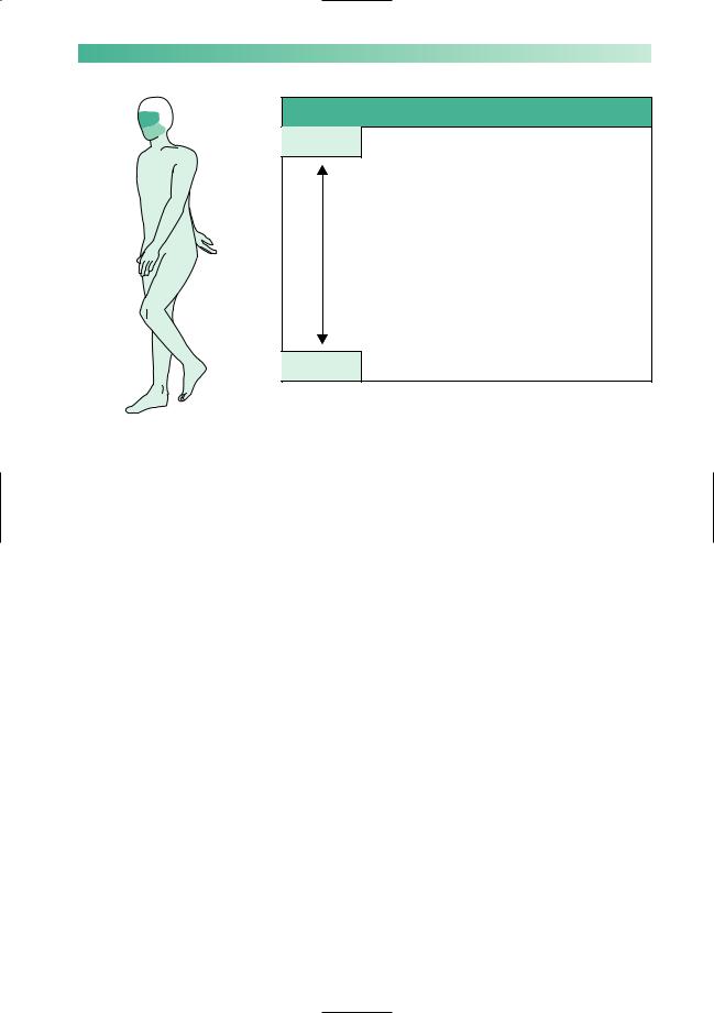
Muscle weakness, with abnormal fatiguability, and improvement after rest, characterize myasthenia gravis. Symptoms tend to be worse at the end of the day, and after repetitive use of muscles for a particular task, e.g. chewing and swallowing may be much more difficult towards the end of a meal than they were at the start. The distribution of muscle involvement is not uniform, as shown in Fig. 10.9.
Termination of axon |
Acetylcholine molecules |
of lower motor neurone |
contained in vesicles |
Voltage-gated channels for release of acetylcholine
Highly convoluted and modified part of the muscle membrane, on the surface of which are acetylcholine receptor sites
Fig. 10.8 Diagram to show a motor end plate in skeletal muscle.
PERIPHERAL NEUROMUSCULAR DISORDERS |
165 |
|
|
Muscles |
Symptoms |
Common |
External ocular |
Double vision and ptosis |
|
Bulbar |
Difficulty in chewing, swallowing |
|
|
and talking |
|
Neck |
Difficulty in lifting head up from |
|
|
the lying position |
|
Proximal limb |
Difficulty in lifting arms above |
|
|
shoulder level, and in standing |
|
|
from low chairs and out of the |
|
|
bath |
|
Trunk |
Breathing problems and difficulty |
|
|
in sitting from the lying position |
Rare |
Distal limb |
Weak hand-grips, ankles and feet |
Fig. 10.9 Frequency of muscle involvement and symptoms in myasthenia gravis.
Confirmation of the diagnosis
Once suspected, the diagnosis of myasthenia gravis may be confirmed by:
1.The Tensilon test. Edrophonium chloride (Tensilon) is a short-acting anticholinesterase, which prolongs the action of acetylcholine at the neuromuscular junction for a few minutes after slow intravenous injection. This produces a transient and striking alleviation of weakness. There may also be an equally exciting bradycardia (reversed by atropine); the test should not be performed lightly in patients who are frail or have heart disease.
2.Detection of serum acetylcholine receptor antibodies. These antibodies are not found in the normal population, but are detected in about 50% of patients with purely ocular myasthenia, increasing up to about 90% of patients with more generalized myasthenia.
3.EMG studies. Sometimes it is helpful to show that the amplitude of the compound muscle action potential, recorded by surface electrodes over a muscle, decreases on repetitive stimulation of the nerve to the muscle.
4.Chest radiography and CT of the anterior mediastinum, to demonstrate an enlargement of the thymus gland. The association of myasthenia gravis with thymic enlargement is not yet fully understood. Of myasthenic patients, 10–15% have a thymoma, and 50–60% show thymic hyperplasia. Both sorts of pathology may enlarge the thymus, which can be clearly shown by suitable imaging procedures.

166
Management of myasthenia gravis
The management of myasthenia gravis includes:
1.The use of oral anticholinesterase drugs, pyridostigmine and prostigmine. These are prescribed at intervals during the day, and work quite effectively. Abdominal colic and diarrhoea, induced by the increased parasympathetic activity in the gut, can be controlled by simultaneous use of propantheline.
2.Immunosuppression by prednisolone or azathioprine. In patients with disabling symptoms inadequately controlled by oral anticholinesterase therapy, suppression of the autoantibody can radically improve muscle strength.
3.Thymectomy. Remission or improvement can be expected in 60–80% of patients after thymectomy, and must be considered in all patients. It may make the use of immunosuppressive drugs unnecessary, which is obviously desirable.
4.Plasma exchange to remove circulating auto-antibody to produce short-term improvement in seriously weak patients.
5.Great care of the myasthenic patient with severe weakness who is already on treatment. The muscle strength of such patients may change abruptly, and strength in the bulbar and respiratory muscles may become inadequate for breathing. The correct place for such patients is in hospital, with anaesthetic and neurological expertise closely to hand. There may be uncertainty as to whether such a patient is undertreated with anticholinesterase (myasthenic crisis), or overtreated so that the excessive acetylcholine at the neuromuscular junction is spontaneously depolarizing the postsynaptic membrane, i.e. depolarization block (cholinergic crisis). Fasciculation may be present when such spontaneous depolarization is occurring. Tensilon may be used to decide whether the patient is underor overdosed, but it is essential to perform the Tensilon test with an anaesthetist present in these circumstances. If the weak state is due to cholinergic crisis, the additional intravenous dose of anticholinesterase may produce further critical paralysis of bulbar or respiratory muscles.
CHAPTER 10
Management of myasthenia gravis
•Anticholinesterase
•Immunosuppression
•Thymectomy
•Plasma exchange
•Crisis management
PERIPHERAL NEUROMUSCULAR DISORDERS |
167 |
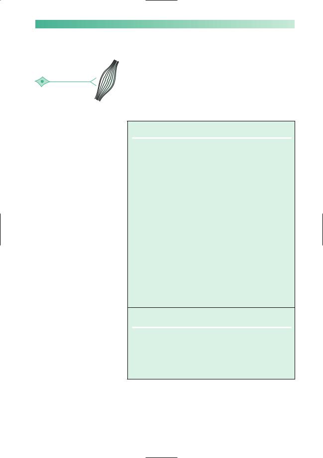
Muscle disease
These are a group of rare diseases in which the primary pathology causing muscle weakness and wasting lies in the X muscles themselves. They are classified in Fig. 10.10 and short
notes about each condition are given in this section.
Fig. 10.10 Classification of muscle diseases.
Inherited
1Muscular dystrophies, whose genetic basis is increasingly understood in terms of gene and gene product identification.
Duchenne |
X-linked recessive gene |
Myotonic dystrophy |
Autosomal dominant gene |
Facio-scapulo-humeral |
Autosomal dominant gene |
Limb girdle |
Not a single entity (variable |
|
inheritance) |
2Muscle diseases in which an inherited biochemical defect is present.
Specific enzyme deficiencies occur which disrupt the pathways of carbohydrate or fat oxidation, often with accumulation of substrate within the muscle cell. The enzyme deficiency may be within the muscle cell cytoplasm, interfering with the utilization of glycogen or glucose, or it may be within the mitochondria of muscle cells (and cells of other organs) blocking the metabolism of pyruvate, fatty acids or individual elements of Krebs cycle.
In other diseases of this sort, there is uncoupling of the electrical excitation of muscle fibres and their contraction.
This is the case in McArdle's syndrome, and in malignant hyperpyrexia where sustained muscle contraction may occur in the absence of nerve stimulation.
Acquired
1 Immunologically mediated inflammatory disease, e.g. polymyositis
dermatomyositis
2 Non-inflammatory myopathy, e.g. corticosteroids thyrotoxicosis

168
Duchenne dystrophy
Duchenne dystrophy is the most serious inherited muscular dystrophy. The X-linked recessive inheritance gives rise to healthy female carriers and affected male children. The affected boys usually show evidence of muscular weakness before the age of 5 years, and die of profound muscle weakness (predisposing them to chest infections), or of associated cardiomyopathy, in late teenage life. In the early stages, the weakness of proximal muscles may show itself by a characteristic way in which these boys will ‘climb up their own bodies with their hands’ (Gower’s sign) when rising from the floor to the standing position. They also show muscle wasting, together with pseudohypertrophy of the calf muscles (which is due to fat deposition in atrophied muscle tissue).
The affected boys have elevated levels of creatine kinase muscle enzyme in the blood, and the clinically unaffected carrier state in female relatives is often associated with some elevation of the muscle enzymes in the blood. The gene locus on the X chromosome responsible for Duchenne dystrophy, and its large gene product, dystrophin, have been identified. Molecular genetic diagnosis of affected patients and female carriers is possible, as is prenatal diagnosis.
This same region of the X chromosome is also implicated in the inheritance of a more benign variant of Duchenne dystrophy (later in onset and less rapidly progressive), known as Becker’s musclar dystrophy.
The combination of family history, clinical examination, biochemical and genetic studies allows the detection of the carrier state, and the prenatal detection of the affected male fetus in the first trimester of pregnancy. Genetic counselling of such families has reached a high degree of accuracy. As a single gene disorder, Duchenne muscular dystrophy is one of the conditions in which gene therapy is being considered.
CHAPTER 10
Key features of Duchenne muscular dystrophy
•Young
•Male
•Generalized weakness
•Muscle wasting
•Calf pseudohypertrophy
•Gower’s sign

PERIPHERAL NEUROMUSCULAR DISORDERS |
169 |
|
|
Myotonic dystrophy |
|
Key features of myotonic |
|
|
|
|
|
dystrophy |
Myotonic dystrophy is characterized by ‘dystrophy’ of several |
|
• Either sex |
organs and tissues of the body, and the dystrophic changes in |
|
muscle are associated with myotonic contraction. |
|
|
• Glum-looking from facial |
|
|
The disease is due to an expanded trinucleotide repeat (see |
|
|
weakness and ptosis |
|
|
box on p. 75). This is inherited as an autosomal dominant, so |
|
|
• Frontal balding |
|
|
men and women are equally affected, usually in early adult life. |
|
|
• Glasses or previous cataract |
|
|
The mutation tends to expand with each generation, especially |
|
|
surgery |
when transmitted from a woman to her child, causing a more |
|
|
|
|
• Hand muscles show |
severe phenotype which is described below. Genetic testing |
|
wasting and myotonia |
allows symptomatic, presymptomatic and prenatal diagnosis, |
|
|
where appropriate. |
|
|
|
|
|
Some impairment of intellectual function, cataracts, prema- |
|
|
ture loss of hair, cardiac arrhythmia and failure, gonadal atro- |
|
|
phy and failure, all feature in patients with myotonic dystrophy, |
|
|
but the most affected tissue is muscle. The facial appearance |
|
|
may be characteristic, with frontal balding, wasting of the tem- |
|
|
poralis muscles, bilateral ptosis and bilateral facial weakness. |
|
|
Muscle weakness and wasting are generalized but the hands are |
|
|
often particularly affected. |
|
|
The myotonia shows itself in two ways: |
|
|
1. The patient has difficulty in rapid relaxation of tightly con- |
|
|
tracted muscle, contraction myotonia, and this is best seen by |
|
|
asking the patient to open the hand and fingers quickly after |
|
|
making a fist. |
|
|
2. Percussion myotonia is the tendency for muscle tissue |
|
|
to contract when it is struck by a tendon hammer, and this is |
|
|
best seen by light percussion of the thenar eminence whilst |
|
|
the hand is held out flat. A sustained contraction of the thenar |
|
|
muscles lifts the thumb into a position of partial abduction |
|
|
and opposition. |
|
|
From its appearance in early adult life, the illness runs a |
|
|
variable but slowly progressive course over several decades. |
|
|
The associated cardiomyopathy is responsible for some of |
|
|
the early mortality in myotonic dystrophy. |
|
|
Some children of females with myotonic dystrophy may |
|
|
show the disease from the time of birth. Such babies may be very |
|
|
hypotonic, subject to respiratory problems (chest muscle in- |
|
|
volvement) and feeding problems (facial muscle involvement). |
|
|
Mental retardation is a feature of these children. Frequently, the |
|
|
birth of such a child is the first evidence of myotonic dystrophy |
|
|
in the family, since the mother’s involvement is only mild. |