

Wilkinson. Essential Neurology 2005
.pdf90
Causes of paraplegia
The four common causes of spinal cord dysfunction are illustrated in Fig. 6.8: trauma, multiple sclerosis, malignant disease and spondylotic degenerative disease of the spine.
Trauma
Road traffic accidents involving motorcycles and cars are the commonest cause, followed by domestic falls, accidents at work and accidents in sport. The order of frequency of injury in terms of neurological level is cervical, then thoracic, then lumbar. Initial care at the site of the accident is vitally important, ensuring that neurological damage is not incurred or increased by clumsy inexperienced movement of the patient at this stage. Unless the life of the patient is in jeopardy by leaving him at the site of the accident, one person should not attempt to move the patient. He should await the arrival of four or five other people, hopefully with a medical or paramedical person in attendance. Movement of the patient suspected of spinal trauma should be slow and careful, with or without the aid of adequate machinery to ‘cut’ the patient out of distorted vehicles. It should be carried out by several people able to support different parts of the body, so that the patient is moved all in one piece.
Multiple sclerosis
An episode of paraplegia in a patient with multiple sclerosis usually evolves over the period of 1–2 weeks, and recovers in a couple of months, as with other episodes of demyelination elsewhere in the CNS. Occasionally, however, the paraplegia may evolve slowly and insidiously (see Chapter 7). Spinal cord lesions due to multiple sclerosis are usually asymmetrical and incomplete.
Malignant disease of the spine
Secondary deposits of carcinoma are the main type of trouble in this category: from prostate, lung, breast and kidney. Often, plain X-rays of the spine will show vertebral deposits within and beside the spine, but sometimes the tumour deposit is primarily meningeal with little bony change. Steroid treatment, surgical decompression, radiotherapy or chemotherapy can sometimes make a lot of difference to the disability suffered by such patients, even though their long-term prognosis is poor.
CHAPTER 6
Causes of spinal cord disease
Common
Trauma
Multiple sclerosis
Malignant disease
Spondylosis
Rare
Infarction Transverse myelitis ‘Bends’ in divers
Vitamin B12 deficiency Syringomyelia
Motor neurone disease Intrinsic cord glioma Radiation myelopathy Arteriovenous malformation Extradural abscess Prolapsed intervertebral
thoracic disc Neurofibroma Meningioma
Atlanto-axial subluxation in rheumatold arthritis
PARAPLEGIA |
91 |
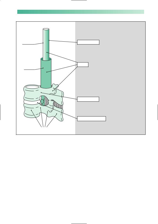
Spinal cord
Meninges
Vertebrae, intervertebral discs and ligaments
Fig. 6.8 The common causes of paraplegia.
Clinical clues of the common causes of a spinal cord lesion
These clues are fallible; MR scan and other imaging techniques should be overused rather than underused, to establish diagnostic certainty
Multiple sclerosis
Evidence of dissemination of lesions in time and space throughout the CNS is essential for the diagnosis of multiple sclerosis. This evidence may lie in the history, on examination, or in the result of investigation, e.g. a delayed visual evoked potential
Trauma
This is usually evident, but may be missed in patients:
 who have previous neurological disease, e.g. multiple sclerosis
 who are unconscious because of an associated head injury or alcohol
 who have other serious injuries which distract medical attention away from the CNS
 if a full neurological examination has been omitted after trauma to the head, neck or spine
Malignant tumour
Malignant disease causing spinal cord compression is usually metastatic, in the spine and/or the meninges. Clinical evidence of a primary malignant tumour, or of metastatic disease elsewhere, will suggest this cause
Spondylotic myelopathy
Spondylotic myelopathy in the cervical region is rare under the age of 50. Segmental symptoms and signs in the arms are common in this condition
Spondylotic myelopathy
Patients with central posterior intervertebral disc prolapse betweenC4andT1,withorwithoutconsitutionallynarrowcanals, make up the majority of this group. The cord compression may be at more than one level. The myelopathy may be compressive or ischaemic in nature (the latter due to interference with arterial supply and venous drainage of the cord in the presence of multiple-level disc degenerative disease in the neck). Decompressive surgery is aimed at preventing further deterioration in the patient, rather than guaranteeing improvement.

92 |
CHAPTER 6 |
Management of recently developed undiagnosed paraplegia
Four principles underly the management of patients with evolving paraplegia.
1.‘Get on with it!’
2.Care of the patient to prevent unnecessary complications.
3.Establish the diagnosis.
4.Treat the specific cause.
‘Get on with it!’
Reversibility is not a conspicuous characteristic of damage to the CNS. It is important to try to establish the diagnosis and treat spinal cord disease whilst the clinical deficit is minor. Recovery from complete cord lesions is slow and imperfect. Hours may make a difference to the outcome of a patient with cord compression.
Care of the patient to prevent unnecessary complications
The parts of the body rendered weak, numb or functionless by the spinal cord lesion need care. Nurses and physiotherapists are the usual people to provide this.
Skin
•Frequent inspection.
•Frequent relief of pressure (by turning).
•Prevention and vigorous treatment of any damage.
Weak or paralysed limbs
•Frequent passive movement and stockings to prevent venous stagnation, thrombosis and pulmonary embolism.
•Frequent passive movement to prevent joint stiffness and contracture, without overstretching.
•Exercise of non-paralysed muscles.
Non-functioning bladder and bowels
•Catheterization.
•Adequate fluids.
•Dietary fibre regulation.
•Laxatives.
•Suppositories.
•Enemas.
*
Tumour at the foramen magnum (asterisk) causing high spinal cord compression.

PARAPLEGIA
High signal in the upper cervical cord due to multiple sclerosis (arrow).
93
Establish the diagnosis
Foremost in establishing the diagnosis is an MR scan of the spine and sometimes other imaging techniques. These investigations will reveal cord-compressing pathology and the need for neurosurgical intervention. Areas of demyelination can also be visualized within the cord.
If no compressive or intrinsic cord lesion is demonstrated by scanning, other investigations may be helpful:
•CSF analysis, visual evoked potentials, MR scan of the brain — multiple sclerosis;
•EMG studies — motor neurone disease;
•haematological tests and serum vitamin B12 estimation — subacute combined degeneration of the cord.
Treat the specific cause
•Trauma: intravenous steroids, restoration of alignment and stabilization by operative and non-operative means.
•Multiple sclerosis: consider use of high dose methylprednisolone.
•Malignant disease: surgical decompression, steroids, radiotherapy, chemotherapy.
•Spondylotic myelopathy: surgical decompression.
•Infarction: nil.
•Bends: hyperbaric chamber.
•Subacute combined degeneration of the cord: vitamin B12 injections.
•Syringomyelia: consider surgery.
•Motor neurone disease: riluzole.
•Benign spinal cord tumours: consider surgery.
•Radiation myelopathy: nil.
•Arteriovenous malformation: embolization or surgery, which may be difficult.
•Extradural abscess: surgery and antibiotics.
•Thoracic disc: surgery, which may be difficult.
•Spinal neurofibroma and meningioma: surgery.
•Atlanto-axial subluxation in rheumatoid arthritis: consider surgery, which is difficult.
Low cervical cord compression due to prolapsed discs (arrows).

94
Management of chronic diagnosed paraplegia
From whatever cause, there is a group of patients who have become severely paraplegic, and will remain so on a long-term basis. Their mobility is going to rely heavily on a wheelchair. Multiple sclerosis accounts for the largest number of such patients in the UK, many of whom are young with much of their lives still ahead. Such patients benefit from education, encouragement and the expertise of nurses, physiotherapists, dietitians, social workers, occupational therapists, housing departments, industrial rehabilitation units, psychologists and doctors. They also need the emotional support of their family and friends. They have to come to terms with a major disability and believe in their value despite the loss of normal function in the lower half of their body.
Attention should be given to the following:
1.Patient education about the level of cord involvement:
•what does and does not work.
2.The loss of motor function:
•wheelchair acceptance and wheelchair skills;
•‘transfers’ on and off the wheelchair;
•physiotherapy: passive to prevent joint contractures; active to strengthen non-paralysed muscles;
•drugs to reduce spasticity: baclofen, dantrolene, tizanidine.
3.Sensory loss:
•care of skin;
•guarding against hot, hard or sharp objects;
•taking the weight of the body off the seat of the wheelchair routinely every 15 or 20 minutes.
4.Bladder:
•reflex bladder emptying, condom drainage;
•intermittent self-catheterization, indwelling catheter;
•cholinergic or anticholinergic drugs as necessary;
•alertness to urinary tract infection.
5.Bowel:
•regularity of diet;
•laxatives and suppositories.
6.Sexual function:
•often an area of great disappointment;
•normal sexual enjoyment, male ejaculation, orgasm, motor skills for intercourse, all lacking;
•fertility often unimpaired in either sex, though seminal emission in males will require either vibrator stimulation of the fraenum of the penis or electro-ejaculation;
•counselling of patient and spouse helps adjustment.
CHAPTER 6
There are lots of ways of helping patients with 'incurable paraplegia'

PARAPLEGIA |
95 |
7.Weight and calories:
•wheelchair life probably halves the patient’s calorie requirements. It is very easy, and counterproductive, for paraplegic patients to gain weight. Eating and drinking are enjoyable activities still left open to them. Heaviness is difficult for their mobility, and bad for the weightbearing pressure areas.
8.Psychological aspects:
•disappointment, depression, shame, resentment, anger and a sense of an altered role in the family are some of the natural feelings that paraplegic patients experience.
9.Family support:
•the presence or absence of this makes a very great difference to the ease of life of a paraplegic patient.
10.Employment:
•the patient’s self-esteem may be much higher if he can still continue his previous work, or if he can be retrained to obtain new work.
11.House adaptation:
•this is almost inevitable and very helpful. Living on the ground floor, with modifications for a wheelchair life, is important.
12.Car adaptation:
•conversion of the controls to arm and hand use may give a great deal of independence.
13.Financial advice:
•this will often be needed, especially if the patient is not going to be able to work. Medical social workers are conversant with house conversion and attendance and mobility allowances.
14.Recreational activity and holidays:
•should be actively pursued.
15.Legal advice:
•this may be required if the paraplegia was the result of an accident, or if the patient’s paraplegia leads to marriage disintegration, which sometimes happens.
16.Respite care:
•this may be appropriate to help the patient and/or his relatives. It can be arranged in several different ways, for example:
—admission to a young chronic sick unit for 1–2 weeks, on a planned, infrequent, regular basis;
—a care attendant lives at the patient’s home for 1–2 weeks, whilst the relatives take a holiday.

96
Syringomyelia
It is worth devoting a short section of this chapter to syringomyelia because the illness is a neurological ‘classic’. It brings together much of what we have learnt about cord lesions, and is grossly over-represented in the clinical part of medical professional examinations. It is a rare condition.
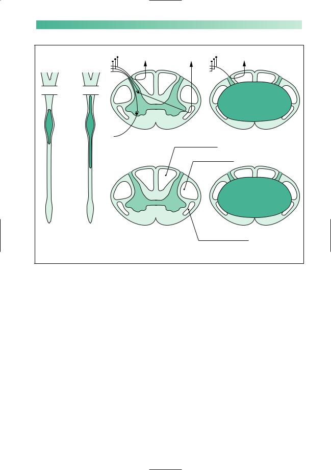
The symptoms and signs are due to an intramedullary (within the spinal cord), fluid-filled cavity extending over several segments of the spinal cord (Fig. 6.9a). The cavity, or syrinx, is most evident in the cervical and upper thoracic cord. There may be an associated Arnold–Chiari malformation at the level of the foramen magnum, in which the medulla and the lowermost parts of the cerebellum are below the level of the foramen magnum. There may be an associated kyphoscoliosis. These associated congenital anomalies suggest that syringomyelia is itself the consequence of malformation of this part of the CNS.
The cavity, and consequent neurological deficit, tend to get larger, very slowly, with the passage of time. This deterioration may occur as sudden exacerbations, between which long stationary periods occur.
The symptoms and signs are the direct consequence of a lesion that extends over several segments within the substance of the cord. There is a combination of segmental and tract signs, as shown in Fig. 6.9b.
Over the length of the cord affected by the syrinx there are segmental symptoms and signs. These are found mainly in the upper limbs, since the syrinx is in the cervical and upper dorsal part of the cord.
•Pain sometimes, but usually transient at the time of an exacerbation.
•Sensory loss which affects pain and temperature, and often leaves the posterior column function intact. Burns and poorly healed sores over the skin of the arms are common because of the anaesthesia. The sensory loss of pain and temperature with preserved proprioceptive sense is known as dissociated sensory loss.
•Areflexia, due to the interruption of the monosynaptic stretch reflex within the cord.
•Lower motor neurone signs of wasting and weakness.
In the legs, below the level of the syrinx, there may be motor
or sensory signs due to descending or ascending tract involvement by the syrinx. Most common of such signs are upper motor neurone weakness, with increased tone, increased reflexes and extensor plantar responses.
CHAPTER 6
The only place where syringomyelia is common is in the clinical part of Finals, and other exams
Low signal cavity within the upper cervical cord, with mild Arnold–Chiari malformation.
Spine
• Kyphoscoliosis
Arms
•Painless skin lesions
•Dissociated sensory loss
•Areflexia
•Weakness and wasting
Legs
• Spastic paraparesis
Plus or minus
•Brainstem signs
•Cerebellar signs
•Charcot joints
PARAPLEGIA |
|
97 |
(a) |
(b) Segmental signs |
|
Midbrain |
|
|
Pons |
|
|
Medulla |
|
|
Cervical |
|
|
cord |
|
|
|
|
Posterior column ≠ |
Thoracic |
Tract signs |
Pyramidal tract Ø |
|
|
|
cord |
|
|
Lumbosacral
cord
Early syrinx |
Late syrinx |
|
confined to |
extending into the |
Spinothalamic tract ≠ |
cervical and |
medulla (syringobulbia), |
|
upper thoracic |
and well down into the |
|
cord |
spinal cord |
|
Fig. 6.9 Diagram to show the main features of syringomyelia. (a) The extent of the cavity in the early and late stages. (b) Segmental and tract signs.
Figure 6.9 is drawn symmetrically, but commonly the clinical expression of syringomyelia is asymmetrical.
Extension of the syrinx into the medulla (syringobulbia), or medullary compression due to the associated Arnold–Chiari malformation, may result in cerebellar and bulbar signs.
The loss of pain sensation in the arms may lead to the development of gross joint disorganization and osteoarthritic change (described by Charcot) in the upper limbs.
There has been a good deal of theorizing about the mechanisms responsible for the cavity formation within the spinal cord in syringomyelia. No one hypothesis yet stands firm. Certainly, a hydrodynamic abnormality in the region of the foramen magnum may exist in those patients with an Arnold–Chiari malformation, and further deterioration may be prevented by surgical decompression of the lower medullary region by removal of the posterior margin of the foramen magnum.

98 |
CHAPTER 6 |
CASE HISTORIES
Case 1
A 63-year-old farmer is admitted in the night with a 3-day history of back pain and weakness in both legs. The admitting doctor notes that weakness is particularly severe in the iliopsoas, hamstrings and tibialis anterior, the lower limb reflexes are normal and the plantar responses are extensor.
Your colleague arranges for the patient to have an MR scan of his lumbosacral spine early the next morning and goes off duty.You are telephoned by the radiologist who informs you that the scan is normal.
a. What should you do now?
Case 2
A 55-year-old instrument maker gradually develops numbness and tingling in the ulnar aspects of both
hands, which worsens over several weeks. He reports that bending his head to look down at his lathe gives him a tingling sensation down both arms. He has started having to hurry to pass urine.
He is a recent ex-smoker with a past history of asthma. He takes an inhaler but no other medication.
On examination of the upper limbs he has normal muscle bulk, tone and power, the biceps and supinator reflexes are absent, the triceps reflexes are brisk, and there is diffuse impairment of light touch and pain appreciation in the hands. In the lower limbs there is increased tone, with no wasting or weakness but brisk jerks and extensor plantar responses.
a.Where in the nervous system does the problem lie?
b.What is the most likely cause?
(For answers, see pp. 258–9.)

7 |
CHAPTER 7 |
Multiple sclerosis |
Multiple sclerosis
•Common in UK
•Not usually severely disabling
•Sufficiently common to be responsible for a significant number of young chronically neurologically disabled people
General comments
After stroke, Parkinson’s disease and multiple sclerosis are the two commonest physically disabling diseases of the CNS in the UK. Multiple sclerosis affects young people, however, usually presenting between the ages of 20 and 40 years, which is quite different from stroke and Parkinson’s disease, which are unusual conditions in patients under 45.
Though potentially a very severe disease, multiple sclerosis does not inevitably lead to disability, wheelchair life, or worse. As with many crippling diseases, the common image of multiple sclerosis is worse than it usually proves to be in practice. This severe image of the disease is not helped by charities (some of which do noble work for research and welfare) who appeal to the public by presenting the illness as a ‘crippler’ in print, in illustration, or in person. A more correct image of the disease has resulted from a greater frankness between patients and neurologists, so that now it is not only patients who have the disease very severely who know their diagnosis. Nowadays, most patients who are suffering from multiple sclerosis know that this is what is wrong with them, and the majority of these patients will be ambulant, working and playing a full role in society. The poor image of the disease leads to considerable anxiety in young people who develop any visual or sensory symptoms from whatever cause, especially if they have some medical knowledge. Most neurologists will see one or more such patients a week, and will have the pleasure of being able to reassure the patients that their symptoms are not indicative of multiple sclerosis.
On the other hand, the disease is common, and enough patients have the disease severely to make multiple sclerosis one of the commonest causes of major neurological disability amongst people under the age of 50 years. Provision for young patients with major neurological disability is not good in this or many other developed countries. There is still plenty that can be done to help patients, and often this is best supervised and coordinated by their doctor.
99