

Wilkinson. Essential Neurology 2005
.pdf150 |
CHAPTER 9 |
Ulnar nerve palsy (Fig. 9.10)
The commonest aetiology is chronic compression, either in the region of the medial epicondyle, or a little more distally where the nerve enters the forearm between the two heads of flexor carpi ulnaris. Sometimes the nerve is compressed acutely in this vicinity during anaesthesia or a period of enforced bedrest (during which the patient supports himself on the elbows whilst moving about in bed). The nerve may be damaged at the time of fracture involving the elbow, or subsequently if arthritic change or valgus deformity are the consequence of the fracture involving the elbow joint.
The patient complains of both motor and sensory symptoms, lack of grip in the hand, and painful paraesthesiae and numbness affecting the little finger and the ulnar border of the palm.
Persistent compression can be relieved by transposing the compressed nerve from behind to the front of the elbow joint. This produces good relief from the unpleasant sensory symptoms, and reasonable return of use of the hand even though there may be residual weakness and wasting of small hand muscles on examination.
Motor loss |
Reflex loss |
Sensory loss |
Flexor carpi ulnaris |
None |
|
Ulnar half of flexor |
|
|
digitorum profundus |
|
|
All the small muscles |
|
|
of the hand except |
|
|
abductor pollicis brevis |
|
|
|
Nerve sensitivity |
|
|
Often quite marked |
|
|
on the medial side |
|
|
of the elbow |
|
Fig. 9.10 Ulnar nerve palsy.

PERIPHERAL NERVOUS SYSTEM |
151 |
Median nerve palsy (Fig. 9.11)
Carpal tunnel syndrome is the commonest clinical expression of median nerve palsy, and is probably the commonest nerve entrapment syndrome. The median nerve becomes chronically compressed within the carpal tunnel, which consists of the bony carpus posteriorly and the flexor retinaculum anteriorly. The carpal tunnel has a narrower cross-sectional area in women than men, and patients with carpal tunnel syndrome have a significantly narrower cross-sectional area in their carpal tunnels than a control population.
Carpal tunnel syndrome is five times more common in women than men. It is more common in patients who have arthritis involving the carpus, and is more frequent in pregnancy, diabetes, myxoedema and acromegaly.
The patient complains of sensory symptoms. Painful paraesthesiae, swollen burning feelings, are felt in the affected hand and fingers, but often radiate above the wrist as high as the elbow. They frequently occur at night and interrupt sleep. They may occur after using the hands and arms. They are commonly relieved by shaking the arms. There are not usually any motor symptoms, except for possible impairment of manipulation of small objects between a slightly numb thumb, index and middle fingers.
Symptomatic control may be established by wearing a wristimmobilizing splint or by the use of hydrocortisone injection into the carpal tunnel. Permanent relief often requires surgical division of the flexor retinaculum, a highly effective and gratifying minor operation.
Motor loss |
Reflex loss |
Sensory loss |
Abductor pollicis |
None |
|
brevis |
|
|
Nerve sensitivity
Sometimes at the
level of the carpus
Fig. 9.11 Median nerve palsy.
152 |
CHAPTER 9 |
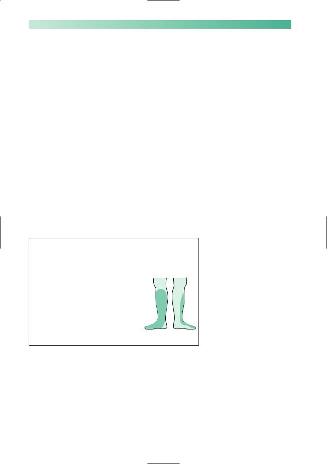
Common peroneal nerve palsy (Fig. 9.12)
The common peroneal (lateral popliteal) nerve runs a very superficial course around the neck of the fibula. It divides into the peroneal nerve to supply the lateral calf muscles which evert the foot, and into the anterior tibial nerve which innervates the anterior calf muscles which dorsiflex the foot and toes. The nerve is liable to damage from trauma, with or without fracture of the fibula. It is highly susceptible to acute compression during anaesthesia or coma, and from overtight or ill-fitting plaster casts applied for leg fractures.
The patient’s predominant complaint is of foot drop, and the need to lift the leg up high when walking. He may complain of loss of normal feeling on the dorsal surface of the affected ankle and foot.
Treatment of common peroneal nerve palsy should be preventative wherever possible. A splint that keeps the ankle at a right angle may assist walking. The condition is not usually helped by any form of surgery.
Common peroneal nerve palsies due to acute compression have a good prognosis.
Motor loss |
Reflex loss |
Sensory loss |
Foot evertors |
None |
|
Foot dorsiflexors |
|
|
Toe dorsiflexors |
|
|
Nerve sensitivity
Sometimes at the
neck of the fibula
Fig. 9.12 Common peroneal nerve palsy.
PERIPHERAL NERVOUS SYSTEM |
153 |
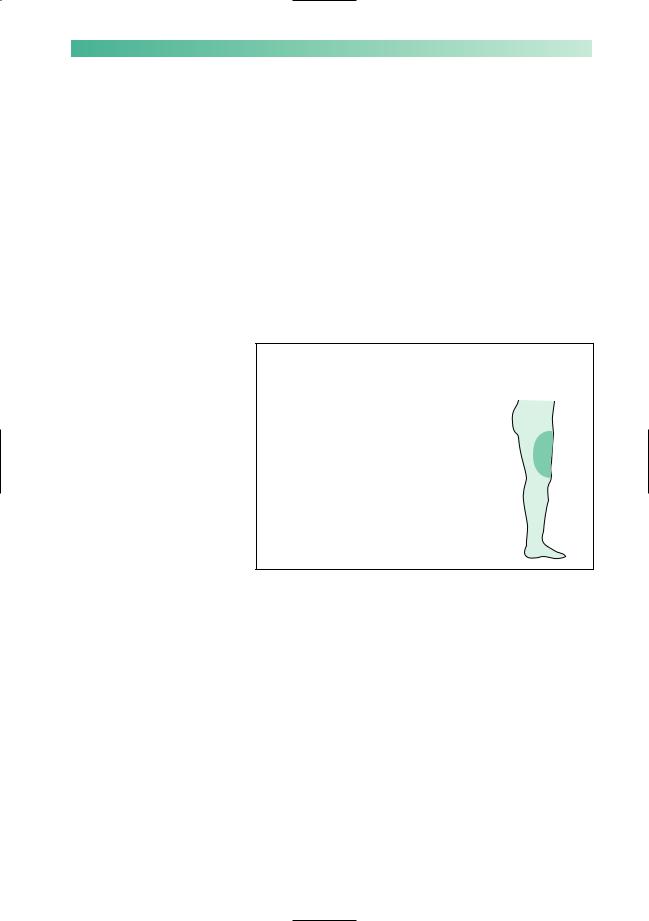
Meralgia paraesthetica (Fig. 9.13)
This irritating syndrome is the consequence of chronic entrapment of the lateral cutaneous nerve of the thigh as it penetrates the inguinal ligament, or deep fascia, in the vicinity of the anterior superior iliac spine.
As the nerve is sensory, there are no motor symptoms. The patient complains of annoying paraesthesiae and partial numbness in a patch of skin on the anterolateral aspect of the thigh. The contact of clothes is slightly unpleasant in the affected area.
No treatment of meralgia paraesthetica may be necessary if the symptoms are mild. Alternatively, nerve decompression or section at the site of compression both give excellent relief.
Motor loss |
Reflex loss |
Sensory loss |
None |
None |
|
Nerve sensitivity
Usually none
Fig. 9.13 Lateral cutaneous nerve of the thigh.

154 |
CHAPTER 9 |
CASE HISTORIES
Which nerve or nerve root is the likely culprit in the following brief histories?
a.A middle-aged man who has difficulty walking because of weak dorsiflexion of the right foot.
b.A young man who cannot use his right hand properly and is unable to dorsiflex the right wrist.
c.A middle-aged lady who is regularly waking at night due to strong tingling and numbness of her hands and fingers.
d.An elderly lady with persistent tingling in her left little finger, and some weakness in the use of the same hand.
(For answers, see p. 261.)
10 |
CHAPTER 10 |
Motor neurone disease, |
peripheral neuropathy, myasthenia gravis and muscle disease
Introduction
Fig. 10.1 The common peripheral neuromuscular disorders.
This chapter is about the common disorders that affect the peripheral nervous system and muscle (Fig. 10.1). They tend to produce a clinical picture of diffuse muscle weakness and wasting, and are sometimes difficult to distinguish from one another.
Motor neurone disease
X 
Generalized wasting, fasciculation and weakness of muscles
Bulbar muscle involvement common Associated upper motor neurone
symptoms and signs
No sensory symptoms and signs Steadily progressive and fatal
Peripheral neuropathy
X
Distal wasting and weakness of muscles
Legs more involved than arms, and bulbar involvement very rare
Associated distal sensory symptoms and signs
Many causes, several of which have specific treatment and are reversible
Myasthenia gravis
X
Muscle weakness without wasting Weakness which varies in severity and
which fatigues
Ocular and bulbar muscles commonly involved
Responds well to treatment
Muscle disease
X
Muscle weakness and wasting, the distribution of which depends on
the type of disease, but with a strong tendency to involve proximal muscles, i.e. trunk and limb girdles
Some inherited and incurable, others inflammatory or metabolic and treatable
155

156 |
CHAPTER 10 |
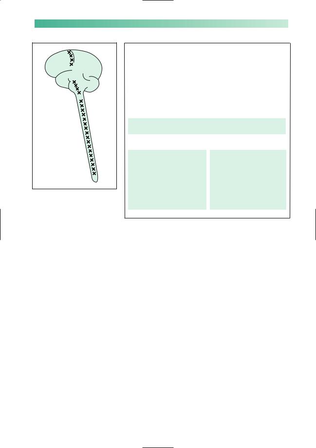
Motor neurone disease
This disease consists of a selective loss of lower motor neurones from the pons, medulla and spinal cord, together with loss of upper motor neurones from the motor cortex of the brain. The process is remarkably selective, leaving special senses, and cerebellar, sensory and autonomic functions intact. Progressive difficulty in doing things because of muscular weakness gradually overtakes the patient.
The cause of the neuronal loss in motor neurone disease (like the selective loss of neurones from other parts of the CNS, in diseases such as Alzheimer’s and Parkinson’s) is not understood.
There is variation in the clinical picture of motor neurone disease from one patient to another, which depends on:
•whether lower or upper motor neurones are predominantly involved;
•which muscles (bulbar, upper limb, trunk or lower limb) are bearing the brunt of the illness;
•the rate of cell loss. Most usually, this is steadily progressive over a few years, but in a minority of cases it may be much
more gradual with long survival.
Motor neurone disease (MND) tends to start either as a problem in the bulbar muscles, or as a problem in the limbs, and initially the involvement tends to be either lower motor neurone or upper motor neurone in nature. This has led to four clinical syndromes at the outset of the illness, which are described in Fig. 10.2.
As the illness progresses and the loss of motor neurones becomes more generalized, there is a tendency for both upper and lower motor neurone signs to become evident in bulbar, trunk and limb muscles. Sometimes, the illness may remain confined to the lower motor neurones, or to the upper motor neurones, but the coexistence of both, in the absence of sensory signs, is the hallmark of motor neurone disease. A limb with weak, wasted, fasciculating muscles, in which the deep tendon reflexes are very brisk, and in which there is no sensory loss, strongly suggests motor neurone disease.
It is the involvement of bulbar and respiratory muscles that is responsible for the inanition and chest infections which account for most of the deaths in patients with motor neurone disease.
The glutamate antagonist riluzole slows progression and prolongs survival, but there is as yet no cure for motor neurone disease. This makes regular medical input more important, not less, and the neurologist should be part of an integrated team of professionals providing advice and support.
X 
Key features of motor neurone disease
•Muscle weakness
•Muscle wasting
•Muscle fasciculation
•Exaggerated reflexes
•No loss of sensation
Bulbar weakness
The medulla oblongata used to be referred to as the ‘bulb’ of the brain. The cranial nerve nuclei 9, 10 and 12 reside in the medulla. This is why weakness of the muscles they supply (mouth, pharynx and larynx) is known as bulbar weakness or bulbar palsy. ‘Bulbar palsy’ and ‘pseudobulbar palsy’ imply that the cause is respectively in the lower
and upper motor neurones supplying these muscles, and have specific physical signs (see pp. 131 and 134–5 and Fig. 10.2). MND characteristically causes a mixture of the two
PERIPHERAL NEUROMUSCULAR DISORDERS |
157 |
||
|
Lower motor neurone |
|
Upper motor neurone |
|
Muscles supplied by the lower cranial nerves |
||
|
|
|
|
|
Bulbar palsy |
|
Pseudobulbar palsy |
|
Weakness, wasting and |
|
Weakness, slowness and |
|
fasciculation of the lower facial |
|
spasticity of the lower |
|
muscles, and muscles moving |
|
facial muscles, jaw, palate, |
|
the palate, pharynx, larynx and |
|
pharynx, larynx and tongue |
|
tongue—most conspicuous in |
|
muscles |
|
the tongue |
|
Exaggerated jaw-jerk |
|
|
|
Emotional lability |
Dysarthria, dysphagia, weight loss and the risk of inhalation pneumonia are the clinical problems facing patients described above
Muscles of the limbs and trunk
Progressive muscular atrophy Weakness, wasting and fasciculation of any of the limb or trunk muscles Often associated with frequent muscle cramps
No sensory loss
Small muscles of the hand frequently involved
Amyotrophic lateral sclerosis Weakness, spasticity, clonus and increased
deep tendon reflexes Any limb, but more commonly in the legs Sphincter control not affected
No sensory loss
Fig. 10.2 The four clinical syndromes with which motor neurone disease may present.
The quality of life for patients can be helped by:
•humane explanation of the nature of the condition to the patient and his family, with the aid of self-help groups;
•sympathy and encouragement;
•drugs for cramp, drooling and depression;
•speech therapy, dietetic advice, and often percutaneous gastrostomy feeding for dysphagia;
•communication aids for dysarthria;
•non-invasive portable ventilators for respiratory muscle weakness;
•provision of aids and alterations in the house (wheelchairs, ramps, lifts, showers, hoists, etc.) as weakness progresses;
•nursing help and respite care;
•gentle and timely discussion and planning of terminal care, often involving hospice staff.
158 |
CHAPTER 10 |
Peripheral neuropathy
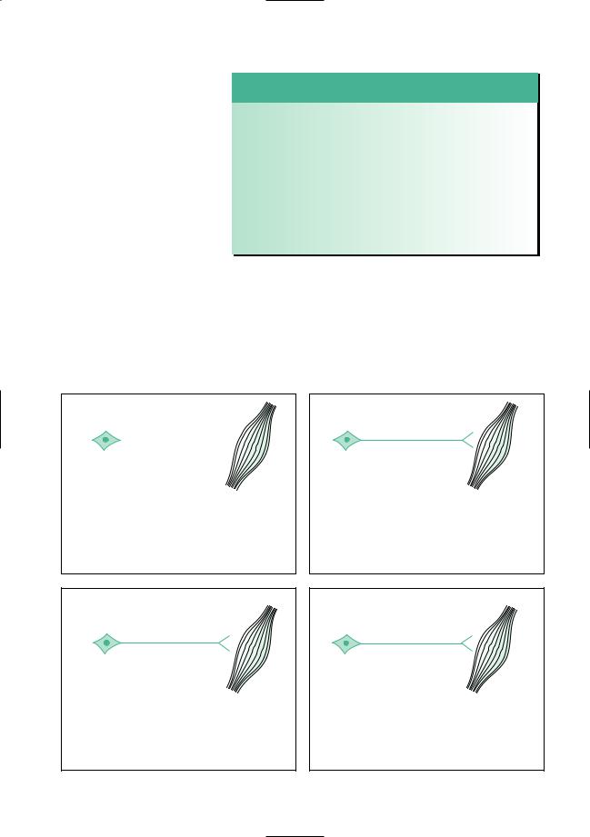
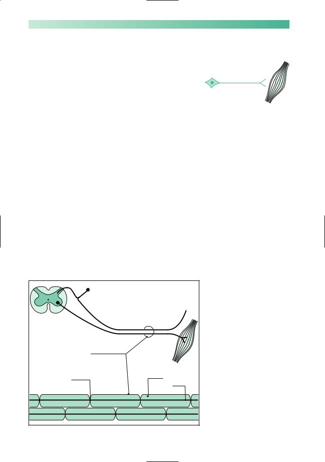
In this section, we are focusing on the axon of the anterior horn cell and the distal axon of the dorsal root ganglion cell. These myelinated nerve fibres constitute the peripheral nerves (Fig. 10.3). Each peripheral nerve, in reality, consists of very many myelinated nerve fibres.
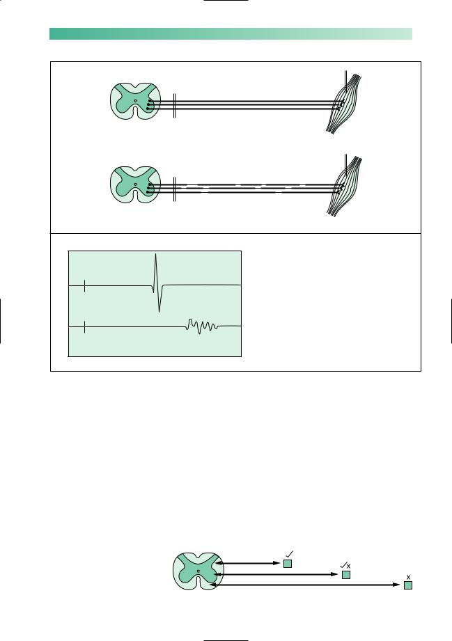
In patients with peripheral neuropathy, there is malfunction in all the peripheral nerves of the body. Two types of pathology may occur. In some cases there may be distal axonal degeneration, explaining the distal distribution of symptoms and signs in the limbs.Alternatively segments of the nerve fibres become demyelinated (Fig. 10.4). The normal saltatory passage of the nerve impulse along the nerve fibre becomes impaired. The impulse either fails to be conducted across the demyelinated section, or travels very slowly in a non-saltatory way along the axon in the demyelinated section of the nerve. This means that a large volley of impulses, which should travel synchronously along the component nerve fibres of a peripheral nerve, become:
•diminished as individual component impulses fail to be conducted;
•delayed and dispersed as individual impulses become slowed by the non-saltatory transmission (Fig. 10.4). Neurotransmission is most impaired in long nerves under such circumstances simply because the nerve impulse is confronted by a greater number of demyelinated segments along the course of the nerve (Fig. 10.5). This is the main reason why the
X
Skin, joints, etc.
Spinal cord
Peripheral nerve
Muscle
Node of |
Myelin |
|
Ranvier |
||
|
Axon
Fig. 10.3 Diagram to show the peripheral nerve components.
PERIPHERAL NEUROMUSCULAR DISORDERS |
159 |
Record
A
Healthy nerve
Stimulate
Spinal cord |
Muscle |
Record
B
Nerve affected by
Stimulate
segmental demyelination
Voltage
A Normal recorded muscle action potential
Stimulate
B The action potential is small (failure of some nerve impulses to
arrive at the muscle), and both delayed and dispersed (slow neurotransmission
Stimulate across segments of demyelinated nerve fibre)
Time
Fig. 10.4 Nerve conduction in healthy and segmentally demyelinated nerve fibres.
symptoms of neuropathy due to segmental demyelination are most evident distally in the limbs, why the legs and feet are more affected than the arms and hands, and why the distal deep tendon reflexes (which require synchronous neurotransmission from stretch receptors in the muscle to the spinal cord and back again to the muscle — the reflex arc) are frequently lost in patients with peripheral neuropathy.
The peripheral nerve pathology may predominantly affect sensory axons, motor axons, or all axons. Accordingly, the patient’s symptoms and signs may be distal and sensory in the limbs, distal and motor in the limbs, or a combination of both. They are shown in greater detail in Fig. 10.6.
Fig. 10.5 Effect of nerve length upon neurotransmission in peripheral neuropathy.