

Wilkinson. Essential Neurology 2005
.pdf100 |
CHAPTER 7 |
The lesion
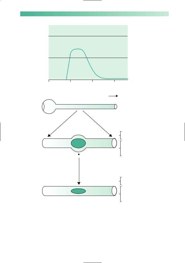
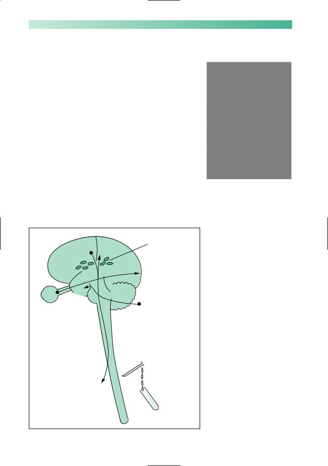
The classical lesion of multiple sclerosis is a plaque of demyelination in the CNS (Fig. 7.1). This means:
1.The lesion is in the CNS, not the peripheral nervous system, i.e. in the cerebrum, brainstem, cerebellum or spinal cord. It must be remembered that the optic nerve is an outgrowth from the CNS embryologically. This explains why multiple sclerosis frequently involves the optic nerves, whereas lesions in the other cranial nerves, spinal nerves and peripheral nerves in the limbs do not occur.
2.In the lesion the main insult is to the myelin sheaths with relative sparing of the axon. Saltatory conduction (from node to node along myelinated nerve fibres) requires healthy myelin sheaths. It cannot occur along the nerve fibres through a plaque of demyelination, and non-saltatory conduction is very slow and inefficient. Neurotransmission is accordingly impaired, depending upon the size of the lesion, for plaques vary considerably in size.
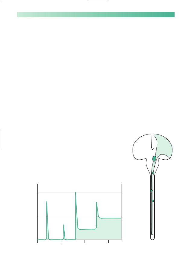
Clinically, the lesion evolves over a few days, lasts for a few days or weeks and gradually settles, as shown in Fig. 7.2. Vision in one eye may deteriorate and improve in this way, or the power in one leg may follow the same pattern. Clearly, the nature of the neurological deficit depends on the site of the plaque of demyelination (in the optic nerve or the pyramidal tract in the spinal cord, in the examples given here).
The evolving pathological lesion underlying the clinical episode is summarized in Fig. 7.2.
Fig. 7.1 An obvious established plaque of demyelination in the myelinated nerve fibres of an optic nerve.
MULTIPLE SCLEROSIS |
101 |
eye |
No |
|
function |
||
theaffected |
Half |
|
in |
||
function |
||
Vision |
||
|
||
|
Full |
|
|
function |
|
|
March |
|
|
Eye |
Acute stage
(1)
(1)
|
|
(2) |
April |
May |
June |
Optic nerve |
|
Brain |
|
Acute destruction of myelin |
|
Inflammatory cells and oedema |
|
No conduction through acutely |
|
demyelinated axons |
|
Impaired conduction through |
Major clinical deficit |
adjacent myelinated axons |
Plaque of demyelinated axons and gliosis
Chronic stage
(2)
No, or very slow, conduction through demyelinated axons
Normal conduction through adjacent healthy myelinated axons
 Little or no clinical deficit depending on the size of the plaque
 Impaired conduction may be detected by sophisticated neurophysiological tests
Fig. 7.2 Diagram to show an episode of demyelination. The optic nerve has been taken as an example in this instance.
102
Dissemination of lesions in time and place
Multiple sclerosis is caused by the occurrence of lesions just described in different parts of the CNS, occurring at different times in a person’s life. This dissemination of lesions in time and place remains the classical and diagnostic characteristic of multiple sclerosis from the clinical point of view.
Lesions may occur anywhere in the CNS. Individual plaques vary in size. One way in which a permanent ongoing disability may evolve in a patient with multiple sclerosis is illustrated in Fig. 7.3, and the common sites of lesions which may occur irregularly during the patient’s life are illustrated in Fig. 7.4.
Another cause for the development of a neurological deficit may be axonal damage occurring in the wake of the primary inflammatory myelin pathology. Evidence is accumulating to suggest that the axons do not escape completely unscathed in the CNS of patients with multiple sclerosis.
The number of lesions that show themselves as attacks or relapses in the clinical history of a patient with multiple sclerosis is much less than the number of lesions that can be found in the patient’s CNS post mortem. This agrees with the fact that MR scanning of the brain and spinal cord at the time of the first clinical episode of demyelination frequently shows the presence of plaques elsewhere in the CNS, especially in the periventricular white matter. Furthermore, it is not uncommon to be able to detect lesions in optic, auditory, sensory and motor CNS pathways by electrical neurophysiological techniques in patients with multiple sclerosis. Often, there are no symptoms or signs accompanying such lesions, indicating unsuspected subclinical involvement of various parts of the CNS.
No function
leg |
Half |
|
Right |
||
function |
||
|
Full function
1995 |
2000 |
2005 |
2010 |
CHAPTER 7
|
2003 |
Right |
Left |
2000
1997
2007
Fig. 7.3 The establishment of a neurological deficit in the right leg by episodes of demyelination along the course of the corticospinal tract over a period of 15 years in a patient with multiple sclerosis.

MULTIPLE SCLEROSIS
Periventricular
white matter
Optic
nerve
Midbrain
Pons
Medulla
Spinal cord
Fig. 7.4 Diagram to show the common sites at which plaques occur in the CNS of patients with multiple sclerosis.
Tell-tale signs of previous optic neuritis
•Slightly impaired acuity
•Slightly impaired colour vision
•Mild afferent pupillary defect
•Slightly pale optic disc
•Delay in the visual evoked potential
103
Common clinical expressions of multiple sclerosis
This section describes what occurs during individual episodes of demyelination in different parts of the CNS. It also describes the common neurological deficits that characterize a patient who has multiple sclerosis moderately severely. Figure 7.4 shows the common sites for plaques of demyelination.
Periventricular white matter
Lesions are very common in this part of the brain. They are seen early in the disease in patients studied by MR brain scanning, and are always found post mortem. They do not give rise to definite symptomatology, however.
Optic nerve
Optic neuritis is a common and typical manifestation of multiple sclerosis. If the lesion is in the optic nerve between the globe of the eye and the optic chiasm, it is sometimes called retrobulbar (behind the globe of the eye) neuritis. If it is right at the front of the optic nerve, the lesion itself is visible with an ophthalmoscope, and is sometimes called papillitis (inflammation of the optic disc). The effect on vision is the same whether the lesion is anterior or posterior in the optic nerve. If anterior, the optic disc is visibly red and swollen, with exudates and haemorrhages. If posterior, the appearance of the optic disc is normal at the time of active neuritis.Asection of the optic nerve is acutely inflamed in all instances of optic neuritis, so that pain in the orbit on eye movement is a common symptom.
The effect on vision in the affected eye is to reduce acuity, and cause blurring, and this most commonly affects central vision. The patient develops a central scotoma of variable size and density. Colour vision becomes faded, even to a point of fairly uniform greyness. In severe optic neuritis, vision may be lost except for a rim of preserved peripheral vision, or may be lost altogether. At this stage, there is a diminished pupil reaction to direct light with a normal consensual response (often called an afferent pupillary defect).
After days or weeks, recovery commences. Recovery from optic neuritis is characteristically very good, taking 4–8 weeks to occur. Five years later, the patient often has difficulty remembering which eye was affected. Occasionally, recovery is slow and incomplete.
104 |
CHAPTER 7 |
Midbrain, pons and medulla
Here episodes of demyelination may cause:
•double vision due to individual cranial nerve dysfunction within the midbrain or pons, or more commonly due to a lesion in the fibre pathways that maintain conjugate movement of the eyes. Lesions of the medial longitudinal fasciculus cause an internuclear ophthalmoplegia, in which there is failure of movement of the adducting eye with preserved movement of the abducting eye, on attempted conjugate deviation of the eyes to one side (see Chapter 8, p. 117);
•facial numbness (cranial nerve 5 within the pons);
•facial weakness (cranial nerve 7 within the pons);
•vertigo, nausea, vomiting, ataxia (cranial nerve 8 within the pons);
•dysarthria and occasional dysphagia (cranial nerves 9, 10 and 12 within the medulla);
•cerebellar dysfunction due to lesions on fibre pathways passing in and out of the cerebellum in the cerebellar peduncles, hence nystagmus, dysarthria, ataxia of limbs and gait;
•motor deficits of upper motor neurone type in any of the four limbs;
•sensory deficits, spinothalamic or posterior column in type, in any of the four limbs.
3
4
5 6 7
8
9
10
12
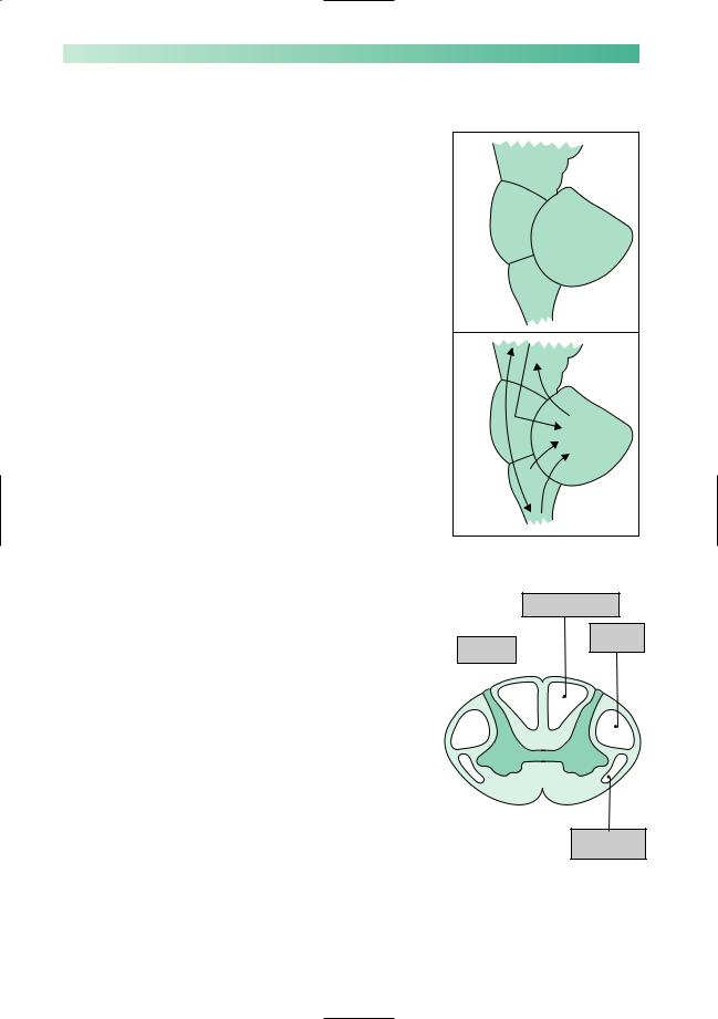
Spinal cord
Lower motor neurone and segmental signs are unusual in multiple sclerosis. Episodes of demyelination in the spinal cord cause fibre tract (upper motor neurone, posterior column, spinothalamic and autonomic) symptoms and signs below the level of the lesion. Since the length of the fibre tracts in the spinal cord are physically longer for leg function than for arm function, there is a greater likelihood of plaques in the spinal cord interfering with the legs than the arms. Episodes of spinal cord demyelination may cause:
•heaviness, dragging or weakness of the arms, trunk or legs;
•loss of pain and temperature sensation in the arms, trunk or legs;
•tingling, numbness, sense of coldness, sense of skin wetness, sense of skin tightness, or a sensation like that which follows a local anaesthetic or a nettle-sting, in the arms, trunk or legs;
•clumsiness of a hand due to loss of position sense and stereognosis;
•bladder, bowel or sexual malfunction.
Posterior column ≠
Pyramidal Autonomic tract Ø tracts Ø≠
Spinothalamic
tract ≠
MULTIPLE SCLEROSIS |
105 |
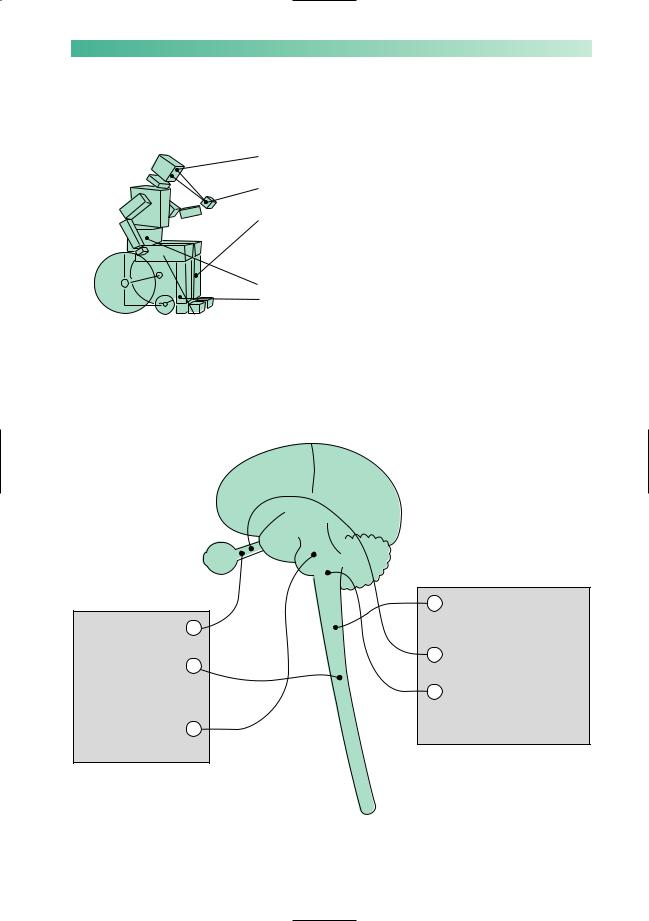
In a patient with established multiple sclerosis, who has suffered multiple episodes of demyelination throughout the CNS (Fig. 7.5), the accumulated ongoing neurological deficit is likely to consist of:
• asymmetrical optic pallor without a major defect in visual acuity;
• a cerebellar deficit causing nystagmus, dysarthria and arm ataxia;
• an upper motor neurone deficit, mild in the arms, moderate in the trunk and most evident in the legs. The weakness of the legs often does not allow ataxia to reveal itself in leg movement and walking;
• impaired sexual, bladder and bowel function;
• a variable amount and variety of sensory loss, more evident in the legs and lower trunk than in the arms.
Doctors probably tend to overfocus on the specific neurological disabilities in a patient with multiple sclerosis. The orientation of the patient and family may be less specific, and more concerned with general lack of mobility and vitality, less robust physical health, and the patient’s limited social roles.
1987 Transient loss of |
1 |
vision in left eye |
|
1997 Numbness and |
2 |
weakness in both legs |
|
with some bladder |
|
disturbance |
|
1998 Double vision |
3 |
and unsteadiness |
|
42000 Weak legs again with incomplete recovery, leg numbness
52004 Transient loss of vision in right eye
62007 Further increase in leg weakness with unsteadiness, ataxia of arms, dysarthria and nystagmus
Fig. 7.5 Diagram to show the classical dissemination of lesions in time and space, and the accumulation of a neurological deficit, in a patient who has multiple sclerosis moderately severely.
106 |
CHAPTER 7 |
Diagnosis
There is no specific laboratory test that confirms the presence of multiple sclerosis. The diagnosis is a clinical one, based upon the occurrence of lesions in the CNS which are disseminated in time and place. The presence of subclinical lesions in the CNS may be detected by:
•various clinical neurophysiological techniques. Such techniques essentially measure conduction in a CNS pathway, detecting any delay in neurotransmission by comparison with normal control data. The visual evoked potential is the one most commonly used;
•imaging techniques. Frequently MR brain scanning reveals multiple lesions, especially in the periventricular regions. The inflammatory nature of the demyelinating lesion may re-
sult in an elevated lymphocyte count and globulin content in the CSF. These changes also lack specificity. Immunoelectrophoretic demonstration of oligoclonal bands in the CSF globulin has come closest to becoming a diagnostic feature of multiple sclerosis, but it is not specific, producing both falsepositive and false-negative results (Fig. 7.6).
MR scan showing multiple areas of high signal in the white matter due to multiple sclerosis.
Slow |
SSEP |
Magnetic |
|
resonance imaging |
|||
CMCV |
delay |
||
of multiple lesions |
|||
|
|
||
|
|
VEP delay |
Eye AEP
delay
Ear
Elevated cell
count and
oligoclonal bands
in CSF
Limbs
Fig. 7.6 Diagram to show the abnormal investigations in patients with multiple sclerosis. None is specific. MR scanning is used. CSF abnormalities are found, especially the presence of oligoclonal bands in the CSF globulin. AEP, auditory evoked potential from ear to temporal cortex; CMCV, central motor conduction velocity from motor cortex to limbs; SSEP, somatosensory evoked potential
from limbs to sensory cortex; VEP, visual evoked potential from eye to occipital cortex.

MULTIPLE SCLEROSIS |
107 |
Aetiology
The cause of multiple sclerosis remains unknown. There appears to be an interaction of environmental factors with some form of genetically determined patient susceptibility.
The evidence for genetic susceptibility is as follows:
•multiple sclerosis is more common in females than males, ratio 1.5:1;
•there is a firm association of multiple sclerosis with certain HLAtypes, particularly DR2;
•there is an increased incidence of multiple sclerosis in close relatives;
•in multiple sclerosis patients who have a twin, identical co-twins are more likely to develop it than non-identical twins.
The evidence for an environmental factor is as follows:
•multiple sclerosis is more common in temperate than in equatorial parts of the world. Migrants moving from highrisk to low-risk areas (e.g. from northern Europe to Israel) under the age of puberty acquire low risk, and vice versa;
•IgG levels are higher in the CSF of patients with multiple sclerosis. Antibodies to measles virus, and to some other viruses, are higher in the CSF of patients with multiple sclerosis.
Management
Mild or early cases
1.Inform the patient and family of the diagnosis.
2.Educate the patient and family about multiple sclerosis.
3.Dispel the concept of inevitable progression to major disability. Make explanatory literature available.
4.Encourage normal attitudes to life, and normal activities. (This advice should be given initially by the consultant neurologist, and two interviews at an interval will nearly always be needed. Subsequent counselling and support by a specialist nurse or the family doctor may be very valuable, depending on the patient’s reaction to the problem.)

108
More serious cases
1.Continued education about the nature of multiple sclerosis.
2.Continued support over the disappointment and uncertainty of having multiple sclerosis.
3.Attention to individual symptoms:
•vision, rarely a major problem. Low visual acuity aids may prove helpful in the minority of patients who need them;
•cerebellar deficit, difficult to help pharmacologically;
•paraplegia — all the problems attendant upon chronic paraplegia (see Chapter 6, pp. 94 and 95) may occur, and require attention;
•pain — may arise from faulty transmission of sensation and may respond to antidepressants (e.g. amitriptyline) or anticonvulsants (e.g. gabapentin);
•fatigue — common and hard to treat, but may respond to antidepressants (e.g. fluoxetine) or yoga.
4.Help from nurses, physiotherapists, occupational therapists, speech therapists and medical social workers, as required.
5.Attention to psychological reactions occurring in the patient or family. Encourage all activities which the patient enjoys and are still possible.
6.Respite care arrangements, as required.
All cases of multiple sclerosis
1.Several immunomodulatory drugs (azathioprine, betainterferon, copaxone, mitoxantrone, etc.) reduce the incidence of relapses somewhat in ambulatory patients with relapsing and remitting multiple sclerosis. They have a much more questionable effect on the development of disability.
2.Corticosteroids, often in the form of high-dose intravenous methyl-prednisolone over 3 days, reduce the duration and severity of individual episodes of demyelination, without influencing the final outcome.
3.Dietary exclusions and most supplements are of no proven advantage. Fish oil supplements may be of benefit. The main dietary requirement is the avoidance of obesity in the enforced sedentary state.
CHAPTER 7

MULTIPLE SCLEROSIS |
109 |
CASE HISTORIES
Case 1
A 37-year-old man presents with double vision, right facial numbness and a clumsy right arm. His symptoms began over the course of a weekend and are starting to improve 3 weeks later. He had an episode of the same symptoms 4 years ago which took 2 months to clear up. His sister has MS.
Examination reveals a right internuclear ophthalmoplegia (i.e. when he looks to the left, the right eye does not adduct and the left eye shows nystagmus), right trigeminal numbness and rightsided limb ataxia.
a.What is the most likely diagnosis?
b.What treatment should he have?
Case 2
A 48-year-old woman has had clinically definite MS for more than 20 years. She had about ten relapses in the
first 15 years, beginning with left optic neuritis. Over the last 5 years her disability has steadily progressed. She is now wheelchair-bound and catheterized. She takes baclofen for leg cramps.
She comes to see you because she is very worried about the slowly increasing tingling and weakness in her hands, which is making it difficult for her to do up buttons or hold a pen. She says that losing the use of her hands would be the final straw. Examination reveals wasting and weakness of the first dorsal interosseus, lumbrical and adductor digiti minimi muscles; the rest of her hand and forearm muscles are reasonably strong. Her reflexes are all
brisk.
a.What is the cause of this problem?
b.What would you advise?
(For answers, see p. 260.)