

Wilkinson. Essential Neurology 2005
.pdfAbbreviations
BG |
basal ganglia |
C |
cerebellum |
CNS |
central nervous system |
CSF |
cerebrospinal fluid |
CT |
computerized tomography |
ECG |
electocardiogram |
EEG |
electroencephalogram |
EMG |
electromyogram |
LMN |
lower motor neurone |
MR |
magnetic resonance |
NMJ |
neuromuscular junction |
S |
sensation |
UMN |
upper motor neurone |
x

1 |
CHAPTER 1 |
Clinical skills, physical |
signs and anatomy
‘There’s something wrong with my left leg, and I’m not walking properly.’ In this chapter we are going to use this clinical problem to remind us:
•how important the details of the history are in making a neurological diagnosis, especially the clear definition of how the problem has evolved with respect to time;
•of the components of the nervous system involved in normal movement, their anatomy (which isn’t complicated), and the physical signs produced by lesions in the various components;
•of the common patterns of neurological malfunction, affecting any part of the body, such as a leg;
•of the importance of the patient’s response to his malfunctioning limb, in defining the size and nature of the whole illness;
•of the reliability of the clinical method in leading us to a diagnosis or differential diagnosis and management plan;
•how important it is to explain things clearly, in language the patient and relatives can understand.
At the end of the chapter are a few brief case histories, given to
illustrate the principles which are outlined above and itemized in detail throughout the chapter.
Our response to a patient telling us that his left leg isn’t working properly must not just consist of the methodical asking of many questions, and the ritualistic performance of a complex, totally inclusive, neurological examination, hoping that the diagnosis will automatically fall out at the end. Nor must our response be a cursory questioning and non-focused examination, followed by the performance of a large battery of highly sophisticated imaging and neurophysiological tests, hoping that they will pinpoint the problem and trigger a management plan.
No, our response should be to listen and think, question and think, examine and think, all the time trying to match what the patient is telling us, and the physical signs that we are eliciting, with the common patterns of neurological malfunction described in this chapter.
1

2 |
CHAPTER 1 |
History
We need all the details about the left leg, all the ways it is different from normal. If the patient mentions wasting, our ideas will possibly start to concentrate on lower motor neurone trouble. We will learn that if he says it’s stiff, our thoughts will move to the possibility of an upper motor neurone or extrapyramidal lesion. If he can’t feel the temperature of the bath water properly with the other (right) leg, it is clear that we should start to think of spinal cord disease. If it makes his walking unsteady; we may consider a cerebellar problem. Different adjectives about the leg have definite diagnostic significance. We will ask the patient to use as many adjectives as he can to describe the problem.
Details of associated symptoms or illnesses need clarification. ‘There’s nothing wrong with the other leg, but my left hand isn’t quite normal’ — one starts to wonder about a hemiparesis. ‘My left hand’s OK, but there’s a bit of similar trouble in the right leg’ — we might consider a lesion in the spinal cord. ‘I had a four-week episode of loss of vision in my right eye, a couple of years ago’ —we might wonder about multiple sclerosis. ‘Mind you, I’ve had pills for blood pressure for many years’ — we might entertain the possibility of cerebrovascular disease. These and other associated features are important in generating diagnostic ideas.
Very important indeed in neurological diagnosis is detail of the mode of onset of the patient’s symptoms. How has the left leg problem evolved in terms of time? Let’s say the left leg is not working properly because of a lesion in the right cerebral hemisphere. There is significant weakness in the left
Years |
|
|
|
|
|
|
|
Benign tumour |
|
|
|
|
|
|
|
Degenerative condition |
|
|
|
Y |
|
|
|
|
|
|
|
|
|
|
|
Months/weeks |
|
|
|
|
M/W |
|
|
Malignant tumour |
|
|
|
|
|
|
|
Chronic inflammation |
|
|
|
|
|
|
|
|
|
|
|
|
|
|
|
|
|
2 |
1 |
0 |
|||
Weeks/days |
|
|
Years |
|
|
||
Demyelination in CNS |
|
|
|
|
|
|
|
|
|
|
|
|
|
||
Acute inflammation |
|
|
|
|
|
|
|
Days/hours |
|
|
|
W/D |
|
|
|
Very acute inflammation |
|
|
|
|
D/H |
|
|
|
|
|
|
|
|
|
|
Instantaneous |
|
|
|
|
I |
|
|
|
|
|
|
|
|
||
Vascular |
|
|
|
|
|
|
|
|
|
|
|
|
|
|
|
Trauma |
2 |
1 |
0 |
||||
|
|
|
|
Weeks |
|
|
|
Duration of symptoms before presentation
Severity of symptoms
Fig. 1.1 The patient’s history indicates the probable pathology.

CLINICAL SKILLS |
3 |
leg, slight weakness in the left hand and arm, some loss of sensation in the left leg and no problem with the visual fields. This same neurological deficit will be present whatever the nature of the pathology at this site. If this part of the brain isn’t working there is an inevitability about the nature of the neurological deficit.
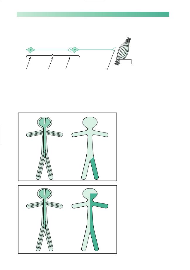
It is the history of the mode of evolution of the neurological deficit which indicates the nature of the pathology (Fig. 1.1).
Components of the nervous system required for normal function; their anatomy; physical signs indicating the presence of a lesion in each component; and the common patterns in which things go wrong
The basic components of the nervous system required for normal movement are shown on the simple diagram below.
Upper motor neurone |
Lower motor neurone |
Muscle
Basal ganglia Cerebellum Sensation |
Neuromuscular junction |
Lesions along the primary motor pathway, UMN–LMN– NMJ–M, are characterized by weakness or paralysis. We will see that the characteristics of the weakness are different in each instance; for example, UMN weakness has different characteristics from LMN weakness. Knowledge of these characteristics is fundamental to clinical neurology.
Normal basal ganglia, cerebellar and sensory function is essential background activity of the nervous system for normal movement. Lesions in these parts of the nervous system do not produce weakness or paralysis, but make movement imperfect because of stiffness, slowness, involuntary movement, clumsiness or lack of adequate feeling.
So we will be questioning and examining for weakness, wasting, stiffness, flaccidity, slowness, clumsiness and loss of feeling in our patient’s left leg. This will help to identify in which component of the nervous system the fault lies.
To make more sense of the patient’s clinical problem, we have to know the basic anatomy of the neurological components, identified above, in general terms but not in every minute detail. This is shown on the next three pages.
4 |
CHAPTER 1 |
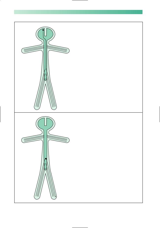
An upper motor neurone involved in left leg movement
Cell body in motor cortex of right cerebral hemisphere
Axon:
 descends through right internal capsule
 crosses from right to left in the medulla
 travels down the spinal cord on the left side in lateral column
 synapses with a lower motor neurone innervating left leg musculature
A lower motor neurone involved in left leg movement
Cell body at the lower end of the spinal cord on the left side
Axon:
 leaves the spine within a numbered spinal nerve
 travels through the lumbosacral plexus
 descends within a named peripheral nerve
 synapses with muscle at neuromuscular junction
CLINICAL SKILLS |
5 |
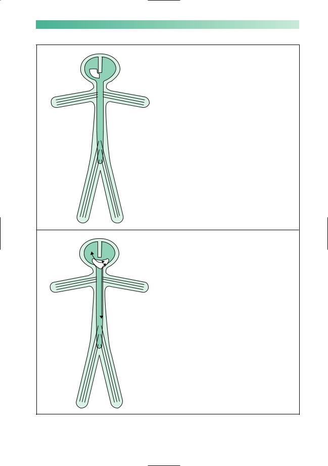
Basal ganglion control of the left leg
The structures involved in extrapyramidal control of the left side of the body reside in the basal ganglia and cerebral peduncle on the right
Basal ganglion function is contralateral
Cerebellar control of the left leg
The left cerebellar hemisphere has two-way connection with the right cerebral hemisphere and the left side of the body, via the cerebellar peduncles, brainstem and spinal cord
Cerebellar function is ipsilateral
6 |
CHAPTER 1 |
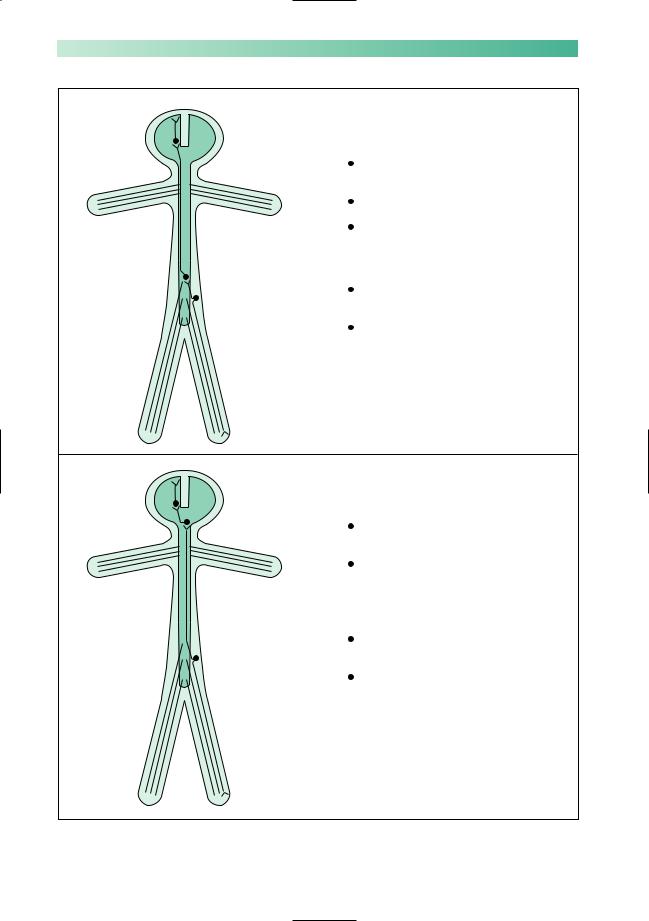
Pain and temperature sensation in the left leg
Third sensory neurone:  cell body in thalamus
axon travels to sensory cortex
Second sensory neurone:
cell body in lumbar spinal cord on the left
axon crosses to the right and ascends to thalamus in lateral column of spinal cord
Dorsal root ganglion cell:
distal axon from the left leg, via peripheral nerve, lumbosacral plexus and spinal nerve
proximal axon enters cord via dorsal root of spinal nerve, and relays with second sensory neurone
Position sense in the left leg
Third sensory neurone:  cell body in thalamus
axon travels to sensory cortex
Second sensory neurone:
cell body in gracile or cuneate nucleus on left side of medulla, axon crosses to the right side of medulla and ascends to thalamus on the right
Dorsal root ganglion cell:
distal axon from left leg, via peripheral nerve, lumbosacral plexus and spinal nerve
proximal axon enters cord, ascends in posterior column on the left to reach the second sensory neurone in left lower medulla
CLINICAL SKILLS |
7 |
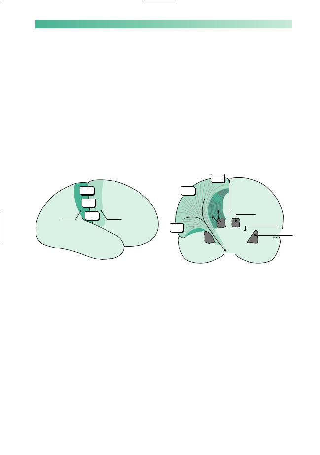
The final piece of anatomical knowledge which is helpful for understanding the neurological control of the left leg, is a little more detail about motor and sensory representation in the brain. The important features to remember here are:
•the motor cortex is in front of the central sulcus, and the sensory cortex is behind it;
•the body is represented upside-down in both the motor and sensory cortex;
•the axons of the upper motor neurones in the precentral motor cortex funnel down to descend in the anterior part of the internal capsule;
•the axons of the 3rd sensory neurone in the thalamus radiate out through the posterior part of the internal capsule to reach the postcentral sensory cortex.
|
|
|
Leg |
|
Leg |
|
Arm |
Postcentral |
Arm |
Precentral |
|
sensory |
Face |
motor |
Thalamus |
cortex |
cortex |
|
Internal capsule
Face
Basal ganglion
Having reviewed the components of the nervous system involved in normal function of the left leg, and their basic anatomy, we now need more detail of:
•the clinical features of failure in each component;
•the common patterns of failure which are met in clinical practice.
The next section of this chapter reviews these features and patterns.
8 |
CHAPTER 1 |
Upper motor neurone
Upper motor neurone |
Lower motor neurone |
X |
|
|
Muscle |
Basal ganglia Cerebellum Sensation |
Neuromuscular junction |
Characteristic of upper motor neurone lesions:
•no wasting;
•increased tone of clasp-knife type;
•weakness most evident in anti-gravity muscles;
•increased reflexes and clonus;
•extensor plantar responses.
X
X
Contralateral monoparesis
Alesion situated peripherally in the cerebral hemisphere, i.e. involving part of the motor homunculus only, produces weakness of part of the contralateral side of the body, e.g. the contralateral leg. If the lesion also involves the adjacent sensory homunculus in the postcentral gyrus, there may be some sensory loss in the same part of the body.
Contralateral hemiparesis
Lesions situated deep in the cerebral hemisphere, in the region of the internal capsule, are much more likely to produce weakness of the whole of the contralateral side of the body, face, arm and leg. Because of the funnelling of fibre pathways in the region of the internal capsule, such lesions commonly produce significant contralateral sensory loss (hemianaesthesia) and visual loss (homonymous hemianopia), in addition to the hemiparesis.
CLINICAL SKILLS
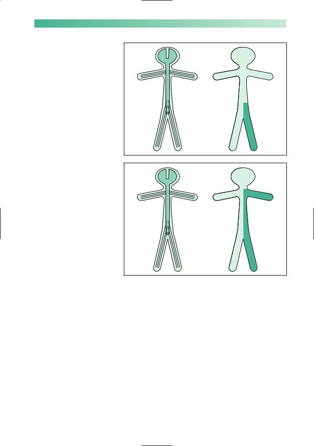
Ipsilateral monoparesis
Aunilateral lesion in the spinal cord below the level of the neck produces upper motor neurone weakness in one leg. There may be posterior column (position sense) sensory loss in the same leg, and spinothalamic (pain and temperature) sensory loss in the contralateral leg. This is known as dissociated sensory loss, and the whole picture is sometimes referred to as the Brown-Séquard syndrome.
Ipsilateral hemiparesis
Aunilateral high cervical cord lesion will produce a hemiparesis similar to that which is caused by a contralateral cerebral
hemisphere lesion, except that the face cannot be involved in the hemiparesis, vision will be normal, and the same dissociation of sensory loss (referred to above) may be found below the level of the lesion.
9
X
X