

Wilkinson. Essential Neurology 2005
.pdf170
Facio-scapulo-humeral dystrophy
Facio-scapulo-humeral dystrophy is generally a benign form of muscular dystrophy. It is due to an unusual dominantly inherited gene contraction near the telomere of chromosome 4, which can usually be detected for diagnostic purposes. It is often mild and asymptomatic. Wasting and weakness of the facial, scapular and humeral muscles may give rise to difficulties in whistling, and in using the arms above shoulder level and for heavy lifting. The thinness of the biceps and triceps, or the abnormal position of the scapula (due to weakness of the muscles which hold the scapula close to the thoracic cage), may be the features that bring the patient to seek medical advice. Involvement of other trunk muscles, and the muscles of the pelvic girdle, may appear with time.
Limb girdle syndrome
Weakness concentrated around the proximal limb muscles has a wide range of causes, including ones which are treatable. Limb girdle weakness should not be regarded as due to dystrophy (and therefore incurable) until thorough investigation has shown this to be the case. Even limb girdle dystrophy is heterogeneous, made up of a large number of pathologically and genetically distinct entities, for example involving many of the proteins which anchor the contractile apparatus of muscle to the muscle membrane. Other important causes of limb girdle syndrome are shown in the box.
Conditions caused by inherited biochemical defects
Muscle diseases in which an inherited biochemical defect is present are rare. Of these conditions, malignant hyperpyrexia is perhaps the most dramatic. Members of families in which this condition is present do not have any ongoing muscle weakness or wasting. Symptoms do not occur until an affected family member has a general anaesthetic, particularly if halothane or suxamethonium chloride is used. During or immediately after surgery, muscle spasm, shock and an alarming rise in body temperature occur, progressing to death in about 50% of cases.
The pathology of this condition involves a defect in calcium metabolism allowing such anaesthetic agents to incur a massive rise in calcium ions within the muscle cells. This is associated with sustained muscle contraction and muscle necrosis. The rise in body temperature is secondary to the generalized muscle contraction.
CHAPTER 10
Limb girdle weakness:
•polymyositis
•myopathy associated with endocrine disease
•metabolic myopathies
•drug-induced myopathies, e.g. steroids
•limb girdle dystrophy

PERIPHERAL NEUROMUSCULAR DISORDERS |
171 |
Polymyositis and dermatomyositis
The muscle problems in polymyositis and dermatomyositis are very similar. There is a mononuclear inflammatory cell infiltration and muscle fibre necrosis. In dermatomyositis, there is the additional involvement of skin, particularly in the face and hands. An erythematous rash over the nose and around the eyes, and over the knuckles of the hands, is most typical. Though all muscles may be involved, proximal limb, trunk and neck muscles are most frequently made weak by polymyositis with occasional involvement of swallowing.
The condition most usually develops subacutely or chronically and is unassociated with muscle tenderness. Problems when trying to use the arms above shoulder level, and difficulty when standing up out of low chairs and the bath, are the most frequent complaints.
Both conditions are autoimmune diseases involving skeletal and not cardiac muscle. Dermatomyositis, especially in older men, can be triggered by underlying malignancy.
Both dermatomyositis and polymyositis respond to immunosuppressive therapy. High-dose steroids, with or without other immunosuppressants, gradually reduced to reasonable long-term maintenance levels, constitute the treatment of choice. Effective control of the disease can be established in the majority of cases.
Acquired non-inflammatory myopathy
Acquired non-inflammatory myopathy can occur in many circumstances (alcoholism, drug-induced states, disturbances of vitamin D and calcium metabolism, Addison’s disease, etc.), but the two common conditions to be associated with myopathy are hyperthyroidism and high-dose steroid treatment.
Many patients with hyperthyroidism show weakness of shoulder girdle muscles. This is usually asymptomatic. Occasionally, more serious weakness of proximal limb muscles and trunk muscles may occur. The myopathy completely recovers with treatment of the primary condition.
Patients on high-dose steroids, especially fluorinated triamcinolone, betamethasone and dexamethasone, may develop significant trunk and proximal limb muscle weakness and wasting. The myopathy is reversible on withdrawal of the steroids, on reduction in dose, or on change to a non-fluorinated steroid.

172 |
CHAPTER 10 |
Investigation of patients with generalized muscle weakness and wasting
The last section of this chapter discusses the common investigations that are carried out in patients with generalized muscle weakness and wasting. Of the four conditions discussed in this chapter, myasthenia gravis does not produce muscle wasting, and is usually distinguishable by virtue of the ocular and bulbar muscle involvement, the abnormal degree of fatiguability, and the response to anticholinesterase. The other three conditions may be quite distinct on clinical grounds too, but investigation is frequently very helpful in confirmation of diagnosis.
Figure 10.11 shows the investigations that are carried out on patients of this sort.
|
Motor |
|
|
|
neurone |
Peripheral |
Muscle |
Test |
disease |
neuropathy |
disease |
Biochemistry |
|
|
|
Creatine kinase |
Normal |
Normal |
Elevated |
Electrical studies |
|
|
|
Electromyography |
Denervation |
Denervation |
Muscle disease |
Motor and |
Normal |
Delayed |
Normal |
sensory nerve |
|
conduction |
|
conduction studies |
|
velocities and |
|
|
|
reduced nerve |
|
|
|
action |
|
Histology |
|
potentials |
|
|
|
|
|
Histochemistry |
|
|
|
Immunofluorescence |
|
|
|
Electron microscopy |
|
|
|
Muscle biopsy |
Denervation |
Denervation |
Specific commentary |
|
|
|
on the nature of the |
|
|
|
muscle disease, |
|
|
|
i.e. dystrophy, |
|
|
|
polymyositis or |
|
|
|
acquired myopathy |
Nerve biopsy |
|
Sometimes |
|
|
|
helpful in |
|
|
|
establishing |
|
|
|
the precise |
|
|
|
cause of |
|
|
|
peripheral |
|
|
|
neuropathy |
|
Molecular |
No help in |
Helpful in |
Helpful in the |
genetics |
conventional |
hereditary |
inherited muscle |
|
MND |
motor and |
diseases |
|
|
sensory |
|
|
|
neuropathy |
|
|
|
|
|
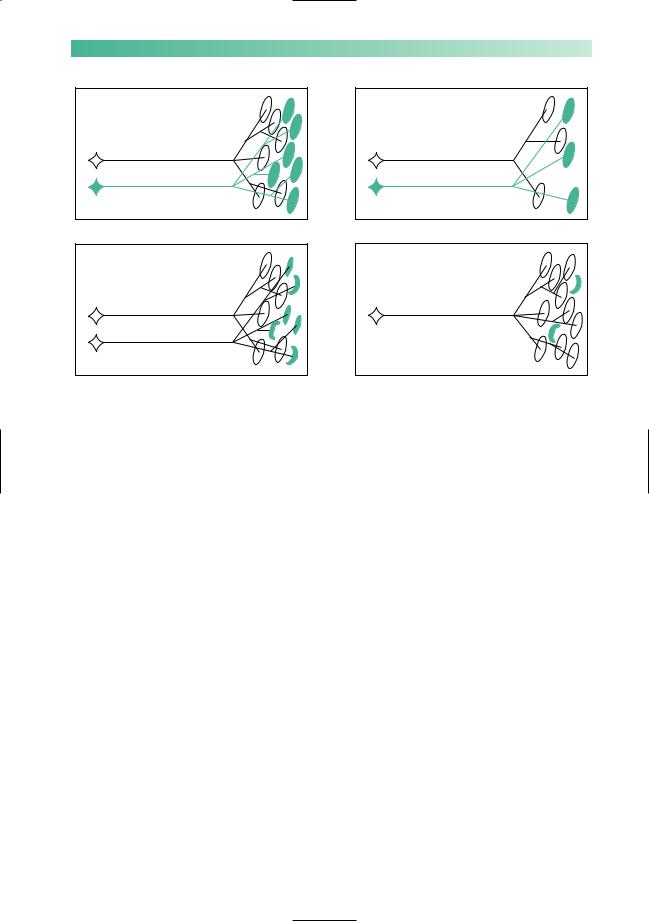
Fig. 10.11 The investigations performed in patients with generalized muscle weakness and wasting.
PERIPHERAL NEUROMUSCULAR DISORDERS
(a) Normal
173 |
(b) Muscle disease
(c) Denervation |
(d) Denervation |
Fig. 10.12 The changes in muscle disease and denervation.
(a)Two normal motor units. Each lower motor neurone supplies several muscle fibres.
(b)In muscle disease (dystrophy, polymyositis or acquired myopathy), there is loss or damage directly affecting muscle fibres. The number of functional muscle fibres decreases. Therefore, in muscle disease, there is a normal number of abnormally small motor units.
(c)Damage to one lower motor neurone, either in the cell body (as in motor neurone disease), or in the axon (as in peripheral neuropathy), results in denervated muscle fibres within the motor unit.
(d)The surviving lower motor neurone produces terminal axonal sprouts which innervate some of the muscle fibres of the damaged motor unit. Therefore, in muscles affected by a denervating disease, there is a reduced number of abnormally large motor units.
It is important that the consequences of denervation and muscle disease on the motor unit are understood, and these are shown in Fig. 10.12. Both electromyography (the recording of muscle at rest and during contraction) and muscle biopsy are able to detect the changes of chronic partial denervation and of primary muscle disease.

174 |
CHAPTER 10 |
CASE HISTORIES
Case 1
A 55-year-old bank manager reports a 12-month history of numbness and burning in his feet. He takes ranitidine for dyspepsia but has no other medical history. He is married with grown-up children. He has no family history of neurological disease. His GP has checked his full blood count (MCV mildly raised at 101, otherwise normal), electrolytes and glucose (normal).
On examination he is generally thin but has no muscle wasting, fasciculation or weakness. His ankle reflexes are absent. Both plantar responses are flexor. He has impaired appreciation of pain and temperature sensation below mid-shin on both sides. He can feel light touch and joint position normally. Romberg’s test is negative (i.e. he can stand to attention with his eyes shut, without falling over).
a.What type of neurological problem does he have?
b.What is the most likely cause?
Case 2
A previously healthy 26-year-old junior doctor asks your advice. She has been choking on drinks for 2 weeks and last night a patient complained about her slurred speech. She is extremely anxious because her uncle died of motor neurone disease. She lives on her own in a hospital flat; she has not registered with
a GP.
On examination she is anxious and breathless. Her speech is nasal and becomes softer as she talks. Her palatal movements are reduced. Her tongue looks normal. Her jaw-jerk is normal. Examination of the other cranial nerves is normal. She has no limb wasting, fasciculation or weakness. Her reflexes are brisk.There is no sensory loss.
a.What is the most likely diagnosis?
b.How would you manage her case?
(For answers, see pp. 261–3.)

11 |
CHAPTER 11 |
Unconsciousness |
Ear
Eye
Face, mouth,
head Limbs and trunk
Fig. 11.1 Diagram to show important factors maintaining consciousness.
Introduction and definitions
Patients who become unconscious make their relatives and their doctors anxious. A structured way of approaching the unconscious patient is useful to the doctor so that he behaves rationally and competently when those around him are becoming alarmed.
Unconsciousness is difficult to define. Most people know what is meant by the word. One way of defining unconsciousness is by asking the reader how he would recognize that a person he had just found was unconscious. Answers to this question would probably include statements like this, ‘in a deep sleep, eyes closed, not talking, not responding to his name or instructions, not moving his limbs even when slapped or shaken’.
In terms of neurophysiology and neuro-anatomy, it is not completely clear on what consciousness depends. Consciousness involves the normally functioning cerebrum responding to the arrival of visual, auditory and somatic afferent stimulation as shown in Fig. 11.1.
The ideal circumstances for normal loss of consciousness, in sleep, are entirely compatible with this concept — eyes closed in a darkened room, where it is quiet, in a bed where the body is comfortable, warm and still.
Abnormal states of unconsciousness occur firstly if there is some generalized impairment of cerebral hemisphere function preventing the brain from responding to normal afferent stimulation, or secondly, if the cerebral hemispheres are deprived of normal afferent stimulation due to pathological lesions in the brainstem, blocking incoming visual, auditory and somatic sensory stimuli. The concept of unconsciousness being the consequence of either a diffuse cerebral problem, or a major brainstem lesion, or both, is useful from the clinical point of view.
A patient may present to the doctor with attacks of unconsciousness between which he feels well, i.e. blackouts, or he may be in a state of ongoing unconsciousness which persists and demands urgent management, i.e. persistent coma. We will consider these two situations separately.
175

176 |
CHAPTER 11 |
Attacks of unconsciousness or blackouts
Here it is most likely that the patient, feeling perfectly well, will consult the doctor about some blackouts which have been occurring. Very often a relative will be with him, since the attack has caused as much anxiety in the witnessing relative as in the patient. It is not common for doctors to witness transient blackouts in patients, for obvious reasons. The value of a competent witness’s account is enormous in forming a diagnosis. Arriving at a firm diagnosis in a patient who has suffered unwitnessed attacks is often much more difficult.
The common causes of blackouts are illustrated in Fig. 11.2.
Generalized cerebral malfunction |
Severe localized brainstem lesion |
|
No blood |
|
|
Vasovagal syncope |
Transient ischaemic attacks in the |
|
vertebro-basilar circulation |
||
Postural hypotension |
||
|
||
Hyperventilation |
|
|
Cardiac dysrhythmia |
Both generalized cerebral and brainstem lesion |
|
Blood no good |
|
|
Hypoxia |
Primary generalized epilepsy |
|
Hypoglycaemia |
|
|
|
Neither generalized cerebral nor brainstem lesion |
Psychologically mediated (hysterical) attacks
Fig. 11.2 Diagram to show the common causes of blackouts.
UNCONSCIOUSNESS |
177 |
Causes of blackouts
Vasovagal syncope
As a consequence of increased vagal and decreased sympathetic activity, the heart slows and blood pools peripherally. Cardiac output decreases and there is inadequate perfusion of
No blood
the brain when the patient is in the upright position. He loses consciousness, falls, becomes horizontal, venous return improves, cardiac output improves and consciousness is restored. The attack is worse if the patient is held upright and it is relieved or prevented by lowering the patient’s head below the level of the heart.
Common features of vasovagal syncope are as follows:
•they are more common in teenage and young adult life;
•they may be triggered by standing for a long time, and by emotionally upsetting circumstances (hearing bad news, hearing or seeing explicit medical details, experiencing minor medical procedures, e.g. venipuncture, sutures);
•the patient has a warning of dizziness, visual blurring, feeling hot or cold, sweating, pallor;
•the patient is unconscious for a short period (30–120 seconds) only, during which time there may be a brief flurry of myoclonic jerks but then the patient is flaccid and motionless;
•the patient feels nauseated and sweaty on recovery, but is back to normal within 15 minutes or so.
Postural hypotension
In circumstances of decreased sympathetic activity affecting the heart and peripheral circulation, the normal cardioacceleration and peripheral vasoconstriction that occurs
No blood
when changing from the supine to the erect position does not occur, and cardiac output and cerebral perfusion are inadequate in the standing position, resulting in loss of consciousness. The situation rectifies itself as described above in vasovagal syncope. The cause of the decreased sympathetic activity is usually pharmacological (overaction of antihypertensive agents or as a side-effect of very many drugs given for other purposes), though it is occasionally due to a physical lesion of the sympathetic pathways in the central or peripheral nervous system.
Postural hypotension should be suspected if the patient:
•is middle-aged or elderly, and is on medication of some sort;
•complains of dizziness or lightheadedness when standing;
•only experiences attacks in the standing position and can abort them by sitting or lying down;
•has a systolic blood pressure which is lower by 30mmHg or more when in the standing position than when supine.
178
Hyperventilation
Patients who overbreathe, wash out carbon dioxide from their blood. Arterial hypocapnia is a very strong cerebral vasoconstrictive stimulus. The patient starts to feel lightheaded, unreal and increasingly dissociated from her surroundings.
The clues to hyperventilation being the cause of blackouts are:
•the patient is young and female;
•the patient is in a state of anxiety;
•the patient mentions that she has difficulty in ‘getting her breath’ as the attack develops;
•distal limb paraesthesiae and/or tetany are mentioned (due to the increased nerve excitability which occurs when the concentration of ionized calcium in the plasma is reduced during respiratory alkalosis);
•the attacks can be culminated by reassurance and rebreathing into a paper bag;
•the symptoms may be reproducible by voluntary hyperventilation.
Cardiac arrhythmia
When left ventricular output is inadequate because of either cardiac tachyarrhythmia or bradyarrhythmia, cardiac output and cerebral perfusion may be inadequate to maintain consciousness. The cardiac arrhythmia is most usually (but not exclusively) caused by ischaemic heart disease. The most classical form of this condition, known as the Stokes–Adams attack, occurs when there is impaired atrioventricular conduction leading to periods of very slow ventricular rate and/or asystole.
Cardiac arrhythmia should be suspected if:
•the patient is middle-aged or elderly;
•the attacks are unrelated to posture;
•there is a history of ischaemic heart disease;
•the patient has noticed palpitations;
•episodes of dizziness and presyncope occur as well as episodes in which consciousness is lost;
•marked colour change and/or loss of pulse have been observed by witnesses during the attacks;
•the patient has a rhythm abnormality at the time of examination;
•ischaemic, rhythm or conduction abnormalities are present in the ECG.
CHAPTER 11
No blood
No blood
UNCONSCIOUSNESS |
179 |
Hypoxia
Hypoxia is a very uncommon cause of attacks of unconsciousness. Even in patients with severe respiratory embarass-
Blood no good
ment, e.g. major asthmatic attack, consciousness is usually retained.
Hypoglycaemia
Except in diabetic patients who are taking oral hypoglycaemic agents or insulin, hypoglycaemia is another very uncommon cause of blackouts. This is because alternative causes of hypo-
Blood no good
glycaemia (e.g. insulinoma of the pancreas) are rare.
Amongst diabetics, hypoglycaemia should be high on the list of possible causes of blackouts.
Hypoglycaemic attacks:
•may be heralded by feelings of hunger and emptiness;
•are associated with the release of adrenaline (one of the body’s homeostatic mechanisms to release glucose from liver glycogen stores in the face of hypoglycaemia). This explains the palpitations, tremor and sweating that characterize hypoglycaemic attacks;
•may not proceed to full loss of consciousness; they may simply cause episodes of abnormal speech, confusion or unusual behaviour;
•may proceed quite rapidly through faintness and drowsiness to coma, especially in children;
•are most conclusively proved by recording a low blood glucose level during an attack, but clearly this is not always possible.
Vertebro-basilar transient ischaemic attacks
Vertebro-basilar transient ischaemic attacks rarely cause loss of consciousness without additional symptoms of brainstem dysfunction. Thrombo-embolic material, derived from the heart or proximal large arteries in the chest and neck, may lodge in the small arteries which supply the brainstem. They may cause ischaemia of the brainstem tissue until lysis or fragmentation of the thrombo-embolic material occurs.
Vertebro-basilar ischaemia is suggested:
•if the patient is middle-aged or elderly;
•if the patient is known to be arteriopathic (i.e. history of myocardial infarction, angina, intermittent claudication or stroke), or has a definite source of emboli;
•if the patient is having transient ischaemic attacks that do not involve loss of consciousness, e.g. episodes of monocular blindness, speech disturbance, hemiplegia, hemianaesthesia, diplopia, ataxia, etc.