

Wilkinson. Essential Neurology 2005
.pdf180
Epilepsy
Chapter 12 deals with epilepsy in detail. Epilepsy may be generalized or focal. In generalized epilepsy the abnormal electrical activity starts in deep midline brain structures and spreads to all parts of the cerebral cortex simultaneously. This gives rise to tonic–clonic and absence seizures in which consciousness is invariably lost.
In focal epilepsy, the abnormal electrical activity is localized to one area of the cerebral cortex. In focal seizures, there is grossly deranged function in that part of the brain where the epileptic activity is occurring, whilst the rest of the brain remains relatively normal. So long as the seizure remains localized, consciousness is maintained. It is only when focal epileptic activity occurs in the temporal lobe, when regions subserving memory are disrupted during the attack, that patients seek help for blackouts which they cannot properly remember. Memory, rather than consciousness, is lost in such attacks.
‘Psychogenic non-epileptic’attacks
Some patients attract attention to themselves, at a conscious or subconscious level, by having blackouts. The attacks may consist of apparent loss of consciousness and falling, sometimes with convulsive movement of the limbs and face. The patient may report no memory or awareness during the attack, or he may acknowledge awareness at a very distant level without any ability to respond to his environment or control his body during the attack.
Such psychologically mediated non-epileptic attacks:
•are more common in teenage and young adult life;
•are associated with self-reported previous physical or sexual abuse;
•may be suggested by the coordinated purposeful kinds of movements which are witnessed in the attacks (shouting, grasping, pelvic thrusting, turning the head from side to side);
•may occur in association with epilepsy. It is easy to understand why a young person with epilepsy might respond to adversity by having non-epileptic attacks rather than developing some other psychosomatic disorder;
•are disabling, very difficult to manage, and potentially dangerous if treated inappropriately with anticonvulsant drugs such as intravenous benzodiazepines.
CHAPTER 11
UNCONSCIOUSNESS |
181 |
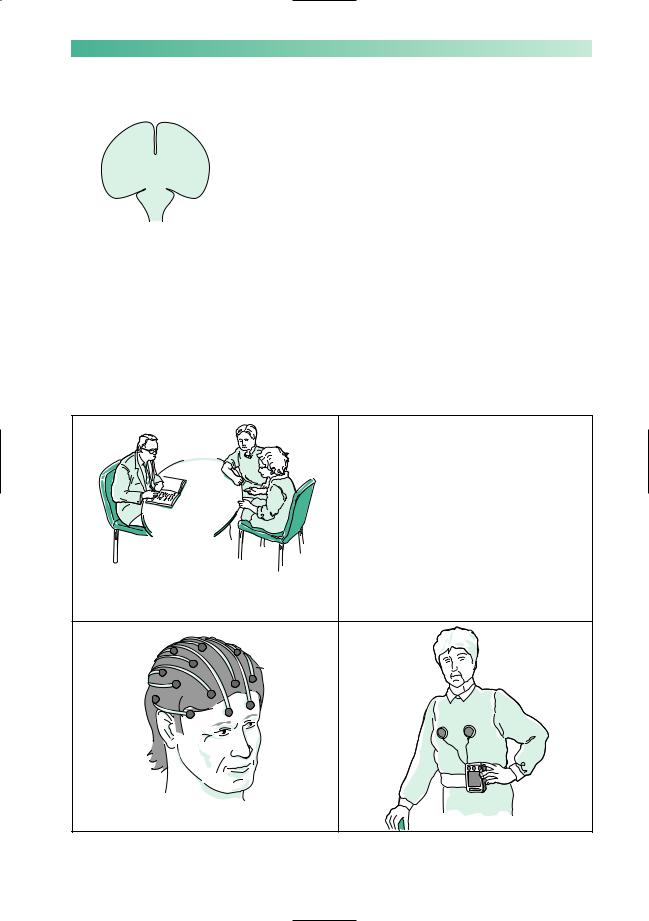
Making the diagnosis in a patient with blackouts
Making the diagnosis is best achieved by giving oneself enough time to talk to the patient so that he can describe all that happens
before, during and after the attacks, and similarly by talking to a Which is it? witness so that he can describe all the observable phenomena of
the patient’s attack. Physical examination of the patient with blackouts is very frequently normal, so it cannot be relied upon to yield very much information of use. Occasionally, it may be necessary to admit the patient to hospital so that the attacks may be observed by medical and nursing staff.
The investigations that may prove valuable in patients with blackouts are:
•EEG and ECG, with prolonged monitoring of the brain and heart by these techniques, if the standard procedures are not diagnostic;
•blood glucose and blood gas estimations (ideally at the time of the attacks) may help to prove either hypoglycaemia or hyperventilation as the basis of the attacks.
Often no tests necessary:
 adolescent with a typical vasovagal syncope
 elderly person on medication with demonstrable postural hypotension
'First you tell me what happened, and then we will ask your husband for all the details too'
Standard EEG |
Ambulatory ECG |
182
Treatment of the common causes of blackouts
Apart from a general explanation of the nature of the attacks to the patient and his family, there are two other aspects of management.
Specific treatment suggestions
Vasovagal syncope: lower head at onset.
Postural hypotension: remove offending drug, consider physical and pharmacological methods of maintaining the standing blood pressure (sleep with bed tilted slightly head up, fludrocortisone).
Hyperventilation: reassurance, and exercises to control breathing.
Cardiac arrhythmia: pharmacological or implanted pacemaker control of cardiac rhythm.
Hypoglycaemia: attention to drug regime in diabetics, removal of insulinoma in the rare instances of their occurrence.
Vertebro-basilar TIAs: treat source for emboli, aspirin. Epilepsy: anticonvulsant drugs.
‘Psychogenic non-epileptic’ attacks: try to establish the reason for this behaviour, and careful explanation to the patient.
Care of personal safety
People who are subject to sudden episodes of loss of consciousness:
•should not drive motor vehicles;
•should consider showering rather than taking a bath;
•may not be safe in some working environments which involve working at heights, using power tools, working amongst heavy unguarded machinery, working with electricity wires;
•may have to curtail some recreational activities involving
swimming or heights.
A firm but sympathetic manner is necessary in pointing out these aspects of the patient’s management.
CHAPTER 11
Treatment
Sensible, but often unpopular restrictions

UNCONSCIOUSNESS |
183 |
•Sudden irresistible need to sleep, for short periods
•Legs give way, when highly amused or angry
Ashort period, lasting hours, of very selective memory loss, other cerebral functions remaining intact
Before leaving the causes of blackouts, there are two rare neurological conditions to mention briefly. One condition predisposes the patient to frequent short episodes of sleep, narcolepsy, and the other gives rise to infrequent episodes of selective loss of memory, transient global amnesia.
Narcolepsy
In this condition, which is familial and very strongly associated with the possession of a particular HLA tissue type, the patient has a tendency to sleep for short periods, e.g. 10–15 minutes. The sleep is just like ordinary sleep to the observer, but is unnatural in its duration and in the strength with which it overtakes the patient. Such episodes of sleep may occur in circumstances where ordinary people feel sleepy, but narcoleptic patients also go to sleep at very inappropriate times, e.g. whilst talking, eating or driving.
The condition is associated with some other unusual phenomena:
•cataplexy: transient loss of tone and strength in the legs at times of emotional excitement, particularly laughter and annoyance, leading to falls without any impairment of consciousness;
•sleep paralysis: the frightening occurrence of awakening at night unable to move any part of the body for a few moments;
•hypnogogic hallucinations: visual hallucinations of faces occurring just before falling asleep in bed at night.
Narcolepsy, and its associated symptoms, are helped by
dexamphetamine, clomipramine and modafinil.
Transient global amnesia
This syndrome, which tends to occur in patients over the age of 50, involves loss of memory for a few hours. During the period of amnesia, the patient cannot remember recent events, and does not retain any new information at all. All other neurological functions are normal. The patient can talk, write and carry out complicated motor functions (e.g. driving) normally. Throughout the episode, the patient repeatedly asks the same questions of orientation. Afterwards, the person is able to recall all events up to the start of the period of amnesia, remembers nothing of the period itself, and has a somewhat patchy memory of the first few hours following the episode.
Transient global amnesia may occur a few times in a patient’s life. Its pathological mechanism remains poorly understood, but may be related to that of migraine aura.
184 |
CHAPTER 11 |
Persistent coma
Assessment of conscious level
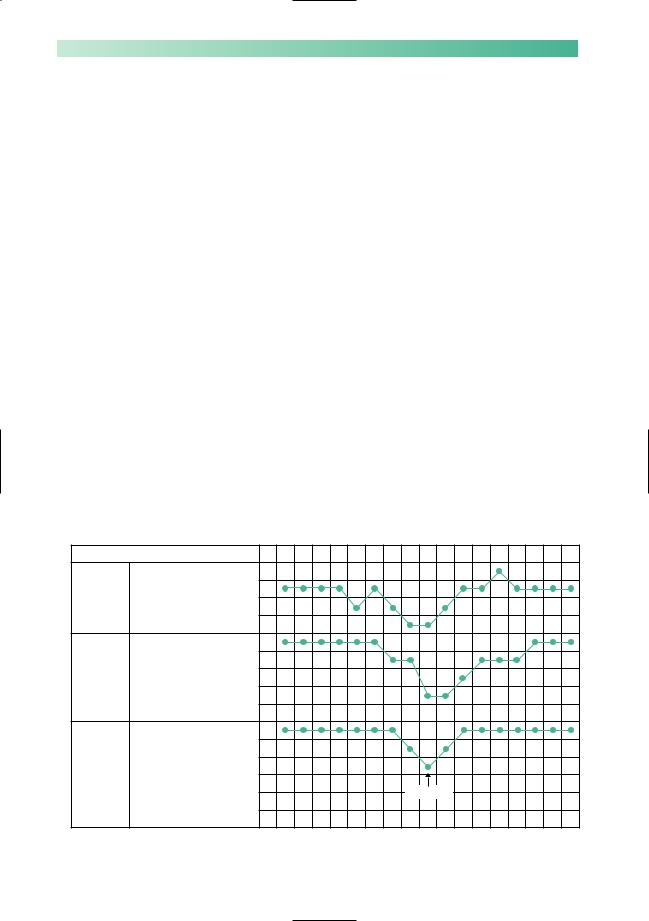
The observations that might be made by someone who finds an unconscious body were mentioned at the beginning of this chapter: ‘in a deep sleep, eyes closed, not talking, not responding to instructions, not moving even when slapped or shaken’. These natural comments have been brought together and elaborated in the Glasgow Coma Scale. This is used very effectively to indicate and monitor a patient’s level of unconsciousness. The Glasgow Coma Scale records the level of stimulus required to make the patient open his eyes and also records the patient’s best verbal and motor responses, as shown in Fig. 11.3. It may be helpful to amplify the scores with a written description of the patient’s precise responses to command or pain, in case subsequent staff are less familiar with the numerical scale.
The responses of the Glasgow Coma Scale depend upon the cerebrum’s response to afferent stimulation. This may be impaired either because of impaired cerebral hemisphere function or because of a major brainstem lesion interfering with access of such stimuli to the cerebral hemispheres. Direct evidence that there is a major brainstem lesion may be evident in an unconscious patient (Fig. 11.4). Dilatation of the pupils and lack of pupillary constriction to light indicate problems in the midbrain, i.e. 3rd cranial nerve dysfunction. Impaired regulation of
|
|
Time |
am |
3 |
4 |
5 |
6 |
7 |
8 |
9 |
10 |
11 |
|
|
Spontaneously |
4 |
|
|
|
|
|
|
|
|
|
|
Eyes |
To speech |
3 |
|
|
|
|
|
|
|
|
|
|
open |
|
|
|
|
|
|
|
|
|||
|
To stimulus |
2 |
|
|
|
|
|
|
|
|
||
|
(E) |
|
|
|
|
|
|
|
|
|||
|
|
|
|
|
|
|
|
|
|
|
|
|
|
|
|
None |
1 |
|
|
|
|
|
|
|
|
|
|
Orientated |
5 |
|
|
|
|
|
|
|
|
|
scale |
Best |
Confused |
4 |
|
|
|
|
|
|
|
|
|
verbal |
Inappropriate words |
3 |
|
|
|
|
|
|
|
|
||
Coma |
response |
Incomprehensible sounds |
2 |
|
|
|
|
|
|
|
|
|
(V) |
|
|
|
|
|
|
|
|
||||
|
|
None |
1 |
|
|
|
|
|
|
|
|
|
|
|
|
|
|
|
|
|
|
|
|
||
|
|
Obey commands |
6 |
|
|
|
|
|
|
|
|
|
|
Best |
Localise stimulus |
5 |
|
|
|
|
|
|
|
|
|
|
Flexion-withdrawal |
4 |
|
|
|
|
|
|
|
|
||
|
motor |
|
|
|
|
|
|
|
|
|||
|
response |
Flexion-abnormal |
3 |
|
|
|
|
|
|
|
|
|
|
(M) |
|
|
|
Treatment |
|
|
|
|
|||
|
Extension |
2 |
|
|
|
|
|
|
|
|||
|
|
|
|
|
|
|
|
|
|
|||
|
|
No response |
1 |
|
|
|
|
|
|
|
|
|
|
|
|
|
Glasgow Coma Scale score at 7am: E1, V2, M4 = 7 |
|
|
|
|||||
Fig. 11.3 Scheme to show the Glasgow Coma Scale. (After Teasdale G. & Jennett B. (1974) Lancet ii, 81–3.)

UNCONSCIOUSNESS
Regulation of pupils
Regulation of Temperature
BP
Pulse
Respiration
Fig. 11.4 Diagram to show important functions of the brainstem.
185
body temperature, blood pressure, pulse and respiration may all indicate trouble in the pons/medulla, where the centres controlling these vital functions exist.
When confronted with a patient in coma, a well-trained doctor will assess:
•vital functions of respiration, temperature control, pulse and blood pressure;
•pupil size and reactivity;
•the patient’s eyes, speech and motor responses according to the Glasgow Coma Scale.
This approach to the assessment of conscious level holds good regardless of the particular cause of coma.
Causes of coma
There is a simple mnemonic to help one to remember the causes of coma (Fig. 11.5). In considering the causes of coma it is helpful to think again in terms of disease processes which impair cerebral hemisphere function generally on the one hand, and in terms of lesions in the brainstem blocking afferent stimulation of the cerebrum on the other. The common causes of coma are illustrated in this way in Fig. 11.6 overleaf.
Fig. 11.5 Asimple mnemonic for the recall of the causes of coma.
|
|
|
A E I O |
U |
A = |
Apoplexy |
Brainstem infarction |
||
|
|
|
Intracranial haemorrhage |
|
E |
= |
Epilepsy |
Post-ictal or inter-ictal coma |
|
|
|
|
Status epilepticus |
|
I |
= |
Injury |
Concussion—major head injury |
|
I |
= |
Infection |
Meningo-encephalitis |
|
|
|
|
Cerebral abscess |
|
O = |
Opiates |
Standing for all CNS depressant drugs, |
||
|
|
|
including alcohol |
|
U = |
Uraemia |
Standing for all metabolic causes for |
||
|
|
|
coma. Quite a useful way of |
|
|
|
|
remembering all possibilities here is |
|
|
|
|
to think of coma resulting from |
|
|
|
|
extreme deviation of normal blood |
|
|
|
|
constituents |
|
|
|
|
Oxygen |
Anoxia |
|
|
|
Carbon dioxide |
Carbon dioxide narcosis |
|
|
|
Hydrogen ions |
Diabetic keto-acidosis |
|
|
|
Glucose |
Hypoglycaemia |
|
|
|
Urea |
Renal failure |
|
|
|
Ammonia |
Liver failure |
|
|
|
Thyroxine |
Hypothyroidism |
|
|
|
|
|

186 |
|
|
CHAPTER 11 |
||
|
|
|
|
|
|
|
|
|
|
Swollen |
|
|
|
|
|
tight |
|
|
|
|
|
|
|
|
Mechanism of coma |
|
|
|
|
|
Generalized impairment |
Major primary pathology |
Unilateral cerebral |
Generalized impairment |
|
|
of cerebral hemisphere |
in the brainstem, |
hemisphere mass lesion, |
of cerebral hemisphere |
|
|
function, leading to a |
depriving the cerebral |
causing downward |
function, associated |
|
|
sub-standard response |
hemispheres of their |
herniation of the medial |
with bilateral cerebral |
|
|
to normal afferent |
normal afferent |
part of the temporal |
hemisphere swelling. |
|
|
stimulation |
stimulation |
lobe through the tent- |
Bilateral medial temp- |
|
|
|
|
orial hiatus, which results |
oral herniation occurs. |
|
|
|
|
in a sideways and down- |
Downward shift of the |
|
|
|
|
ward shift of the brain- |
brainstem occurs at the |
|
|
|
|
stem. This situation of |
level of the midbrain |
|
|
|
|
secondary brainstem |
(tentorial hiatus) and |
|
|
|
|
malfunction is one form |
medulla (foramen |
|
|
|
|
of 'coning', and explains |
magnum). Coma is |
|
|
|
|
such patients' coma. |
due to both generalized |
|
|
|
|
It may progress to |
impairment of cerebral |
|
|
|
|
medullary coning at |
hemisphere function and |
|
|
|
|
foramen magnum level, |
coning at midbrain and |
|
|
|
|
if the mass lesion is left |
medullary levels. |
|
|
|
|
untreated. |
|
|
|
|
|
|
|
|
|
Cause of coma |
|
|
|
|
|
Overdose of CNS |
Brainstem infarction by |
Haematoma |
Brain trauma |
|
|
sedative drugs |
basilar artery |
Abscess |
Meningo-encephalitis |
|
|
Severe alcoholic |
occlusion |
Tumour |
Cerebral anoxia or |
|
|
intoxication |
Brainstem haemorrhage, |
|
ischaemia |
|
|
Diabetic comas |
as occurs in severe |
|
Status epilepticus |
|
|
Renal failure |
hypertension |
|
|
|
|
Hepatic failure |
|
|
|
|
|
|
|
|
|
|
|
Assessment of coma |
|
|
|
|
|
Glasgow Coma Scale |
These patients have a |
These patients have |
These patients have |
|
|
works really well |
multitude of abnormal |
the signs of a unilateral |
the signs of bilateral |
|
|
in these patients, since |
neurological signs, since |
cerebral hemisphere |
cerebral hemisphere |
|
|
there is no focal |
the major brainstem |
lesion and raised intra- |
malfunction and raised |
|
|
neurological damage, |
lesion is causing |
cranial pressure |
intracranial pressure |
|
|
and therefore no later- |
malfunction in the: |
(papilloedema). In |
(papilloedema). They |
|
|
alizing or focal signs. In |
descending motor |
addition the signs of |
too may show signs of |
|
|
severe instances, the |
pathways |
coning (pupillary dilata- |
coning |
|
|
noxious process may |
ascending sensory |
tion and impaired |
|
|
|
involve the brainstem as |
pathways |
regulation of vital |
|
|
|
well as the cerebral |
pathways to and from |
functions) may appear |
|
|
|
hemispheres. Signs of |
the cerebellum |
|
|
|
|
depressed brainstem |
cranial nerve nuclei |
|
|
|
|
function appear . . . |
centres regulating vital |
|
|
|
|
impaired pupils and |
functions |
|
|
|
|
impaired regulation of |
|
|
|
|
|
vital functions |
|
|
|
|
|
|
In these patients, the assessment of eyes, speech and motor responses, |
|
||
|
|
needed for the Glasgow Coma Scale, is somewhat interfered with, because |
|
||
|
|
of the presence of the primary neurological deficit produced by the |
|
||
|
|
primary CNS pathology. In such instances, the best eye, speech and limb |
|
||
|
|
response which can be achieved (in either of the two eyes, or in any of the |
|
||
|
|
four limbs) is the one which is used for the Coma Scale assessment. Despite |
|
||
|
|
this interference the Coma Scale, charted at intervals as shown in Fig. 11.3, |
|
||
|
|
provides a very valuable guide to an unconscious patient's progress |
|
||
Fig. 11.6 Scheme to show the common causes of coma.
UNCONSCIOUSNESS
•Airway
•Level of coma
•Cause of coma
•Cautionoverlumbarpuncture
•Treat cause
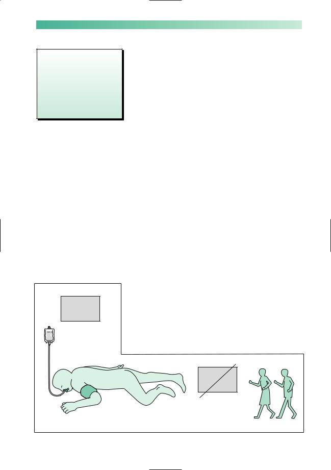
•Routine care of unconscious patient
Fig. 11.7 Diagram to show the routine care of the unconscious patient.
(a)
Chart of observations
187
Investigation and management of a patient in coma
1.Check that the patient’s airway is clear, that breathing is satisfactory, and that the patient’s colour is good.
2.Assess the level of coma, looking at vital functions, pupils and items of the Glasgow Coma Scale as mentioned above.
3.Try to establish the cause of the coma by taking a history from relatives or witnesses, physical examination and appropriate tests. It is obviously important to have the common causes of coma (A, E, I, O, U) clearly in one’s mind whilst questioning, examining and ordering investigations.
4.Remember the danger of lumbar puncture in patients in whom coning is imminent or present (see p. 44). Reduction of the CSF pressure below the foramen magnum may encourage further downward herniation at the tentorial or foramen magnum level, so no lumbar puncture if papilloedema is present, or until a head scan has excluded a mass lesion or any brain swelling.
5.Treat the specific cause of the coma as soon as this is established, e.g. anticonvulsants for epilepsy, antibiotics for meningitis, intravenous glucose for hypoglycaemia, specific antagonists for specific CNS depressant drug poisonings.
6.Establish the routine care of an unconscious patient regardless of the cause (Fig. 11.7).
(a)Observations: assessment every 15–30 minutes of vital functions, pupils and Glasgow Coma Scale, to monitor improvement or deterioration in the patient’s condition.
(b)Airway, ventilation, blood gases.
(c)Blood pressure, to maintain adequate perfusion of the body, particularly of the brain and kidneys.
(d)Fluid and electrolyte balance.
(e)Nutrition and hydration.
(f)Avoidance of sedative or strong analgesic drugs.
(g)General nursing care of eyes, mouth, bladder, bowels, skin and pressure areas, passive limb mobilization to prevent venous stagnation and contractures, chest physiotherapy.
(e) Nutrition by nasogastric tube
(f) |
(g) |
PX morphine
|
(c) Blood |
(b) Airway |
pressure |
(d) Electrolytes
Nurse Physio

188
Prognosis for coma
Patients whose coma has been caused by drug overdose may remain deeply unconscious for prolonged periods and yet have a satisfactory outcome. They may need respiratory support during their coma if brainstem function is depressed, but proceed to make a full recovery.
Prolonged coma from other causes has a much less satisfactory outcome. As an example of this, if one takes a group of unconscious patients, whose coma is not due to drug overdose, who:
•show no eye opening (spontaneous or to voice),
•express no comprehensible words,
•fail to localize painful stimuli,
•remain like this for more than 6 hours,
more than 50% of them will die, and recovery to independent existence will occur in a minority of the rest. This background knowledge must remain in the doctor’s mind when counselling relatives of patients in coma, and when planning the care of patients in prolonged coma.
Some patients who fail to recover from coma enter a state of unawareness of self and environment, in which they can breathe spontaneously and show cycles of eye closure and opening which may simulate sleep and waking. This is known as the vegetative state, which can become permanent. It is difficult, and takes a long time, to determine the precise state of awareness in such unfortunate patients.
Brainstem death
Deeply unconscious patients whose respiration has to be maintained on a ventilator, whose coma is not caused by drug overdose, clearly have a worsening prognosis as each day goes by. Amongst this group of patients, there will be some who have a zero prognosis, whose brainstems have been damaged to such an extent that they will never breathe spontaneously again.
Guidelines exist to help doctors identify patients who have undergone brainstem death whilst in coma which has been supported by intensive care and mechanical ventilation (Fig. 11.8). Testing for brainstem death must be done by two independent, senior and appropriately trained specialists if treatment is to be withdrawn.
CHAPTER 11

UNCONSCIOUSNESS |
|
189 |
|
|
|
|
|
Preconditions |
|
|
|
In coma on ventilator |
|
The patient is deeply comatose, and maintained on a ventilator on account |
|
|
|
of failure of spontaneous respiration |
|
Diagnosis certain |
|
The coma is due to irreversible structural brain damage. The diagnosis is |
|
|
|
certain, and is a disorder which can lead to brainstem death |
|
No drugs |
|
Any of which might be having a reversible effect on the brainstem |
|
No hypothermia |
|
|
|
No metabolic abnormality |
|
|
|
No paralytic drugs |
|
The patient's unresponsiveness is not due to neuromuscular paralytic agents |
|
|
|
|
|
Tests |
|
|
|
Pupils |
1 |
1 Midbrain not working |
|
Doll's head and caloric- |
2 |
Midbrain and pons |
|
induced eye movement |
2 not working |
||
Corneal reflex |
3 |
3 Pons not working |
|
Gag and tracheal reflex |
4 |
4 Medulla not working |
|
Motor responses in cranial |
|
Midbrain, pons and |
|
nerve territory on painful |
|
||
5 |
5 medulla not working |
||
stimulation of the limbs |
|||
|
|||
No respiratory movements |
|
6 Medulla not working |
|
when PaCO2 rises above |
6 |
|
|
6.65kPa (off ventilator) |
|
||
|
|
|
Fig. 11.8 Scheme to show the guidelines that exist to help identify those patients who have undergone brainstem death whilst in coma.