Wilkinson. Essential Neurology 2005
.pdf30
Management of cerebral ischaemia and infarction
There are three aspects of management, which are carried out simultaneously.
Confirmation of diagnosis
ACT brain scan is usually required to exclude with certainty the alternative diagnosis of intracerebral haemorrhage.
Optimization of recovery
Thrombolytic therapy, for example with tissue plasminogen activator, has been shown to improve outcome if given within 3 hours of symptom onset. The great majority of patients come to hospital later than this, and studies to determine the role of thrombolysis in this group are under way. Acute aspirin treatment is of modest but definite benefit, probably because of its effect on platelet stickiness.
There is also good evidence that outcome is also improved by management in a dedicated stroke unit. This effect is probably attributable to a range of factors, including:
•careful explanation to the patient and his relatives;
•systematic assessment of swallowing, to prevent choking and aspiration pneumonia, with percutaneous gastrostomy feeding if necessary;
•early mobilization to prevent the secondary problems of pneumonia, deep vein thrombosis, pulmonary embolism, pressure sores, frozen shoulder and contractures;
•early socialization to help to prevent depression, and help the acceptance of any disability; and treatment of depression when this occurs;
•management of blood pressure, avoiding over-enthusiastic treatment of hypertension in the first 2 weeks or so (when cerebral perfusion of the ischaemic area is dependent upon blood pressure because of impaired autoregulation), moving towards active treatment to achieve normal blood pressure thereafter;
•early involvement of physiotherapists, speech therapists and occupational therapists with a multidisciplinary team approach;
•good liaison with local services for continuing rehabilitation, including attention to the occupational and financial consequences of the event with the help of a social worker.
Prevention of further similar episodes
This means the identification and treatment of the aetiological factors already mentioned. The risk of further stroke is significantly diminished by blood pressure reduction, with a thiazide
CHAPTER 2
Large, wedge-shaped, low density area representing tissue infarction by a right middle cerebral artery occlusion.
Recovery from stroke
•? Thrombolysis
•Aspirin
•Stroke unit
•Attention to swallowing
•Explanation
•Mobilization
•Socialization
•Careful BP management
•Paramedical therapists
•Rehabilitation
STROKE |
31 |
Significant stenosis by atheroma at the origin of the internal carotid artery.
Stroke prevention
•Lower blood pressure with thiazide ± ACE inhibitor
•Statin
•Aspirin
•Warfarin for those with a cardiac source of emboli
•Carotid endarterectomy for ideally selected patients
•Identify and treat ischaemic heart disease and diabetes
•Discourage smoking
diuretic and an angiotensin-converting enzyme (ACE) inhibitor, even in patients with normal blood pressure, and also by statin therapy even if the cholesterol level lies within the normal range.
Investigations should include a full blood count, ESR (if elevated consider vasculitis, endocarditis, atrial myxoma or secondary infection), fasting glucose and lipids, and a search for cardiac sources of emboli. This starts with a careful cardiac examination, ECG and chest X-ray, supplemented by transthoracic or transoesophageal echocardiography if there is any suspicion of a cardiac source.
If the stroke is in the carotid territory, and especially if the patient has made a reasonable recovery and is otherwise healthy, further investigation should be undertaken to see if the patient might benefit from carotid endarterectomy. The patients who benefit most from such surgery are those with localized atheroma at the origin of the internal carotid artery in the neck, producing significant (over 70%) stenosis of the arterial lumen. The severity of stenosis can be established noninvasively by Doppler ultrasound or MR angiography, supplemented if necessary by catheter angiography. The presence or absence of a carotid bruit is not a reliable guide to the degree of stenosis. Well-selected patients in first-class surgical hands achieve a definite reduction in the incidence of subsequent ipsilateral stroke.
Antiplatelet drugs (e.g. aspirin) reduce the risk of further strokes (and myocardial infarction). Anticoagulation with warfarin is of particular benefit in atrial fibrillation and where a cardiac source of embolization has been found.
Despite all this, some patients continue to have strokes and may develop complex disability. Patients with predominantly small vessel disease may go on to suffer cognitive impairment (so-called multi-infarct dementia, described on p. 231) or gait disturbance with the marche à petits pas (walking by means of small shuffling steps, which may be out of proportion to the relatively minor abnormalities to be found in the legs on neurological examination). Patients with multiple infarcts arising from large vessels tend to accumulate physical deficits affecting vision, speech, limb movement and balance. Some degree of pseudobulbar palsy (with slurred speech, brisk jaw-jerk and emotional lability) is common in such cases due to the bilateral cerebral hemisphere involvement, affecting upper motor neurone innervation of the lower cranial nerve nuclei (see p. 134).
32 |
CHAPTER 2 |
Subarachnoid haemorrhage and intracerebral haemorrhage
Anatomy and pathology
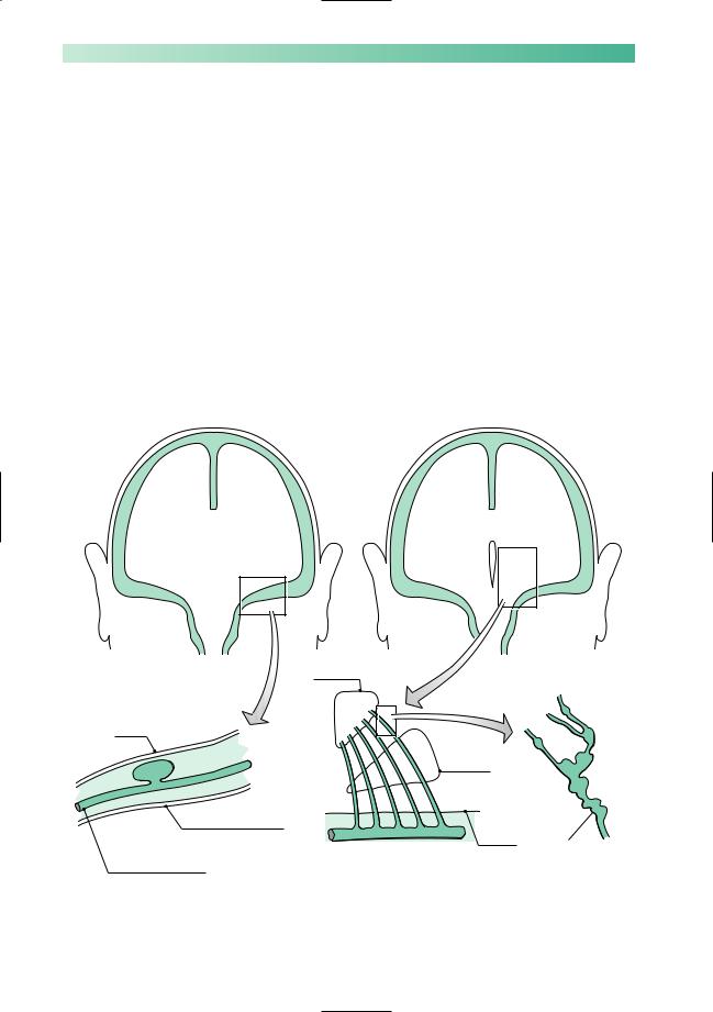
The pathological process here is the sudden release of arterial blood, either into the subarachnoid space around the brain, or directly into the substance of the brain. In subarachnoid haemorrhage the bleeding usually comes from a berry aneurysm arising from one of the arteries at the base of the brain, around the circle of Willis (Fig. 2.5, left). In middle-aged, hypertensive patients, intracerebral haemorrhage tends to occur in the internal capsule or the pons, because of the rupture of long thin penetrating arteries (Fig. 2.5, right). In older patients, intracerebral haemorrhages occur more superficially in the cerebral cortex as a result of cerebral amyloid angiopathy. Arteriovenous malformations of the brain are a rare cause of either subarachnoid or intracerebral haemorrhage.
|
Thalamus |
|
Brain covered |
|
|
by pia |
|
|
|
|
Corpus |
|
|
striatum |
CSF |
|
|
Bone covered by dura |
|
Surface |
|
|
of brain |
Middle cerebral artery |
Middle cerebral artery |
Micro-aneurysms |
bearing an aneurysm |
giving rise to the long |
on the course of a |
|
thin striate arteries |
striate artery in a |
|
|
hypertensive |
|
|
patient |
Fig. 2.5 Left: Berry aneurysm, the common cause of subarachnoid haemorrhage. Right: Micro-aneurysms, the common cause of intracerebral haemorrhage.

STROKE
Subarachnoid haemorrhage
•Sudden, very severe headache
•Neck stiffness/rigidity
±vomiting
loss of consciousness papilloedema neurological deficit
Intracerebral haemorrhage
•Sudden, severe neurological deficit
•Headache
±vomiting papilloedema
33
Neurological symptoms and signs
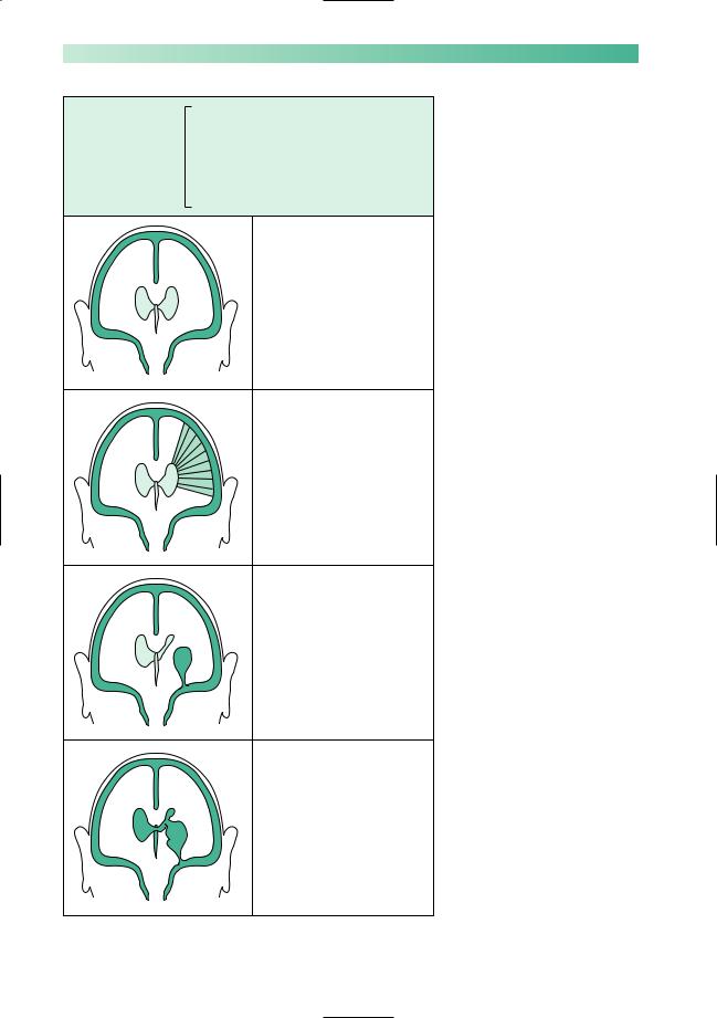
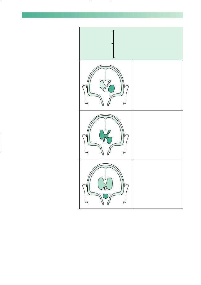
The range of consequences of subarachnoid and intracranial haemorrhage is illustrated in Figs 2.6 and 2.7. Both cause a sudden rise in intracranial pressure, with headache, vomiting and a decrease in conscious level, which may be followed by the development of papilloedema.
In subarachnoid haemorrhage the bleeding irritates the meninges. This causes the characteristic sudden severe headache (‘like being hit on the head with a baseball bat’) and neck stiffness; there is often a brief loss of consciousness at the moment of the bleed. It is the suddenness of onset which helps to differentiate subarachnoid haemorrhage from the headache and neck stiffness of infective meningitis, which come on over a few hours rather than seconds. Migraine can sometimes produce severe headache abruptly but without the severe neck stiffness of subarachnoid haemorrhage.
An intracerebral bleed in the region of the internal capsule will cause sudden severe motor, sensory and visual problems on the contralateral side of the body (hemiplegia, hemianaesthesia and homonymous hemianopia). In the pons, sudden loss of motor and sensory functions in all four limbs, associated with disordered brainstem function, accounts for the extremely high mortality of haemorrhage in this area.
Bleeding into the ventricular system, whether the initial bleed is subarachnoid or intracerebral, is of grave prognostic significance. It is frequently found in patients dying within hours of the bleed.
High blood pressure readings may be found in patients shortly after subarachnoid or intracerebral haemorrhage, either as a response to the bleed or because of pre-existing hypertension. Over-enthusiastic lowering of the blood pressure is not indicated because the damaged brain will have lost its ability to autoregulate. Low blood pressure therefore leads to reduced perfusion of the damaged brain tissue.
34 |
CHAPTER 2 |
1Blood throughout the subarachnoid space, therefore headache and neck stiffness
Common to all cases
of subarachnoid |
|
2 Raised intracranial pressure, therefore the |
|
||
haemorrhage |
|
|
|
|
possibility of depressed conscious level, |
|
|
headache, vomiting, papilloedema |
No brain damage, so no focal neurological deficit
Spasm of a cerebral artery has occurred in the vicinity of an aneurysm, usually a few days after the initial bleed, causing focal ischaemia and neurological deficit
The aneurysm has also bled directly into adjacent brain tissue, forming a haematoma and causing immediate focal neurological deficit
The aneurysm has bled into adjacent brain tissue, and the haematoma has burst into the ventricles. Focal neurological deficit and coma likely
Fig. 2.6 Diagram to show the clinical features of subarachnoid haemorrhage.
STROKE |
|
35 |
|
1 |
Rapid rise in intracranial pressure, therefore |
Common to all cases |
|
the possibility of depressed conscious level, |
|
headache, vomiting, papilloedema |
|
of intracerebral |
|
|
haemorrhage |
2 |
Destruction of brain tissue by the |
|
||
|
|
haematoma producing a focal neurological |
|
|
deficit, appropriate to the site of the lesion |
|
|
In the region of the internal |
|
|
capsule, therefore contralateral |
|
|
hemiplegia, hemianaesthesia |
|
|
and homonymous hemianopia |
Features of an internal capsular bleed, plus rupture of blood into the ventricles with possible subsequent appearance in the CSF. Coma and neck stiffness may result
In the region of the pons, therefore massive neurological deficit (cranial nerve palsies, cerebellar signs and quadriplegia), plus obstructive hydrocephalus making coma extremely likely
Fig. 2.7 Diagram to show the clinical features of intracerebral haemorrhage.

36
Management of subarachnoid haemorrhage
Establish the diagnosis
The first investigation for suspected subarachnoid haemorrhage is an urgent CT brain scan. This will confirm the presence of subarachnoid blood in the great majority of cases. If the scan is normal, a lumbar puncture should be performed to look for blood or xanthochromia (degraded blood) in the CSF. The CSF may be normal if the lumbar puncture is performed within a few hours or more than 2 weeks after the bleed. Alumbar puncture should not be performed if the subarachnoid haemorrhage has been complicated by the development of an intracerebral haematoma, since it may lead to coning (see pp. 42–4).
Prevent spasm and minimize infarction
The calcium channel blocker nimodipine reduces cerebral infarction and improves outcome after subarachnoid haemorrhage. Disturbances of sodium homeostasis are common, and the electrolytes should be monitored regularly.
Prevent re-bleeding
Specialist neurosurgical advice should be sought. Patients who have withstood their first bleed well are offered carotid and vertebral angiography within a few days to establish whether or not a treatable aneurysm is present. Treatment usually involves either surgically placing a clip over the neck of the aneurysm to exclude it from the circulation, or packing of the aneurysm with metal coils, delivered by arterial catheter under radiological guidance, to cause it to thrombose.
Patients with subarachnoid haemorrhages confined to the area in front of the upper brainstem (so-called perimesencephalic haemorrhages) rarely have an underlying aneurysm and have an excellent prognosis without treatment.
Rehabilitation
Many patients who survive subarachnoid haemorrhage (and its treatment) have significant brain damage. A proportion, often young, will not be able to return to normal activities. They will need support from relatives, nurses, physiotherapists, speech therapists, occupational therapists, psychologists and social workers, ideally in specialist rehabilitation units.
CHAPTER 2
CT scan showing high density in the sulci over the right cerebral hemisphere due to subarachnoid blood.
This has come from the large middle cerebral aneurysm arrowed.

STROKE
*
Enhanced CT scan showing high signal in an arteriovenous malformation, with bleeding into the adjacent brain (asterisk) and subarachnoid space.
CT scan showing a recent right parietal haemorrhage and low density from a previous left frontal bleed, due to amyloid angiopathy.
37
Management of intracerebral haemorrhage
Establish the diagnosis
A CT brain scan will confirm the diagnosis and steer you away from thrombolytic therapy which would make the bleeding worse. The location and extent of the bleeding may give a clue to the cause and will determine management.
Lesions in the internal capsule
1.The optimal initial treatment is still a matter of research. It may sometimes be helpful to reduce intracranial pressure, for example with mannitol or by removing the haematoma.
2.Hypertension should be treated gently at first, and more vigorously after a few weeks.
3.Rehabilitation: a major and persistent neurological deficit is to be expected, and all the agencies mentioned under rehabilitation of patients with subarachnoid haemorrhage are likely to be of value.
Lesions in the pons
The mortality and morbidity of lesions in the pons are such as to make active treatment of any sort of questionable medical or ethical merit.
Lesions in the cerebral cortex
If there is a single cortical bleed, especially in a younger patient, then consideration should be given to a search for an underlying arteriovenous vascular malformation. This can be done with MR imaging once the haematoma has resolved. Multiple cortical bleeds in the elderly are usually due to cerebral amyloid angiopathy and are best treated conservatively. There is a high risk of both recurrence and subsequent dementia in this latter group of patients.
The ideal treatment of intracerebral haemorrhage is prophylactic. Intracerebral haemorrhage is one of the major complications of untreated hypertension. There is good evidence to show that conscientious treatment of high blood pressure reduces the incidence of intracerebral haemorrhage in hypertensive patients.

38 |
CHAPTER 2 |
CASE HISTORIES
Case 1
A 42-year-old teacher has to retire from the classroom abruptly on account of the most sudden and severe headache she has ever had in her life. She has to sit down in the staffroom and summon help on the phone.The school secretary calls for an ambulance as soon as she sees the patient, who is normally very robust and is clearly in extreme pain.
On arrival in the hospital A & E department, the patient is still in severe pain. Her BP is 160/100 and she shows marked neck stiffness but no other abnormalities on general or neurological examination. A clinical diagnosis of subarachnoid haemorrhage is made. She is admitted to the medical ward.
a.What investigations would you arrange?
b.How would you proceed now?
Case 2
A 55-year-old publican’s wife is known to suffer from significant hypertension. She is receiving medication from her GP for this, but is not at all compliant. She
smokes, and partakes of a drink. She is very overweight.
She suddenly collapses in the kitchen. She is unrousable except by painful stimuli, is retching and vomiting, and does not move her left limbs at all.
a. What is the most likely diagnosis?
She is taken to hospital by ambulance. Her physical signs in A & E are:
•obese;
•BP 240/140;
•no neck stiffness;
•no response to verbal command, and no speech;
•eyes closed, and deviated to the right when the eyelids are elevated;
•no movement of left limbs to painful stimuli;
•withdrawal of right limbs from painful stimuli;
•extensor left plantar response.
b.What is the most likely diagnosis?
c.What investigations would you arrange?
d.What would you tell her husband?
(For answers, see p. 255.)