

Wilkinson. Essential Neurology 2005
.pdfCHAPTER 3 |
3 |
Brain tumour |
Introduction
Like malignant neoplasms anywhere else in the body, histologically malignant brain tumours carry a poor prognosis. Histologically benign brain tumours are often difficult to remove. This may be the result of a lack of clear boundary between tumour tissue and normal brain substance, e.g. in a low-grade cerebral astrocytoma, or because the tumour lies very close to a part of the brain with important functions, e.g. an acoustic neuroma lying beside the brainstem.
Brain tumours therefore have an unfavourable reputation. It is frustrating that the improvements in our ability to diagnose brain tumours (with better imaging and less invasive biopsy techniques) are only just starting to be accompanied by improvements in our ability to treat them.
Intracranial compartments
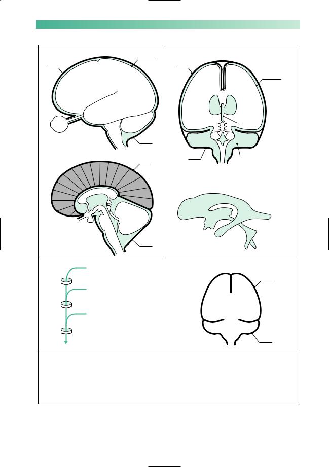
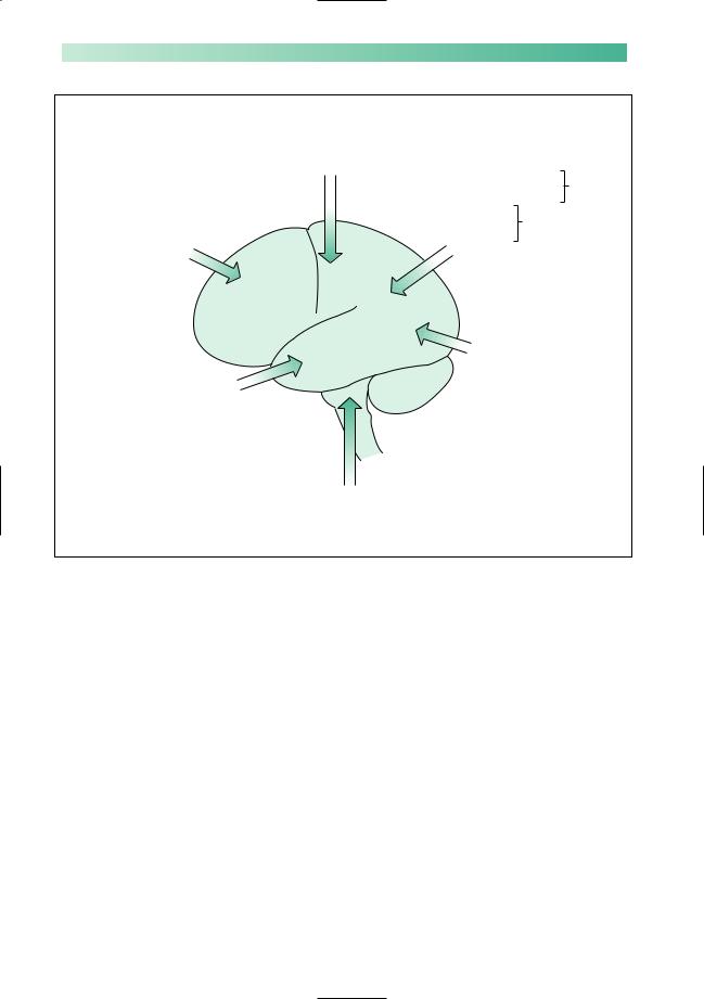
Many of the problems caused by brain tumours arise because the brain lies within a rigid compartmentalized box (Fig. 3.1). The falx cerebri runs vertically from the front to the back of the head. The two compartments on either side each contain a cerebral hemisphere. These are joined together below the front of the falx by the corpus callosum.At the back of the falx, the tentorium cerebelli runs from side to side. Below it is the third compartment, the posterior fossa. This contains the brainstem and the cerebellum. The top of the brainstem (i.e. the midbrain) is continuous with the cerebral hemispheres through a hole in the tentorium, the tentorial hiatus. The bottom of the brainstem (i.e. the medulla) leads to the spinal cord through a hole in the floor of the skull, the foramen magnum.
Figure 3.1 also shows the ventricular system. Cerebrospinal fluid is produced by the choroid plexus in each of the ventricles. It passes down through the ventricular system, leaving the fourth ventricles via the foramina of Luschka and Magendie to enter the subarachnoid space. It then circulates over the surface of the brain and spinal cord before being resorbed.
40
BRAIN TUMOUR |
|
|
|
|
|
|
|
41 |
Lateral aspect of brain |
|
Surface |
Coronal section through brain |
|
||||
|
|
of brain |
(posterior aspect) |
|
|
|
|
|
Dura |
|
|
Dura |
|
|
|
|
Surface |
|
|
|
|
|
|
|
|
|
|
|
|
|
|
|
FC |
|
of brain |
|
|
|
|
LV |
LV |
|
|
|
Eye |
|
|
|
|
|
|
3 |
|
|
|
|
|
|
|
|
|
|
|
|
Dura |
|
TC |
L |
L |
TC |
|
|
|
|
|
|
4 |
|
|
|
Median section through brain |
|
|
|
Dura |
|
M |
|
|
|
Dura |
|
|
|
Cerebellum removed to |
|||
|
|
|
|
|
|
|||
|
|
|
|
|
|
|
reveal posterior aspect |
|
FC |
|
|
|
|
|
|
of midbrain, cut cerebellar |
|
|
|
|
|
|
|
|
peduncles, floor of 4th |
|
|
|
|
Shape of ventricles |
|
|
ventricle, posterior |
||
CC |
|
|
(viewed from left) |
|
|
aspect of medulla and |
||
|
|
|
|
|
|
spinal cord |
||
3 |
C |
TC |
|
|
|
B |
|
|
C |
|
|
F |
|
|
|
|
|
C |
|
|
FM |
|
3 |
|
O |
|
4 |
|
|
|
|
||||
|
|
|
|
|
|
|
|
|
|
M C |
|
|
|
|
T |
4 |
|
|
Dura |
|
|
|
|
|
|
|
|
|
|
|
|
|
|
|
|
(a) |
|
|
(b) |
|
|
|
L |
M |
Flow of CSF |
|
|
The rigid frame containing the brain |
|
||||
Lateral ventricles which contain |
|
|
|
|
|
|
||
choroid plexus |
|
|
|
|
|
|
Dura |
|
Foramina of Munro |
|
|
|
|
|
FC |
||
|
|
|
|
|
|
|||
3rd ventricle which contains |
|
|
|
|
|
|||
|
|
|
|
|
|
|||
choroid plexus |
|
|
|
|
|
|
|
|
Aqueduct |
|
|
|
|
|
|
|
|
4th ventricle which contains |
|
|
|
|
|
|
||
choroid plexus |
|
|
|
|
|
|
|
|
Foramina of Luschka and |
|
|
|
|
|
TC |
|
|
Magendie |
|
|
|
|
|
|
|
Dura |
Subarachnoid space |
|
(c) |
|
|
|
|
||
|
|
|
|
|
|
|||
Key
L= Foramen of Luschka
M= Foramen of Magendie
F |
= Frontal horn |
|
|
Of lateral ventricle |
|
B = Body |
|
||||
|
within each cerebral |
||||
O = Occipital horn |
|
||||
|
hemisphere |
||||
T |
= Temporal horn |
|
|
|
|
LV |
= |
Lateral ventricle |
3 |
= 3rd ventricle |
|
|
Interconnected |
FM = Foramen of Munro |
4 |
= 4th ventricle |
|
|
by aqueduct |
||
|
|
|
|||||
FC |
= |
Falx cerebri |
C |
= Cisterns, i.e. spaces filled by |
|||
CC |
= Corpus callosum |
|
CSF in the subarachnoid space |
||||
TC |
= Tentorium cerebelli |
|
around the base of the brain |
||||
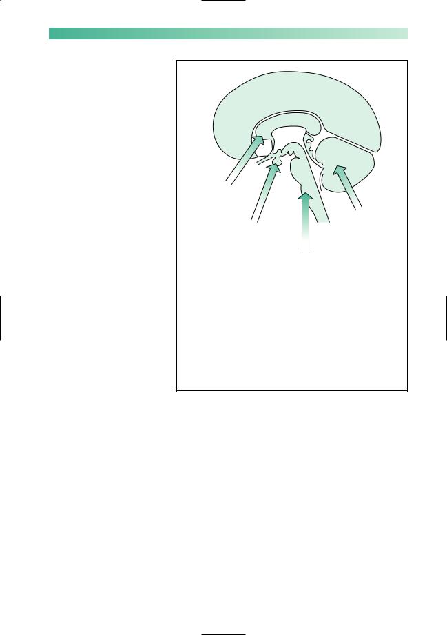
Fig. 3.1 (a) Diagrams to show the lateral aspect of, and a median section through, the brain. (b) Diagrams to show a coronal section through the brain (posterior aspect) and the shape of the ventricles as viewed from the left. (c) Diagram to show the rigid frame containing the brain.
42 |
CHAPTER 3 |
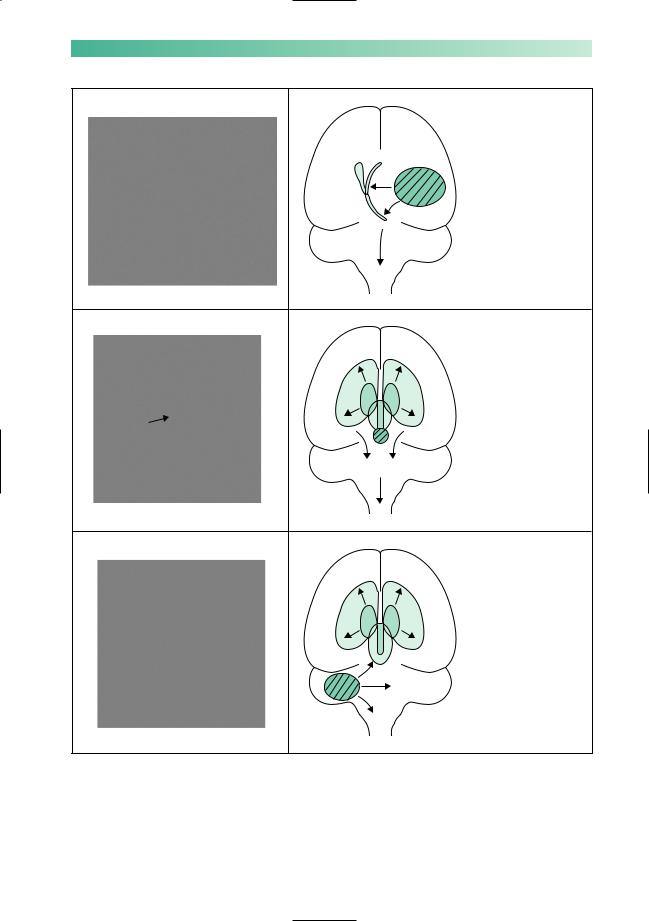
Tentorial herniation, coning and shift
Figure 3.2 shows the influence of mass lesions situated in different parts of the compartmentalized space within the skull. When a mass lesion is making one cerebral hemisphere too large for its compartment (Fig. 3.2a):
•the supratentorial midline structures (corpus callosum and 3rd ventricle) are pushed towards the opposite side of the skull below the falx;
•the infero-medial part of the cerebral hemisphere is pushed through the tentorial hiatus (compressing the midbrain);
•the whole brainstem is pushed downwards so that the lowermost parts of the cerebellum and medulla oblongata become impacted in the foramen magnum.
The movement at the tentorial hiatus is known as tentorial
herniation, and the impaction at the foramen magnum is known as coning of the medulla. They commonly occur simultaneously. The effects on the patient are:
•depression in conscious level (distortion of the reticular formation lying throughout the whole of the brainstem);
•an impairment of ipsilateral 3rd nerve function and dilatation of the pupil (tentorial herniation compressing the midbrain);
•interference with the vital functions of respiration and circulation (compression of the medulla oblongata).
A mass lesion situated in the midline (Fig. 3.2b) causes ob-
struction to the downward flow of CSF through the ventricular system. Under such circumstances, the ventricles above the site of obstruction dilate, and both cerebral hemispheres become too large for their compartments. Bilateral tentorial herniation and coning are likely to occur with the same dangerous clinical consequences.
In the presence of a unilateral posterior fossa mass lesion (Fig. 3.2c), there is movement of the midline posterior fossa structures to one side. This may compress the 4th ventricle sufficiently to block the downward flow of CSF, resulting in ventricular dilatation above the site of obstruction. There will be downward movement and compression at the level of the foramen magnum. At the tentorium cerebelli, there may be upward movement and compression of the midbrain or, if the supratentorial ventricular dilatation becomes very marked there may be downward herniation bilaterally. Depression of conscious level, dilated pupils and impaired vital functions may all result from such a lesion.
Coning
Cause:
•usually downward movement of brainstem
Effects:
•Ø conscious level
•Ø pupils’ reaction to light
•Ø vital functions including breathing
Aggravated:
• by lumbar puncture
BRAIN TUMOUR |
43 |
Brain shift, secondary to a unilateral cerebral hemisphere mass lesion
(a)
Ventricular dilatation and brain shift secondary to a midline mass lesion obstructing the flow of CSF
(b)
Brain shift and ventricular dilatation secondary to a unilateral posterior fossa mass lesion
(c)
Fig. 3.2 (a) Brain shift secondary to a unilateral cerebral hemisphere mass lesion. (b) Ventricular dilatation and brain shift secondary to a midline mass lesion obstructing the flow of CSF. (c) Brain shift and ventricular dilatation secondary to a unilateral posterior fossa mass lesion.
44
Lethal lumbar puncture
Lumbar puncture is dangerous if intracranial pressure is raised due to a mass lesion. It reduces the CSF pressure below the foramen magnum. This can encourage downward brain shifts with tentorial herniation and coning of the medulla. This in turn causes progressive loss of consciousness and impaired control of breathing, which may be ultimately fatal. Lumbar puncture should not, therefore, be performed where there is known to be a mass lesion of sufficient size to cause raised intracranial pressure, or in situations where the possibility of one exists. Examples of these situations include patients with focal deficit (such as a hemiparesis), patients with papilloedema, and patients who are in a coma of unknown cause. In all these situations, the cause should be clarified with brain imaging before a lumbar puncture is contemplated.
On the other hand, where headache and papilloedema are due to a general elevation of intracranial pressure without any mass lesion, e.g. in meningitis and uncomplicated subarachnoid haemorrhage, lumbar puncture is safe and may relieve symptoms.
False localizing signs
We have seen that a mass lesion in one compartment of the brain can induce shift and compression in parts of the brain remote from the primary lesion. Brain tumours that are causing raised intracranial pressure are known to produce false localizing signs, which are no more than clinical evidence of these secondary movements of brain tissue:
•the descent of the brainstem may stretch the 6th cranial nerve to produce a non-localizing lateral rectus palsy:
•the ventricular dilatation above midline CSF obstructive lesions (Fig. 3.2b), or above posterior fossa lesions (Fig. 3.2c), may produce:
—intellectual and behavioural changes suggestive of primary frontal pathology;
—an interference with vertical eye movements (which are programmed in the upper midbrain) because of the dilatation of the posterior part of the 3rd ventricle and aqueduct;
•the impairment of conscious level, pupillary dilatation and depression of vital functions, mentioned already in this chapter, are the most pressing false localizing signs and demand immediate action by doctors in charge of the case.
CHAPTER 3
Practical tip
1.Always measure the CSF pressure at the start of a lumbar puncture
2.Make sure the patient is relaxed and not too tightly curled
3.If the pressure is unexpectedly elevated
(>25 cm of CSF):
•collect the sample in the manometer
•take out the LP needle
•secure IV access in case you need to give mannitol
•start neurological observations at 15-minute intervals
•arrange an urgent brain scan
•consider obtaining neurosurgical advice
False localizing sings
•Sixth nerve palsy
•‘Frontal’ signs
•Impaired up-gaze
•Signs of coning

BRAIN TUMOUR
Clinical features of brain tumours
•Raised intracranial pressure
•Epilepsy
•Evolving neurological deficit
45
Clinical features
There are three groups of symptoms and signs resulting from brain tumours: raised intracranial pressure, epilepsy and an evolving focal neurological deficit.
Raised intracranial pressure
The cardinal features of raised intracranial pressure are:
•headache;
•vomiting;
•papulloedema;
•false localizing signs;
•depression of conscious level;
•signs of tentorial herniation and coning.
Only two further clinical points need to be made about these
features.
1.The headaches of raised intracranial pressure tend not to be extremely severe, they do keep on troubling the patient, they are usually generalized throughout the head, and they tend to be worse in the mornings when the patient wakes. They sometimes wake the patient earlier than his normal waking time. They may be made worse by coughing, straining and bending. Unfortunately, like many things in clinical medicine, none of these features is absolutely specific.
2.Perfusion of the retina and optic disc may become critical in the presence of raised intracerebral pressure and papilloedema. The patient may report transient blurring or loss of vision. Such visual obscurations should stimulate urgent investigation and treatment.
Epilepsy
Focal epilepsy, focal epilepsy progressing on to a generalized tonic–clonic seizure, tonic–clonic seizures with post-ictal focal neurological signs, and tonic–clonic epilepsy without any apparent focal features may all indicate the presence of a tumour in the cerebrum. (Focal epilepsy is discussed in detail on pp. 196–8). Epilepsy is not a feature of posterior fossa tumours.
Epilepsy is not commonly caused by tumours, and less than 50% of cerebral tumours produce epilepsy, but the occurrence of epilepsy in adult life should prompt the possibility of a brain tumour in the doctor’s mind.
46 |
CHAPTER 3 |
Frontal
(Tend to present late since tumours here can become quite large before producing a definite neurological deficit)
Dementia Alteration of mood
Alteration of behaviour Incomplete insight Incontinence Olfactory or optic
nerve malfunction
Central
(Tend to present early) Contralateral limb weakness Contralateral sensory loss Dysphasia if dominant hemisphere
Temporal
Dysphasia if dominant hemisphere Memory impairment
Alteration in mood Alteration in behaviour Contralateral homonymous
upper quadrant visual field defect or neglect (see pp. 113–14)
Lateral posterior fossa e.g. Acoustic neuroma Cranial nerve palsies 8, 7, 5 Ipsilateral cerebellar deficit
Parietal |
If non- |
|
Spatial disorientation |
||
dominant |
||
Dressing dyspraxia |
hemisphere |
Dyslexia |
If dominant |
|
Dysgraphia |
||
hemisphere |
||
Dyscalculia |
||
|
||
Neglect of contralateral limbs |
||
Contralateral homonymous |
||
lower quadrant visual field |
||
defect or neglect (see pp. 113–14) |
||
Occipital
Contralateral homonymous hemianopia
Fig. 3.3 Lateral aspect of the brain, showing the neurological deficits produced by tumours at various sites.
An evolving focal neurological deficit
The presence of a tumour impairs the function of the part of the brain in which it resides. The nature of the evolving focal neurological deficit clearly depends on the site of the lesion. Figures 3.3 and 3.4 depict the principal neurological deficits produced by tumours situated in various parts of the brain. Tumours near the midline (Fig. 3.4) and in the posterior fossa may produce marked features of raised intracranial pressure before there are many localizing signs.
BRAIN TUMOUR |
47 |
Bifrontal and corpus callosum
Dementia
Gait disturbance
Pituitary gland Optic chiasm Hypothalamus
Visual field defects Appetite ≠ or Ø Wakefulness Ø Endocrine effects Cranial nerves 3,
4, 6, 5a
Fig. 3.4 Median section through the brain, showing the neurological deficits produced by tumours at various sites.
Cerebellum
Nystagmus
Dysarthria Limb and gait
ataxia
Brainstem
Cranial nerve palsies 3–12, depending on level of tumour Cerebellar deficit, due
to impaired inflow or outflow from the cerebellum
Long tract, motor and sensory, deficits in limbs and trunk
Impaired vital functions i.e. respiration, thermo-regulation and circulation
The length of history of any of the three main features of a brain tumour is a guide to the nature of the tumour, in terms of malignancy. This is not foolproof, however. Certainly a long history suggests a benign or low-grade malignancy tumour. A short progressive history obviously implies malignancy, but sometimes a benign tumour can be silent for years only to produce a short worrying history when the brain and the intracranial compartments finally become unable to absorb the presence of the enlarging mass lesion.
48
Common brain tumours
Gliomas are seen to appear in both the benign and malignant groups of tumours. Astrocytomas are by far the most common glial tumour; tumours derived from oligodendrocytes, ependyma, neurones, primitive neuroectodermal or other tissues are much rarer. Unless otherwise specified, the words ‘primary brain tumour’ or ‘glioma’ refer to an astrocytoma in clinical practice. Gliomas are classified histologically from grade 1 (benign) to grade 4 (the highly malignant glioblastoma multiforme). Benign gliomas are, unfortunately, much less common than malignant ones and have a tendency to become more malignant with time.
Meningiomas are nearly always benign. They may arise from any part of the meninges, over the surface of the brain, from the falx, or from the tentorium. There is a plane of cleavage between tumour and brain tissue which makes total removal a definite possibility, so long as the tumour is reasonably accessible and unattached to dural venous sinuses, e.g. the sagittal sinus.
Pituitary adenomas produce two principal sets of symptoms: space-occupying effects and endocrine disturbance. When the pituitary gland enlarges in the pituitary fossa, it most commonly expands upwards (suprasellar extension) to compress optic nerves/chiasm/tracts. The classical bitemporal hemianopia resulting from chiasmal compression occurs when the optic chiasm is right above a pituitary extension which is directly upwards. The exact position of the optic chiasm, and the direction of pituitary expansion, do, however, vary from one case to another, so monocular blindness due to optic nerve compression and homonymous hemianopia from optic tract compression are not uncommon in patients with pituitary adenomas. Lateral expansion of pituitary adenomas may compress structures on the lateral wall of the cavernous sinus (cranial nerves, 3, 4, 5a and 6), producing double vision and forehead numbness. Forward and downward expansion of the adenoma results in enormous expansion of the pituitary fossa and occasional erosion through bone into the sphenoidal air sinus.
The endocrine disturbances that accompany the development of a pituitary adenoma are positive if the tumour cells are secretory (prolactin, growth hormone, etc.), and negative if the tumour is preventing normal secretion by the rest of the pituitary gland (varying degrees of panhypopituitarism).
CHAPTER 3
Benign
•Grade 1–2 gliomas
•Meningioma
•Pituitary adenoma
•Acoustic neuroma
Malignant
Primary
• grade 3–4 gliomas
Secondary
• metastatic carcinoma
The glioma (top) is poorly differentiated from the surrounding brain, unlike the meningioma (bottom).
BRAIN TUMOUR |
49 |
Pituitary adenoma (arrows).
Acoustic neuroma (arrow).
Metastases (arrows).
Acoustic neuromas are benign tumours of the Schwann cells along the course of the acoustic nerve, between the cerebellopontine angle and the internal auditory meatus in the petrous temporal bone. First and foremost, they produce progressive unilateral nerve deafness, but by the time of recognition there may well be associated 5th and 7th nerve dysfunction, unilateral cerebellar signs and evidence of raised intracranial pressure. Early diagnosis is highly desirable since a small tumour can be treated with radiotherapy or surgery with fewer complications than a large one which has caused brainstem displacement and raised intracranial pressure.
Common malignant tumours in the brain are either gliomas or metastases, in particular malignant astrocytomas and metastatic carcinoma. Together these constitute well over 60% of all brain tumours. The history is usually short, of raised intracranial pressure, epilepsy or neurological deficit. Not uncommonly, all three groups of symptoms are present by the time of diagnosis. It is not uncommon for a primary carcinoma elsewhere in the body to present with metastatic disease in the brain. If the metastases are multiple, the differentiation from malignant glioma is not difficult, but solitary cerebral metastases are quite common.
Differential diagnosis
The common presenting symptoms of brain tumours are:
•raised intracranial pressure;
•epilepsy;
•a progressive focal neurological deficit.
Clearly, other mass lesions within the head may produce all three features. There may be difficulty in differentiating a malignant tumour from an intracerebral abscess when the history is a short one, and from subdural haematoma when the history is a little longer.
Other causes of raised intracranial pressure include severe arterial hypertension, chronic meningitis and so-called benign intracranial hypertension (BIH). BIH is probably due to reduced venous drainage of the brain, and mainly affects young obese women or patients with a predisposition to venous thrombosis.
Epilepsy and focal epilepsy are more usually caused by epileptogenic scars from previous intracranial disease.
The principal alternative cause of a progressive subacute focal neurological deficit is an ischaemic stroke, which occasionally, instead of developing with characteristic abruptness, comes on in a stuttering way.