

Wilkinson. Essential Neurology 2005
.pdf190 |
CHAPTER 11 |
CASE HISTORIES
Case 1
A 78-year-old man is referred after three blackouts. He feels momentarily ‘dizzy’ and then loses consciousness. He comes round on the floor. On the first occasion he sustained a nasty facial laceration, called an ambulance and was taken to the emergency department, where his examination, full blood count, glucose and ECG were all normal.The second happened as he was finishing his lunch and he slumped out of his chair. He recovered rapidly and did not call an ambulance.The third happened when he was walking his dog but he was able to get home unaided. He has not bitten his tongue on any occasion.
He lives alone and none of the attacks has been witnessed. He smokes 10 cigarettes per day. He takes bendrofluazide for hypertension. He is otherwise well.
You find no abnormalities on examination.
a.What is the differential diagnosis?
b.What further questions would you ask?
c.What tests would you request?
Case 2
A young man is brought to the emergency department by ambulance. He has been found unconscious on waste ground.The ambulance staff cannot provide any other history, but he has a recent prescription for the antibiotic oxytetracycline and the anticonvulsant sodium valproate in his wallet.
He is breathing normally and maintaining normal oxygen saturation. His temperature, pulse and blood pressure are normal.There are no external signs of injury. He does not have neck stiffness.There is no spontaneous eye opening, speech or movement. He makes no response to verbal command but briefly opens his eyes, groans and tries to push your hand away in response to painful stimulation.There is no asymmetry in his motor responses.When you open his eyelids, his eyes are roving slowly from side to side. Limb tone and reflexes are normal with flexor plantar responses. General examination is normal apart from acne. His finger-prick glucose is normal.
a.What is your differential diagnosis?
b.How would you manage his case?
(For answers, see pp. 263–4.)


CHAPTER 12 |
12 |
Epilepsy |
Introduction and definitions
The word epilepsy conjures up something rather frightening and undesirable in most people’s minds, because of the apprehension created in all of us when somebody temporarily loses control of his body, especially if unconsciousness, violent movement and impaired communication are involved. Epilepsy is the word used to describe a tendency to episodes, in which a variety of clinical phenomena may occur, caused by abnormal electrical discharge in the brain, between which the patient is his normal self. What actually happens to the patient in an epileptic attack depends upon the nature of the electrical discharge, in particular upon its location and duration.
The most major form of epilepsy is the generalized tonic– clonic seizure, which involves sudden unconsciousness and violent movement, often followed by coma. This form of seizure has traditionally been called grand mal. Unless otherwise stated, mention of an epileptic attack would generally infer the occurrence of a tonic–clonic seizure.
Amongst patients and doctors a single epileptic attack may be called an epileptic fit, a fit, an epileptic seizure, an epileptic convulsion or a convulsion. The words tend to be used synonymously. This is a somewhat inaccurate use of words since a convulsion (violent irregular movement) is one component of some forms of epilepsy. There is also a confusing tendency for older patients and doctors to use the traditional term petit mal for any form of epilepsy which is not too massive or prolonged. Petit mal originally had a rather restricted and specific meaning which has been lost over time; we describe the modern terminology in the following sections. Patients and their families very often use softer words other than epilepsy, fits, seizures or convulsions, which is quite natural. Within families, words like blackout, episode, funny do, attack, blank spell, dizzy turn, fainting spell, trance, daze and petit mal abound when describing epileptic attacks.
Epilepsy causes attacks which are:
•brief
•stereotyped
•unpredictable
192
EPILEPSY |
193 |
We can see that the use of words is often imprecise here. ‘A minor fit’ may mean one thing to one person and another thing to somebody else, and it may certainly be used by a patient or relative to describe any form of epileptic or non-epileptic attack. We must be tolerant of this amongst patients, but there is always a need to clarify exactly what has happened in an individual attack in an individual patient. Errors in management rapidly appear when the doctor does not know the precise features of each of the patient’s attacks.
The word epilepsy is generally used to indicate a tendency to suffer from epileptic attacks, i.e. more than one attack and perhaps an ongoing tendency. Some medical and non-medical people are upset by the use of the word epilepsy when applied to someone who has only ever had one epileptic attack.
Common forms of epilepsy
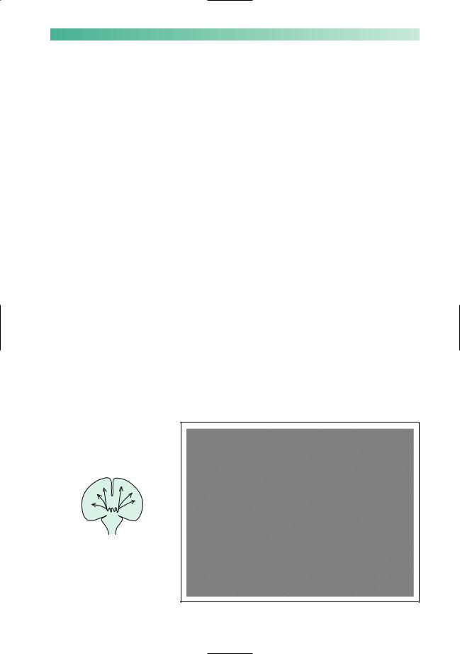
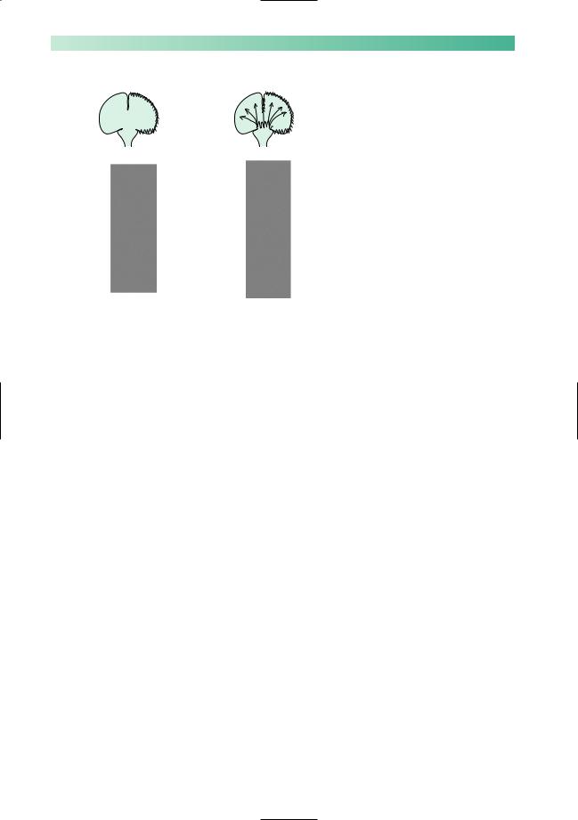
There are two main types of epilepsy: primary generalized and focal. In primary generalized epilepsy, electrical discharges arise within deep midline structures of the brain and spread rapidly and simultaneously to all parts of the cerebral cortex (Fig. 12.1). In focal epilepsy, the electrical discharges start in one part of the cerebral cortex and spread to a greater or lesser extent to other regions (see Figs 12.5 and 12.8).
The distinction between these two main forms of epilepsy is important, because they have different causes and respond to different drugs. Confusion arises because focal seizures can start in one area and eventually spread to all of the cerebral cortex, resulting in a tonic–clonic seizure. This kind of attack is called a secondarily generalized tonic–clonic seizure.
Fig. 12.1 EEG recording typical of primary generalized epilepsy.
194
Primary generalized epilepsy
There are three common manifestations of primary generalized epilepsy:
•primary generalized tonic–clonic seizures;
•absence seizures;
•myoclonic seizures.
Individual patients often have a mixture of these seizure types.
Primary generalized tonic–clonic seizures (Fig 12.2)
•There is no warning or aura.
•Tonic phase. The patient loses consciousness and suddenly stiffens, as all the muscles in his body enter a state of sustained (tonic) contraction. The limbs and neck usually extend. The patient falls stiffly with no attempt to break his fall. He makes a loud groan as air is forced out of the chest through tightened vocal cords. He does not breathe and becomes cyanosed. The tonic phase is brief, usually a matter of seconds, so that observers notice a cry, a fall and stiffening of the body before the clonic phase commences.
•Clonic phase. The sustained muscle contraction subsides into a series of random, disorganized jerks and jitters involving any and all muscles. These convulsive movements are in no way purposeful, coordinated or predictable. The tongue may protrude as the jaw closes, causing tongue biting. Urinary incontinence is frequent. Breathing recommences in the same disorganized way. It is noisy and inefficient and cyanosis usually persists. Saliva accumulates in the mouth which, together with the disorganized respiration, results in froth spilling from the mouth. The patient remains unconscious. The clonic phase typically lasts a minute or two, but can be briefer and can occasionally be very much longer.
•Phase of coma. After the convulsive movements stop, the patient is in coma. Breathing becomes regular and coordinated. The patient’s colour returns to normal so long as the upper airway is clear. The period of time that the patient remains in coma relates to the duration of the previous tonic and clonic phases.
•There follows a state of confusion, headache, restlessness and drowsiness before final recovery. This may last for hours. For a day or two, the patient may feel mentally slow and notice aching pains in the limbs subsequent to the convulsive movement, together with a sore tongue.
CHAPTER 12
No warning
Tonic phase
Loss of consciousness
Tonic contraction
No breathing
Cyanosis
Less than 1 minute
Clonic phase
Still unconscious Convulsive movements Self injury Incontinence
Irregular breathing Cyanosis
Several minutes
Coma
Still unconscious Flaccid
Regular breathing Colour improves
Several minutes
Confusion, headache
Hours
Sore tongue
Aching limbs
Days
Fig. 12.2 Scheme to show typical severe tonic–clonic epilepsy.

EPILEPSY
Photosensitivity
Occasional patients with all the types of primary generalized epilepsy may have seizures triggered by flashing light, for example from the television, computer games or flickering sunshine when driving past a row of trees, and have to avoid such stimuli. Almost all such patients also get spontaneous seizures unrelated to visual stimulation
Whole attack lasts less than 10 seconds
Young person
Sudden onset, sudden end . . .
switch-like
Unaware, still, staring
May occur several times a day
Fig. 12.3 Scheme to show typical absence epilepsy.
195
The description given above is of a severe attack. They may not be as devastating or prolonged. The tonic and clonic phases may be over in a minute and the patient may regain consciousness within a further minute or two. He may feel like his normal self within an hour or so.
During a tonic–clonic epileptic attack, cerebral metabolic rate and oxygen consumption are increased, yet respiration is absent or inefficient with reduced oxygenation of the blood. The brain is unable to metabolize glucose anaerobically, so there is a tendency for accumulation of lactic and pyruvic acid in the brain during prolonged seizure. This hypoxic insult to the brain with acidosis is the probable cause of post-epileptic (post-ictal) coma and confusion.
Absence seizures (Fig. 12.3)
Absence seizures are much less dramatic and may indeed pass unnoticed. The attack is sudden in onset, does not usually last longer than 10 seconds, and is sudden in its ending. The patient is usually able to tell that an episode has occurred only because he realizes that a few moments have gone by of which he has been quite unaware. Conversation and events around him have moved on, and he has no recall of the few seconds of missing time.
Observers will note that the patient suddenly stops what he is doing, and that his eyes remain open, distant and staring, possibly with a little rhythmic movement of the eyelids. Otherwise the face and limbs are usually still. The patient remains standing or sitting, but will stand still if the attack occurs while walking. There is no response to calling the patient by name or any other verbal or physical stimulus. The attack ends as suddenly as it commences, sometimes with a word of apology by the patient if the circumstances have been such as to make him realize an attack has occurred.
Absence seizures usually commence in childhood, so at the time of diagnosis the patient is usually a child or young teenager. It is quite common for absence seizures to occur several times a day, sometimes very frequently so that a few attacks may be witnessed during the initial consultation.
196
Myoclonic seizures (Fig. 12.4)
Myoclonic seizures take the form of abrupt muscle jerks, typically causing the upper limbs to flex and the patient to drop anything that they are holding. The jerks may occur singly or in a brief run. Consciousness is not usually affected. They often happen in the first hour of the day, especially if the patient has consumed alcohol the night before. Myoclonic seizures almost always occur in association with the other generalized seizure types, and their significance in this setting is that they help to confirm a diagnosis of primary generalized epilepsy (most often a specific syndrome called juvenile myoclonic epilepsy). Isolated myoclonic jerks can occur in a wide range of conditions with no relation to epilepsy, including waking and falling asleep in normal people (see Chapter 5, p. 77).
Focal epilepsy
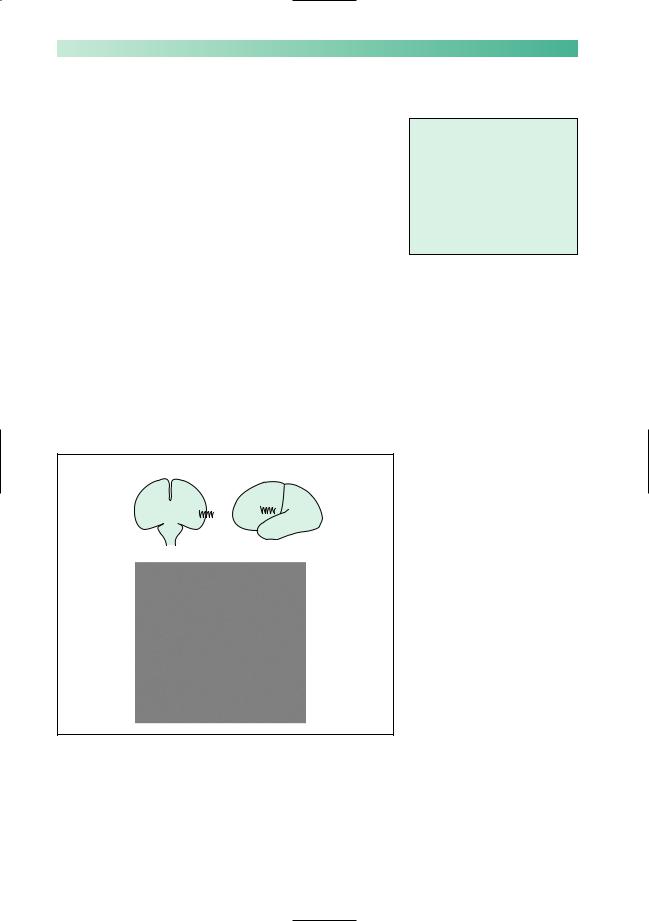
If the primary site of abnormal electrical discharge is situated in one area of the cortex in one cerebral hemisphere, the patient will be prone to attacks of focal epilepsy (Fig. 12.5).
*
*
The phenomena that occur in focal epileptic attacks entirely depend on the location of the epileptogenic lesion. The most obvious form of focal epileptic attack is when the localized epileptic discharge is in part of the motor cortex (precentral gyrus) of one cerebral hemisphere. During the attack, disorganized
CHAPTER 12
One or more brief jerks
Duration up to a few seconds
Consciousness preserved
Coexists with tonic-clonic and absence seizures
Fig. 12.4 Scheme to show typical myoclonic epilepsy.
Fig. 12.5 EEG recording typical of focal epilepsy. In this figure, the focal EEG abnormality might generate focal motor twitching
of the right side of the face. The recordings marked by an asterisk are from adjacent locations of the brain, and show spike discharges.
EPILEPSY |
197 |
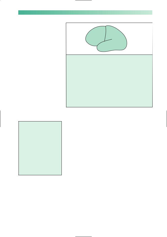
Fig. 12.6 Scheme to show the common forms of focal epilepsy.
Subjective
Déjà vu
Memories rushing through the brain Loss of memory during the attack Hallucination of smell/taste Sensation rising up the body
Objective
Diminished contact with the environment
Slow, confused Repetitive utterances Repetitive movements
(automatisms) Lip-smacking and sniffing
movements
Fig. 12.7 Scheme to show the subjective experiences and objective observations in a patient with temporal lobe epilepsy.
C A B
D
A Focal motor seizures
Strong convulsive movements of one part of the contralateral face, body or limbs
B Focal sensory seizures
Strong, unpleasant, slightly painful, warm, tingling, or electrical sensations in one part of the contralateral face, body or limbs
C Frontal lobe epilepsy
Strong, convulsive turning of the eyes, head and neck towards the contralateral side ('adversive seizure', epileptic activity in the frontal eye fields) or complex posturing with one arm flexed and one arm extended like a fencer (epileptic activity in the supplementary motor area)
D Temporal lobe epilepsy
Strong, disorganized aberration of temporal lobe function (see text and Fig. 12.7)
strong convulsive movement will occur in the corresponding part of the other side of the body. These are focal motor seizures (Fig. 12.6).
Figure 12.6 illustrates the common forms of focal epilepsy.
Focal motor seizures, focal sensory seizures and frontal lobe seizures are quite straightforward. Temporal lobe epilepsy deserves a special mention, not least because it is probably the commonest form of focal epilepsy. It is necessary to remember what functions reside in the temporal lobe in order to understand what might happen when epileptic derangement occurs in this part of the brain.Apart from speech comprehension in the dominant temporal lobe, the medial parts of both temporal lobes are significantly involved in smell and taste function, and in memory (Fig. 12.7).
NB Any indication that a patient’s epilepsy is focal should lead to a search for an explanation of localized cortical pathology, i.e. Why is this area of cerebral cortex behaving in this way? What is wrong with it? In other words, these patients need a brain scan, preferably using MRI.
Focal epileptic discharges may remain focal, but sometimes the abnormal electrical discharge spreads over the surface of the cerebral hemisphere and then triggers generalized epileptic discharges involving all parts of both cerebral hemispheres (Fig. 12.8).
198 |
CHAPTER 12 |
|
|
Fig. 12.8 (a) EEG showing seizure |
|
|
|
discharges that have spread |
|
|
|
across all of the left hemisphere |
|
|
|
(lower eight tracings). (b) Afew |
|
(a) |
(b) |
seconds later the seizure |
|
discharges have become |
|||
|
|
||
|
|
generalized and more intense. |
If the focal discharge begins in one part of the motor cortex, the first symptom might be convulsive twitching of the right side of the face, spreading rapidly into the right arm and then the right leg before affecting the whole body in a secondarily generalized tonic–clonic seizure. This particular kind of attack is known as Jacksonian epilepsy (after the nineteenth-century neurologist Hughlings Jackson).
Any warning or ‘aura’ in the moments before a tonic–clonic seizure indicates that it is a manifestation of focal epilepsy rather than primary generalized epilepsy, with the implication that there is a problem with a localized area of the cerebral cortex which must be investigated. The nature of the aura depends on the location of this area, and it can take any of the forms listed in Figs 12.6 and 12.7. Sometimes the patient himself may not be aware of the warning, if for example it simply consists of turning the eyes and head to the side or extending one limb. This highlights the importance of talking to someone who has seen the patient right through an attack.
The main indication that a tonic–clonic seizure has a focal cause and that the patient does not suffer from primary generalized epilepsy sometimes occurs after the fit rather than before it. Atonic–clonic seizure which is followed by a focal neurological deficit (e.g. weakness of the right side of the body), lasting for an hour or two, strongly indicates a localized cortical cause for the fit (in or near the left motor cortex in this example). The tell-tale post-epileptic unilateral weakness is known as Todd’s paresis. It may be apparent to the patient, but is commonly recognized only by a doctor examining the patient shortly after the tonic–clonic seizure has ceased.

EPILEPSY |
199 |
Febrile convulsions
The young, immature brain of children is less electrically stable than that of adults. It appears that a child’s EEG is made more unstable by fever.
Tonic–clonic attacks occurring at times of febrile illnesses are not uncommon in children under the age of 5 years. Such attacks are usually transient, lasting a few minutes only. In the majority of cases, the child has only one febrile convulsion, further convulsions in subsequent febrile illnesses being unusual rather than the rule.
A febrile convulsion generates anxiety in the parents. The attack itself is frightening, especially if prolonged, and its occurrence makes the parents wonder if their child is going to have a lifelong problem with epilepsy.
The very small percentage of children with febrile convulsions who are destined to suffer from epileptic attacks unassociated with fever later in life are identifiable to a certain extent by the presence of the following risk factors:
•a family history of non-febrile convulsions in a parent or sibling;
•abnormal neurological signs or delayed development identified before the febrile convulsion;
•a prolonged febrile convulsion, lasting longer than 15 minutes;
•focal features in the febrile convulsion before, during or after
the attack.
Those who do go on to have epilepsy in later life usually have a pathology called mesial temporal sclerosis affecting one hippocampus, giving rise to focal epilepsy with temporal lobe seizures. They may be cured by having the abnormal hippocampus surgically resected.