

Wilkinson. Essential Neurology 2005
.pdf140
Prolapsed intervertebral discs are most common between C4 and T1 in the cervical spine and between L3 and S1 in the lumbosacral spine. In the cervical region, there is not a great discrepancy between the level of the cervical spinal cord segment and the cervical vertebra of the same number, i.e. the C5 segment of spinal cord, the C5 nerve roots and the C4/5 intervertebral foramen, through which the C5 spinal nerve passes, are all at much the same level (see Fig. 6.1, p. 83). If the patient presents with a C5 neurological deficit, therefore, it is very likely that it will be a C4/5 intervertebral disc prolapse.
Figures 6.1 and 9.3 show that this is not the case in the lumbar region. The lower end of the spinal cord is at the level of the L1 vertebra. All the lumbar and sacral nerve roots have to descend
CHAPTER 9
Common nerve roots to be compressed by prolapsed intervertebral discs:
In the arm C5 |
In the leg L4 |
C6 |
L5 |
C7 |
S1 |
C8 |
|
Vertebral body
Theca, i.e. spinal dura mater
Intervertebral
disc
Sacrum
Spinal cord
Lumbosacral nerve roots forming the cauda equina, in the subarachnoid space, within the theca
Spinal nerve, with dural sleeve, leaving the spinal canal via an intervertebral foramen
Fig. 9.3 Posterior view of the cauda equina. NB The pedicles, laminae and spinous processes of the vertebrae, and the posterior half of the theca, have been removed.
PERIPHERAL NERVOUS SYSTEM |
141 |
over a considerable length to reach the particular intervertebral foramen through which they exit the spinal canal. These nerve roots form the cauda equina, lying within the theca. Each nerve root passes laterally, within a sheath of dura, at the level at which it passes through the intervertebral foramen. Posterolateral disc prolapses are likely to compress the emerging spinal nerve within the intervertebral foramen, e.g. an L4/5 disc prolapse will compress the emerging L4 root. More medially situated disc prolapses in the lumbar region may compress nerve roots of lower numerical value, which are going to exit the spinal canal lower down. This is more likely to happen if the patient has a constitutionally narrow spinal canal. (Some individuals have wide capacious spinal canals, others have short stubby pedicles and laminae to give a small cross-sectional area for the cauda equina.) It cannot be assumed therefore that an L5 root syndrome is the consequence of an L5/S1 disc prolapse; the trouble may be higher up. A more centrally prolapsed lumbar disc may produce bilateral leg symptoms and signs, involving more than one segment, often associated with sphincter malfunction due to lower sacral nerve root compression.
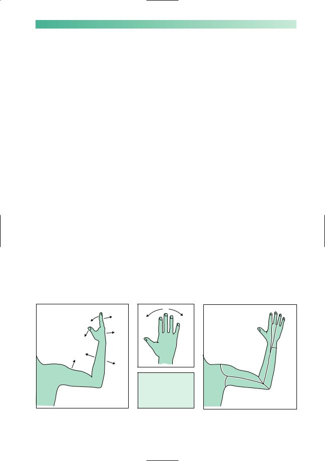
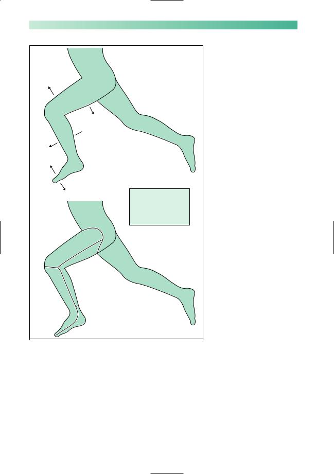
Figures 9.4 and 9.5 show the segmental value of the movements, reflexes and skin sensation most frequently involved in cervical and lumbar disc disease. From these diagrams, the area of pain and sensory malfunction, the location of weakness and wasting, and the impaired deep tendon reflexes can all be identified for any single nerve root syndrome. (Note that Figs 9.4 and 9.5 indicate weak movements, not the actual site of the weak and wasted muscles, which are of course proximal to the joints being moved.)
|
T1 |
|
C7 |
C7 |
|
|
|
C8 |
|
|
|
C7 |
|
|
C8 |
C8 |
|
|
|
|
|
C6 |
|
C5/6 |
|
|
|
|
|
|
|
C5 |
|
|
|
C7/8 |
|
C5 |
T1 |
|
|
||
|
Biceps jerk C5/6 |
|
|
|
Triceps jerk C7/8 |
T2 |
|
|
|
|
|
|
Supinator jerk C5/6 |
|
|
Fig. 9.4 Segmental nerve supply to the upper limb, in terms of movements, tendon reflexes and skin sensation.
142 |
CHAPTER 9 |
L2/3
L4/5/S1
 L5/S1
L5/S1
L3/4
L4/5
S1/2
L2/3
S2
L4/5
S1
Knee jerk L3/4
(NB no jerk for L5)
Ankle jerk S1/2
Fig. 9.5 Segmental nerve supply to the lower limb, in terms of movements, tendon reflexes and skin sensation.
There are four main intervertebral disc disease syndromes.
1.The single, acute disc prolapse which is sudden, often related to unusually heavy lifting or exertion, painful and very incapacitating, often associated with symptoms and signs of nerve root compression, whether it affects the cervical or lumbar region.
2.More gradually evolving, multiple-level disc herniation in association with osteo-arthritis of the spine. Disc degeneration is associated with osteophyte formation, not just in the main intervertebral joint between body and body, but also in the

PERIPHERAL NERVOUS SYSTEM |
143 |
intervertebral facet joints. Figure 9.2 shows how osteoarthritic changes in the intervertebral facet joint may further encroach upon the space available for the emerging spinal nerve in the intervertebral foramen. This is the nature of nerve root involvement in cervical and lumbar spondylosis.
3.Cervical myelopathy (Chapter 6, see p. 91) when 1, or more commonly 2 above, causes spinal cord compression in the cervical region. This is more likely in patients with a constitutionally narrow spinal canal.
4.Cauda equina compression at several levels due to lumbar disc disease and spondylosis, often in association with a constitutionally narrow canal, may produce few or no neurological problems when the patient is at rest. The patient may develop sensory loss in the legs or weakness on exercise. This syndrome is not common, its mechanism is ill-understood, and it tends to be known as ‘intermittent claudication of the cauda equina’.
Disc disease is best confirmed by MR scanning of the spine at the appropriate level.
Most acute prolapsed discs settle spontaneously with analgesics. Patients with marked signs of nerve root compression, with persistent symptoms or with recurrent symptoms, are probably best treated by microsurgical removal of the prolapsed material.
Cervical and lumbar spondylosis are difficult to treat satisfactorily, even when there are features of nerve root compression. Conservative treatment, analgesics, advice about bodyweight and exercise, and the use of collars and spinal supports are the more usual recommendations.
Symptomatic cauda equina compression is usually helped by surgery. The benefit of surgical treatment for spinal cord compression in the cervical region is less well proven.
Herpes zoster
Any sensory or dorsal root ganglion along the entire length of the neuraxis may be the site of active herpes zoster infection. The painful vesicular eruption of shingles of dermatome distribution is well known. Pain may precede the eruption by a few days, secondary infection of the vesicles easily occurs, and pain may occasionally follow the rash on a long-term basis (postherpetic neuralgia). The dermatome distribution of the shingles rash is one of the most dramatic living neuro-anatomical lessons to witness.
The healing of shingles is probably not accelerated by the topical application of antiviral agents. In immunocompromised patients aciclovir should be given systemically. It is not clear that antiviral treatment prevents post-herpetic neuralgia.

144
Spinal tumours
Pain in the spine and nerve-root pain may indicate the presence of metastatic malignant disease in the spine. More occasionally, such pains may be due to a benign tumour such as a neurofibroma. The root pain may be either unilateral or bilateral. It is known as girdle pain when affecting the trunk, i.e. between T3 and L2. Segmental neurological signs in the form of lower motor neurone weakness, deep tendon reflex loss, and dermatome sensory abnormality may be evident, but the reason for early diagnosis and management is to prevent spinal cord compression, i.e. motor, sensory and sphincter loss below the level of the lesion (Chapter 6, see pp. 90–2).
Brachial and lumbosacral plexus lesions
Lesions of these two nerve plexuses are not common, so they will be dealt with briefly. Of the two, brachial plexus lesions are the more common. In both instances, pain is a common symptom, together with sensory, motor and deep tendon reflex loss in the affected limb.
Spinal nerves from C5 to T1 contribute to the brachial plexus, which runs from the lower cervical spine to the axilla, under the clavicle, and over the first rib and lung apex. Lesions of the brachial plexus are indicated in Fig. 9.6.
Spinal nerves from L2 to S2 form the lumbosacral plexus, which runs downwards in the region of the iliopsoas muscle, over the pelvic brim to the lateral wall of the pelvis. The common pathology to affect this plexus is malignant disease, especially gynaecological cancer in women.
CHAPTER 9
PERIPHERAL NERVOUS SYSTEM |
145 |
Trauma
Often very extensive damage
Usually a young man after a motorcycle injury
Disappointing recovery
Malignancy
Particularly apical lung cancer involving the lower elements of the plexus, known as the Pancoast tumour
As a consequence of metastases or of radiotherapy for breast cancer
Cervical rib
Lower elements of the plexus (C8, T1) are compressed as they pass over the rib to reach the axilla
There may be associated vascular insufficiency in the hand, due to subclavian artery compression
The 'rib' may be bone, or a fibrous band running from the transverse process of C7 vertebra
More common in women
Symptoms aggravated by carrying anything heavy
Brachial neuritis
Uncommon patchy lesion of brachial plexus causing initial pain, followed by weakness, wasting, reflex and some sensory loss
Good prognosis
Fig. 9.6 Lesions of the brachial plexus.

146
Peripheral nerve lesions
Individual peripheral nerves in the limbs may be damaged by any of five mechanisms.
1.Trauma: in wounds created by sharp objects such as knives or glass (e.g. median or ulnar nerve at the wrist), by inaccurate localization of intramuscular injections (e.g. sciatic nerve in the buttock), or by the trauma of bone fractures (e.g. radial nerve in association with a midshaft fracture of the humerus).
2.Acute compression: in which pressure from a hard object is exerted on a nerve. This may occur during sleep, anaesthesia or coma in which there is no change in the position of the body to relieve the compression (e.g. radial nerve compression against the posterior aspect of the humerus, common peroneal nerve against the lateral aspect of the neck of the fibula).
3.Iatrogenically: following prolonged tourniquet application (e.g. radial nerve in the arm), or as a result of an ill-fitting plaster cast (e.g. common peroneal nerve in the leg).
4.Chronic compression: so-called entrapment neuropathy, which occurs where nerves pass through confined spaces bounded by rigid anatomical structures, especially near to joints (e.g. ulnar nerve at the elbow or median nerve at the wrist).
5.As part of the clinical picture of multifocal neuropathy. There are some conditions that can produce discrete focal lesions in individual nerves, so that the patient presents with more than one nerve palsy either simultaneously or consecutively (e.g. leprosy, diabetes and vasculitis).
The speed and degree of recovery from injury or compression obviously depends on the state of the damaged nerve. No recovery will occur if the nerve is severed, unless it is painstakingly stitched together soon after injury by surgery. Damage which has injured the nerve sufficiently to cause axonal destruction will require regrowth of axons distally from the site of injury, a process which tends to be slow and incompletely efficient. Damage which has left the axons intact, and has only injured the myelin sheaths within the nerve, recovers well. Schwann cells reconstitute myelin quickly around intact axons.
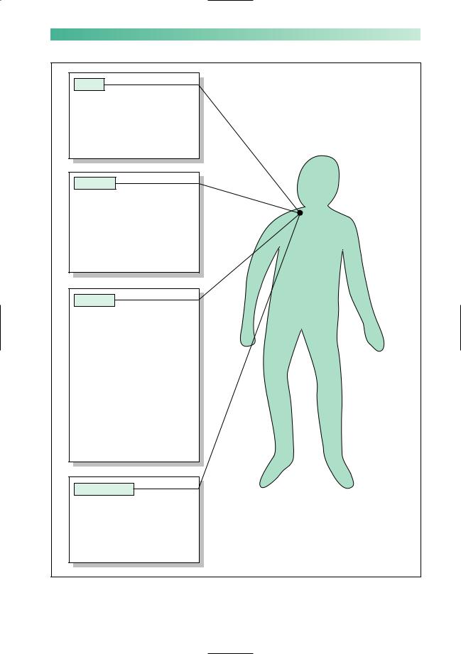
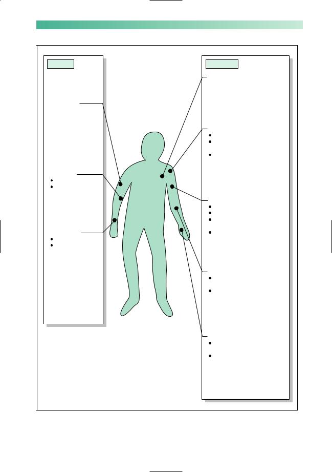
Some peripheral nerve palsies are more common than others. Figures 9.7 and 9.8 show the common and uncommon nerve lesions in the upper and lower limbs, respectively. Brief notes about the uncommon nerve palsies are shown. The remainder of this chapter deals with the three common nerve palsies in the upper limbs and the two common ones in the legs.
CHAPTER 9
PERIPHERAL NERVOUS SYSTEM |
147 |
Common |
Uncommon |
Long thoracic nerve
 Paralysis of serratus anterior
 Winging of the scapula when arms held forward
Radial nerve
 Mid humerus
 Acute compression or trauma
|
Axillary or circumflex nerve |
|
Damaged by shoulder dislocation |
|
Weak deltoid, i.e. shoulder |
|
abduction |
|
Sensory loss just below shoulder |
Ulnar nerve |
|
Elbow |
|
Chronic compression |
|
|
Musculocutaneous nerve |
|
Damaged by fracture of humerus |
|
Weak biceps, i.e. elbow flexion |
|
Sensory loss down lateral |
Median nerve |
forearm |
Absent biceps jerk |
|
Wrist |
|
Chronic compression |
|
|
Posterior interosseous nerve |
|
Site of occasional chronic |
|
compression |
|
Like radial nerve palsy, except |
|
brachioradialis and wrist |
|
extensors intact, and no |
|
sensory loss |
|
Deep palmar branch of ulnar nerve |
|
|
|
Site of occasional chronic |
|
compression |
|
Like ulnar nerve palsy, except |
|
little finger abduction intact, |
|
and no sensory loss |
Fig. 9.7 Peripheral nerve palsies of the upper limb.
148 |
CHAPTER 9 |
Common |
Uncommon |
Obturator nerve
 Damaged by trauma in labour and by pelvic cancer
 Weak hip adduction
 Sensory loss down inner aspect of thigh
Lateral cutaneous nerve of thigh
 Inguinal ligament
  Chronic compression
Sciatic nerve
 Damaged by acute compression during coma, by misplaced intramuscular injections, and by hip dislocation and surgery
 Weak knee flexion, and all muscles below the knee
 Sensory loss throughout foot and over lateral calf
 Absent ankle jerk
Common peroneal |
Femoral nerve |
Damaged by trauma, |
|
nerve |
haematoma, psoas abscess |
Neck of fibula |
Weak quadriceps, i.e. knee |
Trauma, acute or |
extension |
chronic compression |
Sensory loss on front of thigh |
|
Absent knee jerk |
Posterior tibial nerve
 Damaged by trauma and tibial fractures
 Weak foot and toe plantar flexion
 Sensory loss on sole of foot
  Absent ankle jerk
Fig. 9.8 Peripheral nerve palsies of the lower limb.
PERIPHERAL NERVOUS SYSTEM |
149 |
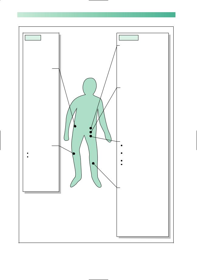
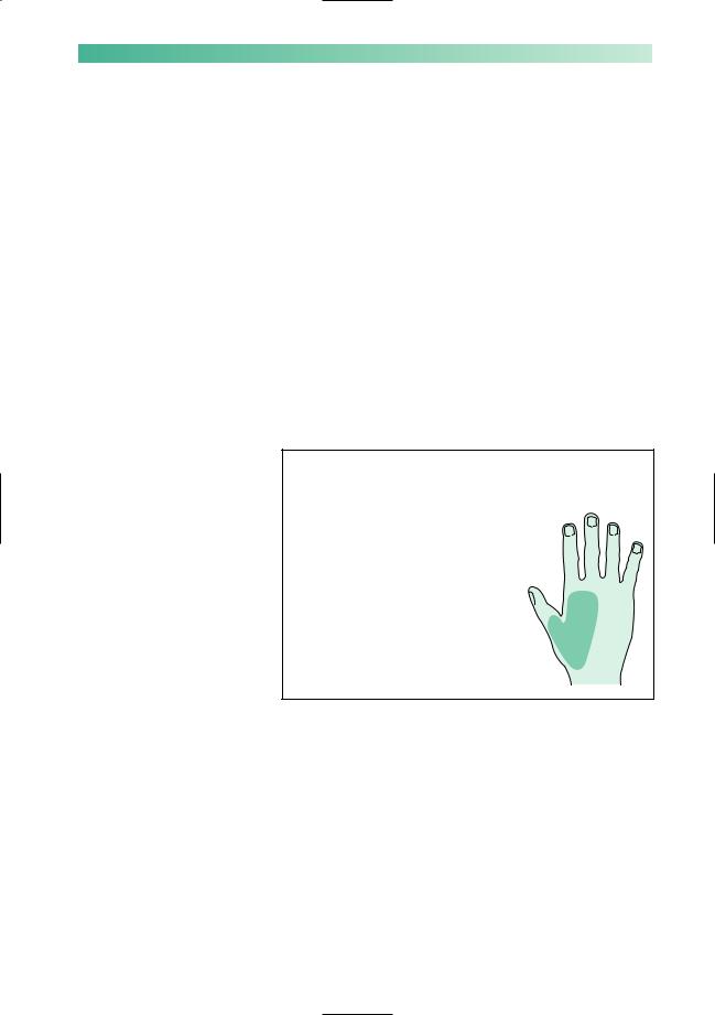
Radial nerve palsy (Fig. 9.9)
The nerve is most usually damaged where it runs down the posterior aspect of the humerus in the spiral groove. This may occur as a result of acute compression, classically when a patient has gone to sleep with his arm hanging over the side of an armchair (Saturday night palsy!). It may occur in association with fractures of the midshaft of the humerus.
The predominant complaint is of difficulty in using the hand, because of wrist drop. The finger flexors and small hand muscles are greatly mechanically disadvantaged by the presence of wrist, thumb and finger extensor paralysis. There are usually no sensory complaints.
The prognosis is good after acute compression, and more variable after damage in association with a fractured humerus. The function of the hand can be helped by the use of a special ‘lively’ splint which holds the wrist, thumb and fingers in partial extension.
Motor loss |
Reflex loss |
Sensory loss |
Brachioradialis |
Absent brachioradialis |
|
Wrist extensors |
(supinator) jerk |
|
Finger extensors |
|
|
Thumb extensors |
|
|
and abductor |
|
|
|
Nerve sensitivity |
|
|
Usually none |
|
Fig. 9.9 Radial nerve palsy.