

Wilkinson. Essential Neurology 2005
.pdf20
Diagnosis, explanation and planning
A diagnosis, or differential diagnosis, for the patient’s left leg problem is established, and a good feel for the level of patient concern has been achieved. Tests to confirm the diagnosis often need to be arranged before the diagnosis is finally established. Careful explanation of the differential diagnosis, and careful explanation of the tests to the patient, often with another family member present, is important. Diagnostic certainty may be achieved nowadays as a result of sophisticated scanning, neurophysiological tests and laboratory investigations, but some patients are apprehensive about such investigations and others are very apprehensive about what diagnosis may emerge as a result of them. The need for excellent communication and patient explanation reaches its height when the final diagnosis and management plan are discussed with the patient (and family member). Plenty of opportunity should be given for the patient and family to express their feelings at this stage.
The following five points are helpful from the communication point of view. The doctor should show in an open and friendly way, that:
1.there is always enough time;
2.there is always enough concern;
3.enough privacy is always available for the patient to speak freely and openly;
4.there is always an opportunity to talk to the patient’s family;
5.he can talk to the patient and family in language they can easily understand.
Investment of such time and effort with a patient who has a neurological illness is always worthwhile. The more the patient and family trust, like and respect the doctor, the greater will be their confidence in the diagnosis and their compliance with the management. It is clearly a shame if a bright doctor has established the correct explanation of our patient’s left leg problem, but communicates very poorly and establishes little rapport with the patient. The patient may be far from satisfied, and seek help elsewhere.
Patience with patients
Time
Concern
Privacy
Relatives
Language
CHAPTER 1
CLINICAL SKILLS |
21 |
Some typical case histories
Let’s create a few different neurological scenarios which might develop from a patient presenting with a malfunctioning left leg, to show the range of different outcomes.
A semi-retired builder of 68 years, smoker, has noticed
gradually progressive weakness in the left leg for 6–8 weeks. Both he and his wife are worried, mainly
because they have an imminent 4-week trip to visit their son and family in Australia.
General examination is normal.
Neurological examination reveals mild UMN signs in the left arm and major UMN signs in the left leg.
A chest X-ray shows a mass at the right hilum and a CT brain scan shows two mass lesions, one (apparently producing no problem) in the left frontal region, and one in the region of the precentral motor
cortex high up in the right fronto-parietal region. Bronchoscopy confirms that the right hilar lesion is a bronchial carcinoma.
In discussion, it transpires that the patient had a strong notion that this was what was wrong from fairly early on, confirmed for him when he was asked to have a chest X-ray; that he would like to take advantage of the transient improvement produced by large-dose steroids, reducing the oedema around the brain lesions; and that, although not rich, he could afford hospital care or urgent flights home from Australia if required.They would continue with their planned visit, and cope as best they could when the inevitable worsening of his condition occurred, hopefully when they have returned home.
A widow of 63 years, who has been on treatment for high blood pressure for some years, developed sudden weakness of the left leg whilst washing up at 9 a.m. She fell over and had to call the doctor by shuffling across the floor of the house to the telephone. Now, 3
days later, there has been moderate recovery so that she can walk but she feels far from safe.
Her father had hypertension and died after a stroke. General examination reveals a BP of 200/100, a
right carotid arterial bruit, a right femoral arterial bruit and hypertensive retinopathy.
Neurological examination reveals mild UMN signs in the left arm and major UMN signs in the left leg.
Chest X-ray and ECG both confirm left ventricular hypertrophy.
Blood tests are all normal.
CT brain scan shows no definite abnormality. Carotid doppler studies show critical stenosis of the
lower end of the right internal carotid artery. A small stroke, from which she seems to be
recovering satisfactorily, is the diagnosis discussed with her.The patient is happy to see the physiotherapist to help recovery. She is worried about the degree of recovery from the point of view of driving, which isn’t safe in her present car (which has a manual gear shift). She understands she is predisposed to future strokes because of her hypertension and carotid artery disease. She is prepared to take preventative drug treatment in the form of aspirin, blood pressure pills and a statin. She doesn’t smoke. She wants to have a good talk to her doctor son before submitting herself to carotid endarterectomy, although she understands the prophylactic value of this operation to her.

22 |
CHAPTER 1 |
A golf course groundsman of 58 years gives a history of lack of proper movement of the left leg,
making his walking slower. It has been present for 6 months and has perhaps worsened slightly. Most of his work is on a tractor so the left leg problem hasn’t really
affected him at work. He feels he must have a nerve pinched in his left leg somewhere.
General examination is normal.
Neurological examination reveals a rather fixed facial expression, tremor of the lightly closed eyes, a little cogwheel rigidity in the left arm with slow fine movements in the left fingers. In the left leg there is a slight rest tremor and moderate rigidity. He walks in a posture of mild flexion, with reduced arm swinging on
the left and shuffling of the left leg. His walking is a little slow.
He is profoundly disappointed to hear that he has Parkinson’s disease. He has never had any previous illness, and somebody in his village has very severe Parkinson’s disease indeed.
Several consultations are required to explain the nature of Parkinson’s disease, the fact that some people have it mildly and some severely, that effective treatment exists in the form of tablets, and that a very pessimistic viewpoint isn’t appropriate or helpful to him.
Gradually he’s coming round to the idea and becoming more optimistic. Levodopa therapy is producing significant improvement. Literature produced by the Parkinson’s Disease Society has helped his understanding of the illness.
A woman of 24 years presents with a 3-week history of heaviness and dragging of the
left leg. She has had to stop driving because of left leg weakness and clumsiness. For a week she hasn’t been able to
tell the temperature of the bath water with the right leg, though she can with the weak leg. She has developed a little bladder frequency and urgency. She has had to stop her job as a riding school instructor.
Three years ago she lost the vision in her left eye for a few weeks, but it recovered well. Doctors whom she saw at the time talked about inflammation of the optic nerve.
She is engaged to be married in a few month’s time.
General examination is normal.
Neurological examination reveals no abnormalities in the cranial nerves or arms. She has moderate UMN
signs in the left leg, loss of sense of position in the left foot and toes, and spinothalamic sensory loss (pain and temperature) throughout the right leg. She drags her left leg as she walks.
She understands that she now, almost certainly, has another episode of inflammation, this time on the left hand side of her spinal cord, similar in nature to the optic nerve affair 3 years ago.
She accepts the offer of treatment with high-dose steroids for 3 days, to help to resolve the inflammation. She is keen to return to work.
The neurologist knows that he has got quite a lot more work to do for this girl. He has to arrange for the investigations to confirm his clinical opinion that she has multiple sclerosis. He will then have to see her (and her fiancé, if she would like it) and explain that multiple sclerosis is the underlying explanation for the symptoms. He will have to do his best to help her to have an appropriate reaction to this information. Both she and her fiancé will need information and support.
CLINICAL SKILLS |
23 |
A man of 46 years, scaffold-erector, knows that his left foot is weak. It has
been present for a few months. He has lost spring at the ankle, and the left foot is weak when taking all his weight on ladders and scaffold. He’s had back pain, on and off, for many years like a
lot of his work mates. He doesn’t get paid if he’s not working.
General examination is normal except for some restriction of forward flexion of the lumbar spine.
Neurological examination reveals wasting and weakness of the left posterior calf muscles (i.e. foot and toe plantar-flexors), an absent left ankle jerk, and impaired cutaneous sensation on the sole and lateral aspect of the left foot.
Scanning confirms the presence of a large prolapsed intervertebral disc compressing the left S1 nerve root.
He is offered referral to a neurosurgeon. His concerns are:
•Will the operation work (i.e. restore better function to the left leg)?
Yes — more likely than not, but only over several months, even up to a year.
•How much time off work?
Probable minimum of 6–8 weeks and then light duties for a further 6–8 weeks.
•Should he be thinking of changing his job? Not essential, but an excellent idea if a good opportunity turned up.
A 38-year-old unkempt alcoholic presents with a left
foot drop so that he cannot lift
up the foot against gravity, and
as he walks there is a double strike as his left foot hits the ground, first with the toe and
then with the heel. He’s very frequently intoxicated, and he can’t remember how, or precisely when, the foot became like this.
General examination reveals alcohol in his breath, multiple bruises and minor injuries all over his body, no liver enlargement, but generally poor nutritional state.
Neurological examination reveals weakness of the left foot dorsiflexion, left foot eversion, left toe dorsiflexion and some altered cutaneous sensation down the lower anterolateral calf and dorsal aspect of the foot on the left.
A left common peroneal nerve palsy, secondary either to compression (when intoxicated) or to trauma, at the neck of the left fibula, is explained as the most probable diagnosis.Arrangements are made for a surgical appliance officer to provide a foot-support, the physiotherapists to assess him, and for neurophysiological confirmation of the diagnosis.
The patient defaults on all these and further appointments.


2 |
CHAPTER 2 |
Stroke |
Introduction
Stroke causes sudden loss of neurological function by disrupting the blood supply to the brain. It is the biggest cause of physical disability in developed countries, and a leading cause of death. It is also common in many developing countries.
The great majority of strokes come on without warning. This means that for most patients the aims of management are to limit the damage to the brain, optimize recovery and prevent recurrence. Strategies to prevent strokes are clearly important. They concentrate on treating the vascular risk factors that predispose to stroke, such as hypertension, hyperlipidaemia, diabetes and smoking.
The two principal pathological processes that give rise to stroke are occlusion of arteries, causing cerebral ischaemia or infarction, and rupture of arteries, causing intracranial haemorrhage (Fig. 2.1). Haemorrhage tends to be much more destructive and dangerous than ischaemic stroke, with higher mortality rates and a higher incidence of severe neurological disability in survivors. Ischaemic stroke is much more common, and has a much wider range of outcomes.
Fig. 2.1 (a) CT scan showing a |
|
|
right middle cerebral artery |
|
|
occlusion as a large wedge of low |
|
|
density; the blocked artery itself |
|
|
is bright. (b) CT scan showing a |
|
|
large left internal capsule |
(a) |
(b) |
haemorrhage. |
25
26 |
CHAPTER 2 |
Cerebral ischaemia and infarction
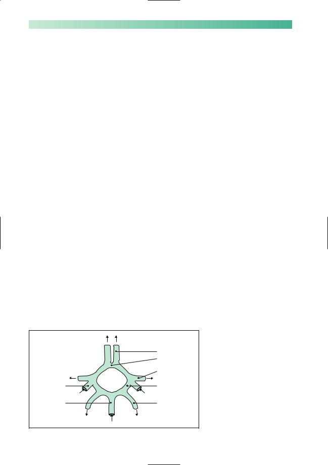
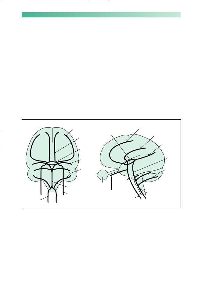
Reduction in the flow of blood to any part of the brain first causes ischaemia, a reversible loss of function, and then, if the reduction is severe or prolonged, infarction with irreversible cell death. The blood supply to the anterior parts of the brain (and to the eyes) comes from the two carotid arteries, which branch in the neck to give rise to the internal carotid arteries; these branch again in the head to give rise to the anterior and middle cerebral arteries. The posterior parts of the brain are supplied by the two vertebral arteries, which join within the head to form the basilar artery, which in turn gives rise to the posterior cerebral arteries (Figs 2.2 and 2.3).
The internal carotid and basilar arteries connect at the base of the brain through the circle of Willis. This anastomosis allows some cross-flow if one of the supply arteries is occluded, but the extent of this varies enormously from patient to patient. Beyond the circle of Willis, the cerebral arteries are best thought of as end-arteries. Restoration of normal perfusion in tissue made ischaemic by occlusion of one of these end-arteries cannot rely on blood reaching the ischaemic area through anastomotic channels. Recovery of function in the ischaemic tissue depends much more upon lysis or fragmentation of the occluding thrombo-embolic material.
The usual cause of occlusion of one of the cerebral arteries is acute thrombus formation at the site of an atheromatous plaque. The thrombus can occlude the vessel locally or throw off emboli which block more distal arteries. This process is particularly common at the origin of the internal carotid artery, but can occur anywhere from the aorta to the cerebral artery itself. Aless common cause of occlusion is embolism from the heart. In younger patients, dissection of the carotid or vertebral artery (in which a split forms between the layers of the artery wall, often after
|
Anterior cerebral |
|
Anterior |
|
communicating |
|
Middle cerebral |
Internal carotid |
Posterior |
|
communicating |
Basilar |
Posterior cerebral |
Fig. 2.2 Circle of Willis.
STROKE |
27 |
minor neck trauma) can either occlude the vessel or allow a thrombus to form and embolize distally (Fig. 2.3).
Patients with hypertension or diabetes may occlude smaller arteries within the brain through a pathological process which may have more to do with degeneration in the artery wall than atheroma and thrombosis. This small vessel disease may cause infarcts a few millimeters in diameter, termed lacunar strokes, or a more insidious illness with dementia and gait disturbance.
If complete recovery from an ischaemic event takes place within minutes or hours, it is termed a transient ischaemic attack (TIA). Where recovery takes longer than 24 hours the diagnosis is stroke. The pathophysiology of the two conditions, and the implications for investigation and treatment, are the same. In both situations, the history and examination help to establish the cause (with a view to secondary prevention) and assess the extent of the damage (to plan rehabilitation).
Middle |
|
|
Anterior |
cerebral |
Circle of |
|
cerebral |
Anterior |
|
Middle |
|
Willis |
|
||
|
cerebral |
||
cerebral |
|
|
|
|
|
Posterior |
|
|
|
|
|
Circle of |
|
|
cerebral |
Willis |
|
|
|
Posterior |
|
|
Basilar |
|
|
|
|
cerebral |
|
|
|
Cerebellar |
|
|
Cerebellar |
|
|
Internal |
|
Basilar |
Eye |
carotid |
|
|
|
||
|
|
|
|
Internal |
Ophthalmic |
Vertebral |
|
carotid |
|
Common |
|
Vertebral |
|
carotid |
|
|
|
|
|
Fig. 2.3 Arteries supplying the brain.

28 |
CHAPTER 2 |
Symptoms and signs of the cause of cerebral ischaemia and infarction
The common conditions which give rise to cerebral ischaemia and infarction are listed below.
1. Atheroma in either the large neck arteries or the cerebral arteries close to the brain. There may be a history of other atheromatous disease:
•previous angina pectoris or heart attack;
•intermittent claudication of the legs;
•previous TIAor stroke.
There may be a history of vascular risk factors:
•hypertension;
•diabetes;
•hyperlipidaemia;
•family history of atheromatous disease;
•smoking.
Examination may reveal evidence of these risk factors or evidence of atheroma, with bruits over the carotid, subclavian or femoral arteries, or absent leg pulses.
2.Cardiac disease associated with embolization:
•atrial fibrillation;
•mural thrombus after myocardial infarction;
•aortic or mitral valve disease;
•bacterial endocarditis.
Leg
Trunk
Arm
Hand
Face Reading
Writing
Speech Arithmetic
Vision
Eye
Cranial nerve nuclei Pathways to and from cerebellum Ascending and descending tracts
Less common conditions associated with cerebral ischaemia or infarction
Non-atheromatous arterial disease
•carotid or vertebral artery dissection
•arteritis
•subarachnoid haemorrhage
Unusual cardiac conditions giving rise to emboli
•non-bacterial endocarditis
•atrial myxoma
•paradoxical embolism (via a patent foramen ovale)
Unusual embolic material
•fat
•air
Increased blood viscosity or coagulability
•polycythaemia
•antiphospholipid antibodies
•perhaps other thrombophilic disorders
Venous infarction (where there is impaired perfusion of brain tissue drained by thrombosed or infected veins)
•sagittal sinus thrombosis
•cortical thrombophlebitis
Fig. 2.4 Localization of function within the brain.
STROKE |
29 |
|
|
|
|
r |
|
|
|
io |
|
|
|
r |
|
|
|
e |
|
|
|
t |
|
|
|
|
n |
|
|
|
|
A |
|
|
|
|
Middle
|
|
r |
|
rio |
|
ste |
|
|
Po |
|
|
Vertebro-basilar
Anterior |
Middle |
|
|
|
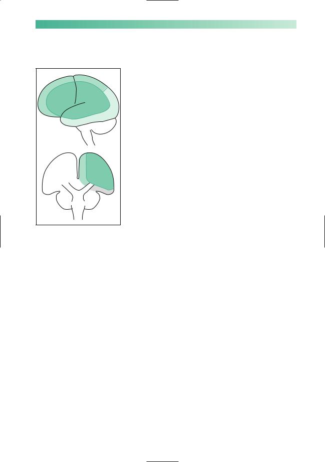
Posterior |
Vertebro-basilar
Neurological symptoms and signs of cerebral ischaemia and infarction
The loss of function that the patient notices, and which may be apparent on examination, depends on the area of brain tissue involved in the ischaemic process (Fig. 2.4).
The follow suggest middle cerebral artery ischaemia:
•loss of use of the contralateral face and arm;
•loss of feeling in the contralateral face and arm;
•dysphasia;
•dyslexia, dysgraphia, dyscalculia.
The following suggests anterior cerebral artery ischaemia:
• loss of use and/or feeling in the contralateral leg.
The following suggests posterior cerebral artery ischaemia:
• contralateral homonymous hemianopia.
Involvement of face, arm and leg with or without a homonymous hemianopia suggests:
• internal carotid artery occlusion.
The ophthalmic artery arises from the internal carotid artery just below the circle of Willis (see Fig. 2.3). The following suggests ophthalmic artery ischaemia:
• monocular loss of vision.
Combinations of the following suggest vertebrobasilar artery ischaemia:
•double vision (cranial nerves 3, 4 and 6 and connections);
•facial numbness (cranial nerve 5);
•facial weakness (cranial nerve 7);
•vertigo (cranial nerve 8);
•dysphagia (cranial nerves 9 and 10);
•dysarthria;
•ataxia;
•loss of use or feeling in both arms or legs.
The following suggest a small but crucially located lacunar stroke due to small vessel ischaemia:
•pure loss of use in contralateral arm and leg;
•pure loss of feeling in contralateral arm and leg.