

Wilkinson. Essential Neurology 2005
.pdf50
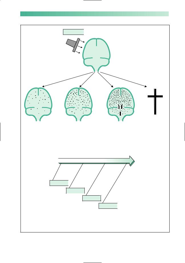
Investigation
Brain imaging
Brain tumours can be detected using CT X-ray scanning or MR scanning. The choice of technique used will often depend upon local facilities. CT is cheaper, more widely available and (when used with contrast enhancement) capable of detecting the majority of tumours. MR scanning is superior in many ways, especially in detecting small tumours and tumours in the base of the skull and the posterior fossa. MR also allows the images to be presented in a range of planes, which helps with surgical planning.
Admission to hospital
If the clinical presentation and results of scanning suggest a brain tumour, admission to hospital for further investigation will be indicated in most cases, especially if there is progressive neurological deficit or evidence of raised intracranial pressure. This will be an extremely stressful experience for the patient and relatives. Reassurance, sympathy and encouragement together with adequate explanation are required.
Other tests
The brain imaging procedures mentioned above sometimes require the support of:
•radiological and other imaging techniques elsewhere in the body if metastatic disease seems likely;
•carotid or vertebral angiography if the neurosurgical team needs this information prior to surgery;
•haematological and biochemical investigation if cerebral abscess, granuloma, metastatic disease or pituitary pathology are under consideration.
(NB Lumbar puncture is contra-indicated in suspected cases of cerebral tumour.)
CHAPTER 3

BRAIN TUMOUR
Management of brain tumours
•Admission to hospital
•Scanning
•No lumbar puncture
•Dexamethasone
•Surgery
•Radiotherapy
•Anticonvulsants
51
Management
If the patient shows marked features of raised intracranial pressure and scanning displays considerable cerebral oedema, dexamethasone may be used with significant benefit. The patient will be relieved of unpleasant, and sometimes dangerous, symptoms and signs, and the intracranial state made much safer if neurosurgical intervention is to be undertaken.
Surgical management
Complete removal
Meningiomas, pituitary tumours not susceptible to medical treatment, acoustic neuromas and some solitary metastases in accessible regions of the brain can all be removed completely. Sometimes, the neurosurgical operation required is long and difficult if the benign tumour is relatively inaccessible.
Partial removal
Gliomas in the frontal, occipital and temporal poles may be removed by fairly radical debulking operations. Sometimes, benign tumours cannot be removed in their entirety because of tumour position or patient frailty.
Biopsy
If at all possible, the histological nature of any mass lesion in the brain should be established. What looks like a glioma or metastasis from the clinical and radiological points of view occasionally turns out to be an abscess, a benign tumour or a granuloma. If the mass lesion is not in a part of the brain where partial removal can be attempted, biopsy by means of a needle through a burrhole usually establishes the histological diagnosis. The accuracy and safety of this procedure may be increased by use of stereotactic surgical techniques. Histological confirmation may not be mandatory where there is strong collateral evidence of metastatic disease.
Histological confirmation may be postponed in patients presenting with epilepsy only, in whom a rather small mass lesion in an inaccessible part of the brain is revealed by a scan. Sequential scanning, initially at short intervals, may be the most reasonable management plan in such patients.

52
Shunting and endoscopic surgery
Midline tumours causing ventricular dilatation are routinely treated by the insertion of a shunt into the dilated ventricular system. The shunt tubing is tunnelled under the skin to drain into the peritoneal cavity. This returns intracranial pressure to normal, and may completely relieve the patient’s symptoms. Sometimes it is possible and desirable to remove the tumour or treat the hydrocephalus using intracranial endoscopic procedures instead.
Additional forms of treatment
Radiotherapy
Middle-grade gliomas, metastases and incompletely removed pituitary adenomas are the common intracranial tumours which are radiosensitive. The posterior fossa malignant tumours of childhood and lymphoma are also sensitive to radiotherapy. Radiotherapy commonly follows partial removal or biopsy of such lesions, and continues over a few weeks whilst the preoperative dose of dexamethasone is being gradually reduced.
Chemotherapy
Chemotherapy can be useful as primary treatment for lymphoma and as adjunctive therapy for oligodendroglioma and some high-grade astrocytomas.
Anticonvulsants
Control of epilepsy may be an important part of the management of a patient with a supratentorial brain tumour.
Dexamethasone
Taken in large and constant dosage, dexamethasone may be the most humane treatment of patients with highly malignant gliomas or metastatic disease. Used in this way, dexamethasone often allows significant symptomatic relief so that the patient can return home and enjoy a short period of dignified existence before the tumour once more shows its presence. At this point, dexamethasone can be withdrawn and opiates used as required.
CHAPTER 3

BRAIN TUMOUR |
53 |
Prognosis
The fact that the majority of brain tumours are either malignant gliomas or metastases, which obviously carry a very poor prognosis, hangs like a cloud over the outlook for patients with the common brain tumours.
The table below summarizes the outlook for patients with the common brain tumours. It can be seen that such pessimism is justified for malignant brain tumours, but not for the less common benign neoplasms.
|
|
|
|
|
|
Tumour |
Treatment |
Outcome |
|
|
|
|
|
|
|
Meningioma |
Surgical removal if possible |
Residual disability common |
|
|
|
and/or radiotherapy if not |
Recurrence rate (1% per year) |
|
|
Glioma |
|
|
|
|
Lower grades |
Watch and wait or |
Prolonged survival, usually with |
|
|
|
biopsy and radiotherapy or |
residual disability, but recurrence is |
|
|
|
partial removal ± radiotherapy |
very common and often higher grade |
|
|
|
± chemotherapy |
|
|
|
High grade |
Partial removal and radiotherapy |
Few patients survive 1 year |
|
|
|
± chemotherapy or palliative care |
|
|
|
Lymphoma |
Biopsy and chemotherapy |
Improving: median survival about 2 years |
|
|
Pituitary adenoma |
Medical therapy for prolactinoma |
Excellent |
|
|
|
or |
|
|
|
|
surgical removal ± radiotherapy |
|
|
|
Acoustic neuroma |
Watch and wait |
Deafness ± facial weakness are |
|
|
|
or radiotherapy |
common; survival is excellent |
|
|
|
or surgical removal |
|
|
|
|
|
|
|
|
|
|
|
|

54 |
CHAPTER 3 |
CASE HISTORIES
Case 1
A 65-year-old widow presents with a 6-month history of unsteadiness. She has started to veer to the left. She has been well prior to this, apart from a longstanding hearing problem and surgery for colon cancer 5 years ago. On examination she has an ataxic gait, slight leftwards nystagmus, an absent left corneal reflex and marked left-sided deafness.There is no papilloedema.
a. What do you think is the cause of her symptoms?
Case 2
A 45-year-old oil company executive returns from secondment in Nigeria because of ill health. Over the
last 3 months he has become slow and erratic, making frequent mistakes at work and getting lost on his way home. His memory has become poor and he has had difficulty in finding his words. His appetite has faded and he has lost weight. In the last 2 weeks he has become unsteady on his feet and incontinent of urine.
On examination he is thin. He has no fever but his axillary and inguinal lymph nodes are slightly enlarged. He is drowsy. He had bilateral papilloedema. He has difficulty cooperating with a neurological examination and becomes increasingly irritated when you persist.
a. What do you think is the cause of his symptoms?
(For answers, see p. 256.)

4 |
CHAPTER 4 |
Head injury |
The causes
Road traffic accidents involving car drivers, car passengers, motorcycle drivers and pillion-seat riders, cyclists, pedestrians and runners constitute the single greatest cause of head injury in Western society. Compulsory speed limits, car seat-belts, motorcycle and cycle helmets, stricter control over driving after drinking alcohol, and clothing to make cyclists and runners more visible have all proved helpful in preventing road accidents, yet road accidents are still responsible for more head injuries than any other source.
Accidents at work account for a significant number of head injuries, despite the greater use of protective headgear. Sport, especially boxing and horse riding, constitutes a further source for head injury, prevented to some extent by the increasing use of protective headgear. Accidents around the home account for an unfortunate number of head injuries, especially in young children who are unable to take proper precautions with open windows, ladders, stairs and bunk-beds. Child abuse has to be remembered as a possible cause of head injury.
It is a sad fact that head injury, trivial and severe, affects young people in significant numbers. Approximately 50% of patients admitted to hospital on account of head injury in the UK are under the age of 20 years. Accidents in the home, sports accidents, and accidents involving motorcycles, cars and alcohol, account for this emphasis on youth.
The outcome from head trauma depends upon the age and pre-existing health of the patient at the time of injury, and upon the type and severity of the primary injury to the brain. In the care of the individual patient, there is little that the doctor can do about these factors. They have had their influence by the time the patient is delivered into his hands. Outcome does also depend, however, upon minimizing the harm done by secondary insults to the brain after the injury. Prevention of secondary brain injury is the direct responsibility of those caring for the patient. It is one of the main messages of this chapter.
55

56
The effect in pathological terms
Primary brain injury
The brain is an organ of relatively soft consistency contained in a rigid, compartmentalized, unyielding box which has a rough and irregular bottom. Sudden acceleration, deceleration or rotation certainly allow movement of the brain within the skull. If sudden and massive enough, such movement will cause tearing of nerve fibres and petechial haemorrhages within the white matter, and contusions and lacerations of the cortex, especially over the base of the brain.
If the trauma has been severe, the diffuse damage to the brain described above may cause generalized brain swelling, just like the swelling that occurs with injury to any other organ. The brain, however, is contained within a rigid box, unlike other organs of the body. Diffuse brain swelling may result in the brain becoming too large in volume for the space allocated to it. As discussed in Chapter 3 (p. 42), this may lead to tentorial herniation, midbrain compression, impaction of the lower medulla and cerebellar hemispheres in the foramen magnum, secondary brainstem pathology and death (Fig. 4.1).
Severe rotational head injuries may cause primary brainstem injury, which may itself be fatal.
Secondary brain injury
In the circumstances of head injury, the diffusely damaged brain is extremely vulnerable to four other insults, all of which produce further brain swelling (Fig. 4.1). By causing further swelling, each of the four insults listed below tend to encourage the downward cascade towards brainstem failure and death:
1.Arterial hypotension —from blood loss at the time of injury, possibly from the scalp but more probably from an associated injury elsewhere in the body. Hypotension and hypoxia, in isolation but especially in combination, will cause hypoxic– ischaemic brain damage with swelling.
2.Arterial hypoxia —because of airway obstruction, associated chest injury or an epileptic fit.
3.Infection —head injuries in which skull fracture has occurred may allow organisms to enter the skull via an open wound, or from the ear or nose. Infection causes inflammatory oedema.
4.Intracranial haematoma —the force of injury may have torn a blood vessel inside the skull (either in the brain substance or in the meninges) so that a haematoma forms. This causes further compression of the brain by taking up volume within the rigid skull.
CHAPTER 4
HEAD INJURY |
57 |
Primary injury 
Mild |
Moderate |
Severe |
Lethal |
|
|
|
Death |
Minimal |
Sufficient |
Severe |
Brainstem |
diffuse |
diffuse |
diffuse |
failure |
damage |
damage |
damage |
secondary |
|
to cause |
and brain |
to herniation |
|
generalized |
swelling, |
and coning |
|
brain swelling |
tentorial |
|
|
|
herniation |
|
|
|
and coning |
|
|
Secondary brain injury |
|
|
Hypotension 
Hypoxia
Infection
Haematoma 
The four main additional insults, any of which may produce further brain swelling, and push the patient in the direction of secondary brainstem damage and death
Fig. 4.1 Asummary of the pathological effects that may occur as a consequence of head injury.

58
The effect in clinical terms
The clinical instrument that is used to monitor the effects of head injury on patients requiring admission to hospital is the neurological observation chart created by competent, trained nursing staff.
This chart includes the patient’s responsiveness using the Glasgow Coma Scale (GCS), recording the patient’s best eye, verbal and motor responses, together with his vital signs of pupil size and reactivity, blood pressure, pulse, respiratory rate and temperature. The observations are made at intervals appropriate to the patient’s clinical condition —every 15 minutes in critically ill patients.
The Glasgow Coma Scale is described and illustrated in Chapter 11 (p. 184, Fig. 11.3). The changes in the charted observations of best eye, verbal and motor responses are used to signal changes in the patient’s brain condition with great sensitivity. The vital signs of pupil size and reactivity, blood pressure, pulse, respiratory rate and temperature are, essentially, indicators of brainstem function.
Figure 4.2 shows the clinical states that accompany mild, moderate and severe degrees of diffuse cerebral damage after head injury. The adverse influence of hypotension, hypoxia, infection and intracranial haematoma will become apparent in the patient’s chart observations, possibly accompanied by an epileptic fit. (An epileptic fit may be the cause or effect of a deteriorating intracranial situation, undesirable because of the associated anoxia and effects on intracranial pressure.) The appearance of abnormal brainstem signs indicates that the additional insult is adversely affecting the brain very seriously.
Figure 4.2 also shows the clinical clues, and definite signs, that confirm the presence of one or more of the four additional insults. Ideally, the clues should make the doctor so alert to the possibility of the insult becoming operational that the problem is rectified before the patient starts to decline significantly in his Glasgow Coma Scale observations, and certainly before evidence of impaired brainstem function appears. This is what is meant by the prevention of secondary brain injury.
CHAPTER 4

HEAD INJURY |
|
|
|
|
|
|
|
59 |
|||
|
|
|
|
|
|
|
|
|
|
|
|
|
|
|
|
|
|
|
|
|
|
|
|
|
Primary injury |
|
|
|
|
|
|
|
|
|
|
|
|
|
Concussion, |
|
|
Persistent |
|
Persistent coma |
|||
|
|
|
|
|
|
||||||
|
|
|
or concussion |
|
|
coma after |
|
after the |
|||
|
Mild |
plus some |
Moderate |
the accident |
Severe |
accident, poor |
|||||
|
retrograde |
with fairly |
scores on the |
||||||||
|
|
|
and post- |
|
|
good scores |
|
Glasgow Coma |
|||
|
|
|
traumatic |
|
|
on the |
|
Scale, and |
|||
|
|
|
amnesia |
|
|
Glasgow |
|
evidence of |
|||
|
|
|
|
|
|
|
Coma Scale, |
|
failing brainstem |
||
|
|
|
|
|
|
|
and no signs |
|
function |
||
|
|
|
|
|
|
|
of brainstem |
|
|
|
|
|
|
|
|
|
|
|
malfunction |
|
|
|
|
|
|
|
|
Secondary brain injury |
|
|
|
||||
|
|
|
The four additional insults will show their adverse influence |
|
|
|
|||||
|
|
|
on the brain by : |
|
|
|
|
|
|
|
|
|
|
|
declining performance in Glasgow Coma Scale observations |
|
|
||||||
|
|
|
an epileptic fit |
|
|
|
|
|
|
|
|
|
|
|
impaired brainstem function observations |
|
|
|
|||||
|
|
|
|
|
|
|
|
|
|
|
|
|
Hypotension |
|
|
Hypoxia |
|
|
Infection |
|
|
Haematoma |
|
|
Clinical clues |
|
|
|
|
|
|
|
|
|
|
|
Large scalp laceration |
Patient found face |
Open scalp wound |
|
Factors known to be |
||||||
|
i.e. external blood loss |
down, unconscious |
over skull fracture |
|
associated with the |
||||||
|
Associated major |
|
Upper airways |
Leakage of CSF from |
development of an |
||||||
|
injury to chest, |
|
obstruction whilst |
scalp wound, nose |
|
intracranial |
|||||
|
abdomen, pelvis, |
|
unconscious |
or ear |
|
haematoma, whether |
|||||
|
limbs, i.e. external |
|
Severe associated |
Inadequate inspection, |
extradural, subdural |
||||||
|
and internal blood |
|
facial injury |
cleaning, or |
|
or intracerebral : |
|||||
|
loss |
|
Aspiration of blood |
debridement of |
|
Skull fracture |
|||||
|
History that the |
|
or vomit into |
open scalp wound |
|
Impaired conscious |
|||||
|
patient was propped |
trachea |
|
|
over fracture site |
|
level (even |
||||
|
upright after injury |
Prolonged epileptic |
Skull fracture found |
|
disorientation) i.e. |
||||||
|
with known blood |
|
fit |
|
|
on CT scan in |
|
a fully orientated |
|||
|
loss and probable |
|
Associated injury to |
region of wound, |
|
patient with no |
|||||
|
hypotension |
|
chest wall or lungs |
nose or ear |
|
skull fracture is |
|||||
|
|
|
|
Respiratory |
Intracranial air seen |
|
very unlikely to |
||||
|
|
|
|
depression by |
on CT scan |
|
develop a |
||||
|
|
|
|
alcohol or drugs |
|
|
|
haematoma |
|||
|
Clinical evidence |
|
|
|
|
|
|
|
|
|
|
|
Development of shock |
Noisy obstructed |
Purulent discharge |
|
CT scan |
||||||
|
Low blood pressure |
breathing |
|
|
from scalp wound |
|
|
|
|||
|
Rapid pulse |
|
Abnormal chest |
Proved infection |
|
|
|
||||
|
Sweating |
|
movement |
of CSF |
|
|
|
||||
|
|
|
|
Abnormal respiratory |
|
|
|
|
|
||
|
|
|
|
rate |
|
|
|
|
|
|
|
|
|
|
|
Abnormal chest |
|
|
|
|
|
||
|
|
|
|
X-ray |
|
|
|
|
|
|
|
|
|
|
|
Abnormal blood |
|
|
|
|
|
||
|
|
|
|
gases |
|
|
|
|
|
|
|
|
|
|
|
|
|
|
|
|
|
|
|
Fig. 4.2 Asummary of the clinical effects of head injury.