

Wilkinson. Essential Neurology 2005
.pdf130 |
CHAPTER 8 |
Glossopharyngeal (9), vagus (10) and hypoglossal (12) nerves
These three lower cranial nerves are considered in one section of this chapter for two reasons.
1.Together they innervate the mouth and throat for normal speech and swallowing.
2.They are commonly involved in disease processes together, to give rise to the clinical picture of bulbar palsy.
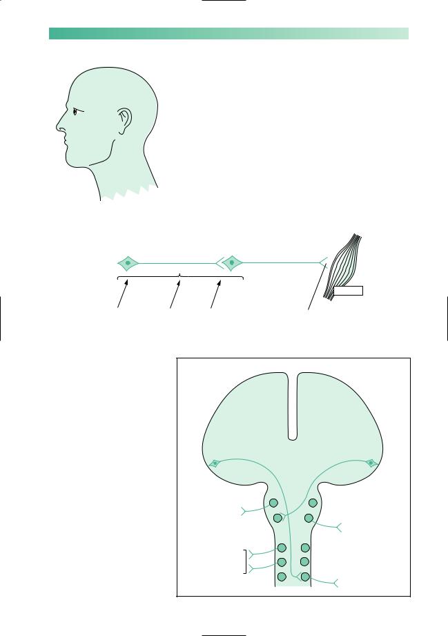
More specifically, the glossopharyngeal nerve supplies the palate and pharynx, the vagus nerve supplies the pharynx and larynx, and the hypoglossal nerve supplies the tongue. Taste perception in the posterior third of the tongue is a function of the glossopharyngeal nerve. Both the glossopharyngeal and vagus nerves (especially the latter) have an enormous autonomic function, as shown in Fig. 8.14. Innervation of the vocal cords by the long, thin, recurrent laryngeal nerves (from the vagus) exposes them to possible damage as far down as the subclavian artery on the right, and the arch of the aorta on the left.
Midbrain
Pons
|
9 |
|
|
Medulla |
|
|
|
|
|
||
|
10 |
|
|
Spinal cord |
|
Palate |
|
|
|
||
12 |
|
|
|
||
Larynx |
|
|
|
||
Pharynx |
|
|
|
|
|
Tongue |
|
|
|
|
|
+ vast autonomic |
salivary glands |
|
|
||
heart |
neck |
|
|||
innervation of |
great vessels in |
Fig. 8.14 Highly diagrammatic |
|||
thorax |
|||||
|
|
||||
|
|
|
|||
abdomen
representation of cranial nerves 9, 10 and 12.
CRANIAL NERVE DISORDERS |
131 |
Bulbar palsy
When there is bilateral impairment of function in the 9th, 10th and 12th cranial nerves, the clinical syndrome of bulbar palsy evolves. The features of bulbar palsy are:
•dysarthria;
•dysphagia, often with choking episodes and/or nasal regurgitation of fluids;
•dysphonia and poor cough, because of weak vocal cords;
•susceptibility to aspiration pneumonia.
Even though the vagus nerve has a vast and important autonomic role, it is uncommon for autonomic abnormalities to feature in the diseases discussed in this section.
Common conditions affecting 9th, 10th and 12th nerve function
Figure 8.15 illustrates the common conditions which affect 9th, 10th and 12th nerve function. The use of the word ‘common’ is relative, since none of the conditions are very common.
Fig. 8.15 Common conditions that affect cranial nerves 9, 10, and 12. *Because these conditions involve 9th, 10th and 12th nerve function bilaterally, they are the common causes of bulbar palsy.
Palate |
Motor neurone disease* |
Larynx |
Cerebrovascular disease |
Pharynx |
Syringobulbia |
Tongue |
Erosive tumours of the |
|
skull base |
|
Guillain–Barré syndrome* |
|
Recurrent laryngeal nerve |
|
palsy |
|
Myasthenia gravis* |

132
Motor neurone disease
When motor neurone disease is causing loss of motor neurones from the lower cranial motor nuclei in the medulla, the bulbar palsy can eventually lead to extreme difficulty in speech (anarthria) and swallowing. Inanition and aspiration pneumonia are commonly responsible for such patients’ deaths. The tongue is small, weak or immobile, and fasciculating (Chapter 10, see pp. 155–7).
Infarction of the lateral medulla
Infarction of the lateral medulla, following posterior inferior cerebellar artery occlusion, is one of the most dramatic cerebrovascular syndromes to involve speech and swallowing. Ipsilateral trigeminal, vestibular, glossopharyngeal and vagal nuclei may be involved, along with cerebellar and spinothalamic fibre tracts in the lateral medulla.
Guillain–Barré syndrome
Patients with Guillain–Barré syndrome, acute, post-infectious polyneuropathy (Chapter 10, see p. 163), may need ventilation via an endotracheal tube or cuffed tracheostomy tube. This may be necessary either because of neuropathic weakness of the chest wall and diaphragm, or because of bulbar palsy secondary to lower cranial nerve involvement in the neuropathy.
Recurrent laryngeal nerve palsy
The recurrent laryngeal nerves are vulnerable to damage in the neck and mediastinum, e.g. aortic aneurysm, malignant chest tumours, malignant glands and surgery in the neck (especially in the region of the thyroid gland). Aunilateral vocal cord palsy due to a unilateral nerve lesion produces little disability other than slight hoarseness. Bilateral vocal cord paralysis is much more disabling, with marked hoarseness of the voice, a weak ‘bovine’ cough (because the cords cannot be strongly adducted) and respiratory stridor.
Myasthenia gravis
Bulbar muscle involvement in myasthenia gravis is quite common in this rare condition. The fatiguability of muscle function, which typifies myasthenia, is frequently very noticeable in the patient’s speech and swallowing (Chapter 10, see pp. 164–6).
CHAPTER 8
CRANIAL NERVE DISORDERS |
133 |
Dysarthria
|
The speech disturbance in patients with dysarthria is a purely |
|
mechanical one caused by defective movement of the lips, |
|
tongue, palate, pharynx and larynx. Clear pronunciation of |
|
words is impaired due to the presence of a neuromuscular |
X |
lesion. |
Speaking is a complex motor function. Like complex move- |
|
|
ment of other parts of the body, normal speech requires the in- |
|
tegrity of basic components of the nervous system, mentioned |
|
in Chapter 1, and illustrated again in Fig. 8.16. There are charac- |
|
teristic features of the speech when there is a lesion in each ele- |
|
ment of the nervous system identified in Fig. 8.16. These are the |
Slurred speech |
different types of dysarthria. |
Upper motor neurone |
Lower motor neurone |
Muscle
Basal ganglia Cerebellum Sensation |
Neuromuscular junction |
Fig. 8.16 Basic components of the nervous system required for normal movement.
Fig. 8.17 The upper and lower motor neurones involved in speech.
5 5
Jaw muscles
7 7
Facial muscles
9 9
Larynx muscles
10 10
Pharynx muscles
12 12
Tongue muscles
134 |
CHAPTER 8 |
It is important to remember the availability of communication aids for patients with severe dysarthria. These may be quite simple picture or symbol charts, alphabet cards or word charts. More ‘high tech’ portable communication aids that incorporate keyboards and speech synthesizers are also very valuable for some patients.
Upper motor neurone lesions
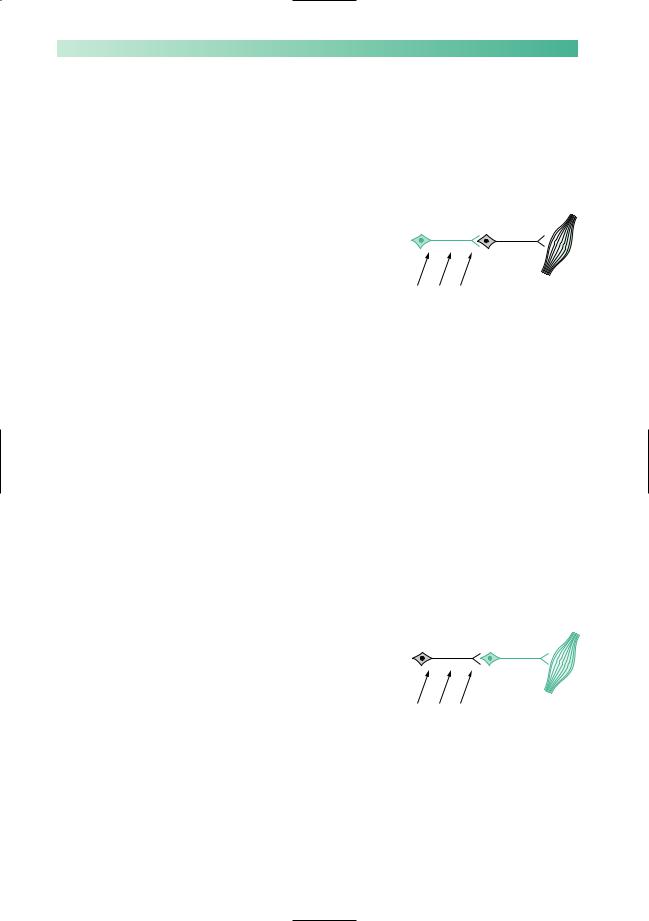
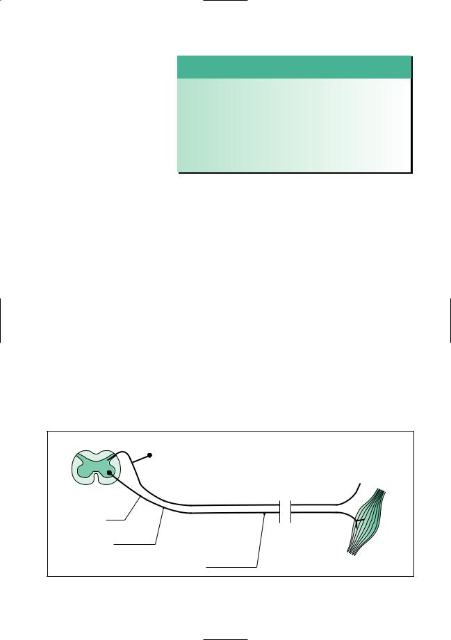
The upper motor neurones involved in speech have their cell bodies at the lower end of the precentral (motor) gyrus in each cerebral hemisphere. From the motor cortex, the axons of these cells descend via the internal capsule to the contralateral cranial nerve nuclei 5, 7, 9, 10 and 12, as shown in Fig. 8.17.
Aunilateral lesion does not usually produce a major problem of speech pronunciation. There is some slurring of speech due to facial weakness in the presence of a hemiparesis.
Bilateral upper motor neurone lesions, on the other hand, nearly always produce a significant speech disturbance. Weakness of the muscles supplied by cranial nerves 5–12 is known as bulbar palsy if the lesion is lower motor neurone in type (see the next section in this chapter). It is known as pseudobulbar palsy if the weakness is upper motor neurone in type. Patients who have bilateral upper motor neurone weakness of their lips, jaw, tongue, palate, pharynx and larynx, i.e. patients with pseudobulbar palsy, have a characteristic speech disturbance, known as a spastic dysarthria. The speech is slow, indistinct, laboured and stiff. Muscle wasting is not present, the jaw-jerk is increased, and there may be associated emotional lability. The patient is likely to be suffering from bilateral cerebral hemisphere cerebrovascular disease, motor neurone disease or serious multiple sclerosis.
Lower motor neurone lesions, and lesions in the neuromuscular junction and muscles
The lower motor neurones involved in speech have their cell bodies in the pons and medulla (Fig. 8.17), and their axons travel out to the muscles of the jaw, lips, tongue, palate, pharynx and larynx in cranial nerves 5–12.
Asingleunilateralcranialnervelesiondoesnotusuallyproduce a disturbance of speech, except in the case of cranial nerve 7. A severe unilateral facial palsy does cause some slurring of speech.
Multiple unilateral cranial nerve lesions are very rare. Bilateral weakness of the bulbar muscles, whether produced
by pathology in the lower motor neurones, neuromuscular junction or muscles, is known as bulbar palsy. One of the predominant features of bulbar palsy is the disturbance of speech. The other main features are difficulty in swallowing and incom-
BG C S
Speech is slow, indistinct, laboured and stiff in patients with pseudobulbar palsy
BG C S
Speech is quiet, indistinct and nasal in patients with bulbar palsy

CRANIAL NERVE DISORDERS |
135 |
BG C S
Parkinsonian patients have quiet, indistinct, monotonous speech
BG C S
Speech is slurred, and irregular in volume and timing
petence of the larynx leading to aspiration pneumonia. The speech is quiet, indistinct, with a nasal quality if the palate is weak, poor gutterals if the pharynx is weak, and poor labials if the lips are weak. (Such a dysarthria may be rehearsed if one tries to talk without moving lips, palate, throat and tongue.)
Motor neurone disease, Guillain–Barré syndrome and myasthenia gravis all cause bulbar palsy due to lesions in the cranial nerve nuclei, cranial nerve axons and neuromuscular junctional regions of the bulbar muscles, respectively (see Chapter 10 and Fig. 8.15).
Basal ganglion lesions
The bradykinesia of Parkinson’s disease causes the characteristic dysarthria of this condition. The speed and amplitude of movements are reduced. Speech is quiet and indistinct, and lacks up and down modulation. A monotonous voice from a fixed face, both voice and face lacking lively expression, is the typical state of affairs in Parkinson’s disease.
Patients with chorea may have sudden interference of their speech if a sudden involuntary movement occurs in their respiratory, laryngeal, mouth or facial muscles.
Cerebellar lesions
As already mentioned, the dysarthria of patients with cerebellar disease often embarrasses them because their speech sounds as if they are drunk. There is poor coordination of muscular action, of agonists, antagonists and synergists. There is ataxia of the speaking musculature, very similar to the limb ataxia seen in patients with cerebellar lesions. Speech is irregular, in both volume and timing. It is referred to as a scanning or staccato dysarthria.
Drugs that affect cerebellar function (alcohol, anticonvulsants), multiple sclerosis, cerebrovascular disease and posterior fossa tumours are some of the more common causes of cerebellar malfunction.

136 |
CHAPTER 8 |
CASE HISTORIES
Can you identify the most likely problem in each of these brief histories, and suggest a treatment?
a.‘I could see perfectly well last week.The right eye is still OK but the left one is getting worse every day. I can’t see colours with it and I can’t read small print. It hurts a bit when I look to the side.’
b.‘I see two of everything, side by side, but only when I look to the right.Apart from that, I’m fine.’
c.‘I’m getting terrible pains on the left of my face. I daren’t touch it but it’s just at the corner of my mouth, going down into my chin. It’s so sharp, it makes me jump.’
d.‘I’ve had an ache behind my ear for a couple of days but this problem started yesterday. I’m
embarrassed, I look so awful. I can’t close my left eye, it keeps running. My mouth is all over to the right, I’m slurring my words, and making a real mess when I drink.’
e.‘I keep bumping into doorways and last week I drove into a parked car: I just didn’t realize it was there. I think my eyesight is perfect but the optician said I needed to see a doctor straight away.’
f.‘I keep getting terribly dizzy. Rolling over in bed is the worst: everything spins and I feel sick. It’s the same when I turn my head to cross the road. I am too frightened to go out.’
(For answers, see pp. 260–1.)
9 |
CHAPTER 9 |
Nerve root, nerve plexus |
and peripheral nerve lesions
Introduction
In this chapter, we are considering focal pathology in the peripheral nervous system. This means a study of the effect of lesions between the spinal cord and the distal connections of the peripheral nerves with skin, joints and muscles (as shown in Fig. 9.1). We shall become familiar with focal disease affecting nerve roots and spinal nerves, nerve plexuses and individual peripheral nerves. Focal disease infers a single localized lesion, affecting one nerve root or one peripheral nerve. Diffuse or generalized diseases affecting these parts of the nervous system, e.g. a peripheral neuropathy affecting all the peripheral nerves throughout the body, are the subject of Chapter 10.
Focal lesions of the lower cervical and lower lumbar nerve roots are common, as are certain individual peripheral nerve lesions in the limbs. Accurate recognition of these clinical syndromes depends on some basic neuro-anatomical knowledge. This is not formidably complicated but possession of a few hard anatomical facts is inescapable.
Spinal cord
Skin, joints, etc.
Nerve roots and spinal nerve
Nerve plexus
Peripheral nerve |
|
(broken lines |
|
indicate length) |
Muscle |
Fig. 9.1 Schematic diagram of the peripheral nervous system.
137

138
Nerve root lesions
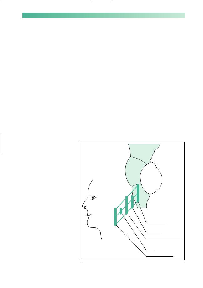
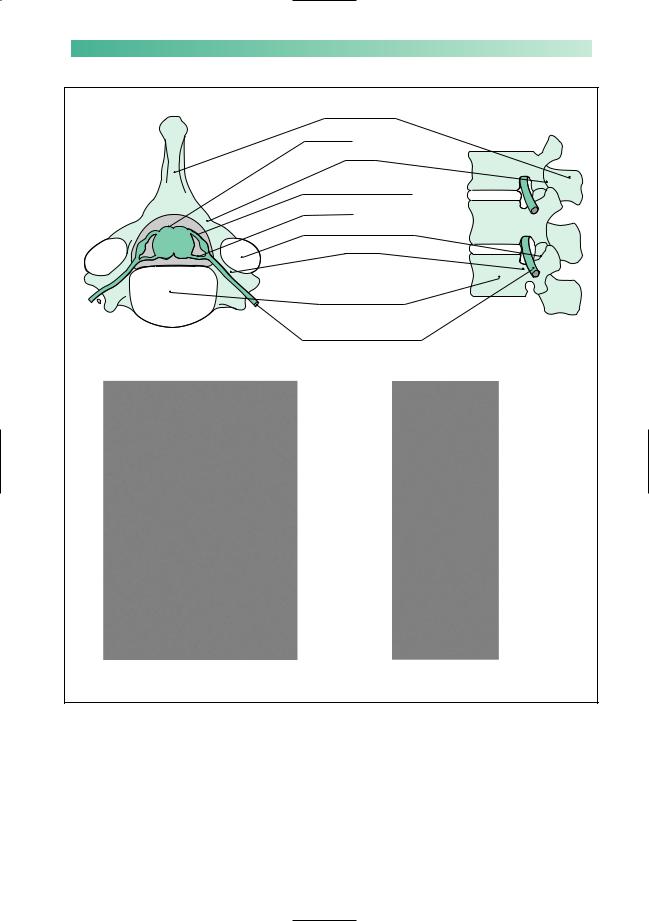
Figure 9.2 is a representation of the position of the nerve roots and spinal nerve in relation to skeletal structures. The precise position of the union of the ventral and dorsal nerve roots, to form the spinal nerve, in the intervertebral foramen is a little variable. This is why a consideration of the clinical problems affecting nerve roots embraces those affecting the spinal nerve. A nerve root lesion, or radiculopathy, suggests a lesion involving the dorsal and ventral nerve roots and/or the spinal nerve.
The common syndromes associated with pathology of the nerve roots and spinal nerves are:
•prolapsed intervertebral disc;
•herpes zoster;
•metastatic disease in the spine.
Less common is the compression of these structures by a
neurofibroma.
Prolapsed intervertebral disc
When the central, softer material, nucleus pulposus, of an intervertebral disc protrudes through a tear in the outer skin, annulus fibrosus, the situation is known as a prolapsed intervertebral disc. This is by far the most common pathology to affect nerve roots and spinal nerves. The susceptibility of these nerve elements to disc prolapses, which are most commonly posterolateral in or near the intervertebral foramen, is well shown in Fig. 9.2.
The typical clinical features of a prolapsed intervertebral disc, regardless of the level, are:
1.Skeletal:
•pain, tenderness and limitation in the range of movement in the affected area of the spine;
•reduced straight leg raising on the side of the lesion, in the case of lumbar disc prolapses.
2.Neurological:
•pain, sensory symptoms and sensory loss in the dermatome of the affected nerve root;
•lower motor neurone signs (weakness and wasting) in the myotome of the affected nerve root;
•loss of tendon reflexes of the appropriate segmental value;
•since most disc prolapses are posterolateral, these neurological features are almost always unilateral.
CHAPTER 9
PERIPHERAL NERVOUS SYSTEM |
139 |
Spinous process
Spinal cord
Lamina
Dorsal root and ganglion
Ventral root
Intervertebral facet joint
Pedicle
Body of vertebrae, separated from each other by intervertebral discs
Spinal nerve passing through the intervertebral foramen
*
(a) |
(b) |
Fig. 9.2 Diagrams showing the superior aspect of a cervical vertebra, and the lateral aspect of the lumbar spine. Disc prolapse in the cervical region can cause cord and/or spinal nerve root compression (scan a). Disc prolapse in the lumbar region (scan b) can cause nerve root compression, but the spinal cord ends alongside L1 (asterisk) and is unaffected. In either region, additional degenerative changes in the facet joints may aggravate the problem.