

Книги по МРТ КТ на английском языке / Thomas R., Connelly J., Burke C. - 100 cases in radiology - 2012
.pdfANSWER 23
Figure 23.1 shows the abdomen. Two tubes passing over the umbilicus are umbilical artery and vein catheters. They can be distinguished by their course, the umbilical artery catheter passes inferiorly to join the internal iliac artery before passing superiorly. The tip should lie between T6 and T9 vertebral level to avoid major branches. The umbilical venous catheter passes superiorly to the left portal vein, passing through the ductus venosus into the inferior vena cava (IVC). The tip should lie within the upper IVC or at the border with the right atrium. In Figure 23.2, the tip of a nasogastric (NG) tube is seen overlying the stomach. Aspiration and radiographs are typically used to check the position.
The appearances on the first image (Figure 23.1) are non-specific but gas-filled small and large bowel in a symptomatic premature infant should ring alarm bells.
The second image (Figure 23.2) shows dilatation of the bowel (greater than the width of the L1 vertebral body). Bowel wall gas lucencies (pneumatosis intestinalis), particularly in the right lower quadrant and portal venous gas (lucency over the liver) is seen. No evidence of perforation is seen to warrant immediate surgical intervention.
Subsequent images (Figure 23.3) showed further distension and free gas outside the bowel.
(a) |
(b) |
Figure 23.3 (a) Anterior–posterior (AP) and (b) lateral abdominal views with the patient supine showing free gas (arrows) above the liver and around the bowel loops, indicating perforation.
The main differential diagnosis is necrotizing enterocolitis and other forms of sepsis. Other differentials include Hirschprung’s disease (aganglionic distal colon/rectum), bowel obstruction (such as small bowel atresia, meconium ileus, meconium plug) and ischaemia, particularly in congenital cardiac disease.
Initial management is usually conservative including antibiotics and repeated imaging. Surgery may be required for perforation or failure of medical management.
72

Necrotizing enterocolitis has a complex aetiology. Immaturity of the gut mucosa and immune response, coupled with ischaemia/hypoxia, are felt to contribute with premature and low birth weight babies at highest risk. Other risk factors more apparent in term infants include sepsis, cyanotic congenital heart disease, polycythaemia and gastroschisis. Long-term complications include strictures and short bowel syndrome.
KEY POINTS
•A low threshold for suspecting necrotizing enterocolitis in preterm infants is advisable.
•Radiograph findings may be non-specific, although comparing successive images may indicate persistent bowel dilatation, thickening or pneumatosis.
•Management ranges from conservative to surgery if perforation is evident.
73
This page intentionally left blank
CASE 24: YOUNG CHILD WITH PAINFUL ARM
History
This 1-year-old girl has been in the paediatric department for a few weeks for treatment of streptococcal sepsis after chickenpox. Around the time of admission the child had swelling over the left upper arm but no abnormality was seen on plain radiograph and there was no collection on ultrasound. After treatment on the paediatric intensive care ward she improved but then started to get intermittent fevers and worsening of the left upper arm swelling.
Examination
The child is irritable and off her feeds. There is tachycardia and pyrexia. Cardiovascular and respiratory examinations are normal. The abdomen is soft and non-tender. Her left arm is not moving, red and swollen over the upper aspect. She complains when you handle or move the arm.
You arrange a plain radiograph and compare it with the previous image (Figure 24.1a,b).
(a) |
(b) |
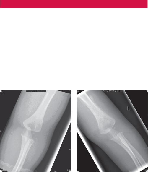
Figure 24.1 (a) Initial radiograph of left elbow. (b) Radiograph taken 14 days later.
Questions
•Describe the changes seen.
•What other imaging would you arrange?
75

ANSWER 24
The first radiograph (Figure 24.1a) is essentially normal (there may be some soft tissue swelling). The second radiograph (14 days later) (Figure 24.1b) shows thick periosteal reaction along the shaft of the humerus. Patchy lucency is seen within the distal humerus. The appearance is consistent with an aggressive process. Given the acute onset and sepsis, this is likely to be acute osteomyelitis that is an inflammation of bone caused by an infecting organism. The differential for periosteal reaction in an infant could include infiltrative tumour such as leukaemia or neuroblastoma, (non-accidental) trauma, prostaglandin therapy or vitamin deficiency such as rickets (not uncommon) and scurvy (rare).
Osteomyelitis has a bimodal distribution. In children it typically occurs by haematogenous spread with insidious onset. Typical bacteria are Staphylococcus aureus, group A
Streptococcus (in this case), Haemophilus influenzae and Enterobacter species. Patients with sickle cell disease are particularly at risk of S. aureus and Salmonella species. The bacteria pass into the metaphysis of the most rapidly growing tubular bones via nutrient vessels where they lodge and cause inflammation, vascular congestion and increased pressure. There may also be some thrombosis. This is followed by a suppurative phase where pus forms subperiosteal abscesses. Increased pressure and thrombosis compromises the blood supply causing bone necrosis and sequestrum formation (fragments of necrotic bone) over a period of days. A layer of new bone, or an involucrum, forms around the raised periosteum. With treatment there is a resolution phase with remodelling of bone. A skin sinus can form in the absence of treatment or as a complication. Other complications in children arise due to damage of the growth plate with deformity or shortening of the developing bone. Haematologically spread osteomyelitis in adults is typically centred in a vertebra.
Direct osteomyelitis is associated with traumatic inoculation, more focal and more typical in adults, particularly those who are at increased risk due to peripheral neuropathy and immune compromise such as diabetic patients. Typical bacteria are S. aureus, Enterobacter species and Pseudomonas species. The development is much the same as described but more focal.
Other imaging that is helpful is a three-phase bone scan that images the whole skeleton and identifies further sites of disease. A magnetic resonance (MR) scan is useful to confirm the diagnosis and examine the soft tissues and bone marrow for abscess formation and complications.
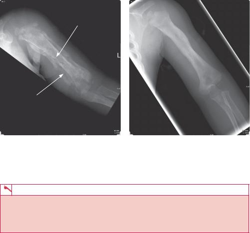
Figure 24.2a,b shows subsequent plain radiographs of the sequestrum phase and resolution.
76
Necrotic bone-sequestrum
Possible abscess
(a) (b)
Figure 24.2 Subsequent plain radiographs showing (a) the sequestrum phase and (b) resolution.
KEY POINTS
•Osteomyelitis in children is more commonly caused by haematogenous spread; in adults it is more usually by traumatic inoculation.
•The radiological appearance is aggressive with periosteal reactions and irregular bone resorption, necrosis and reformation.
77
This page intentionally left blank
CASE 25: ACUTE EPIGASTRIC PAIN
History
This 57-year-old woman presented to the accident and emergency department with sudden onset of epigastric pain with vomiting and retching. There is a background history of grumbling epigastric discomfort with loss of appetite and weight but no bowel disturbance. There is no other medical history of note.
Examination
She looks unwell with mild pyrexia and dehydration. The pulse is 96/minute and regular, blood pressure is 122/80 lying and 104/72 standing. The chest examination is otherwise normal. Abdominal examination shows mild distension with no significant scars but it is rigid to palpitation, dull to percussion and very reduced bowel sounds are heard.
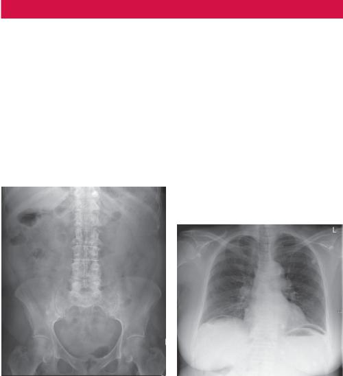
Abdominal and erect chest radiographs are obtained (Figure 25.1).
(a) |
(b) |
Figure 25.1 (a) Abdominal and (b) chest radiographs.
Questions
•What abnormality is present on the X-ray?
•What is your differential diagnosis?
•What further imaging would you like to do?
79
ANSWER 25
This is a case of acute abdomen. The first priority is to resuscitate and stabilize the patient and as the patient is middle aged and relatively fit, she may be compensating quite well. The abdominal X-ray shows undilated small and large bowel. No classic signs of free gas outside the bowel wall (Rigler’s sign), triangular gas pockets or gas outlining the falciform ligament are seen on the abdominal radiograph.
A chest X-ray is part of the investigation of acute abdomen and is obtained after about 5 minutes with the patient sitting as upright as possible. This allows any free intraabdominal gas to rise, giving the characteristic free gas under the diaphragm sign. This chest radiograph shows free gas below the diaphragm (pneumoperitoneum). Appearances may be deceptive and gas within an organ may look like a perforation. The right upper quadrant is a good place to look as the liver normally abuts the diaphragm and any gas will be obvious. Occasionally the bowel will occupy this space, a condition called Chiladiti’s syndrome. Look for characteristic bowel wall folds.
Differential diagnosis for free gas under the diaphragm includes:
•iatrogenic causes – normal appearance after a surgical or laparoscopic procedure or perforation from a surgical anastomosis or after endoscopy;
•perforation due to gastroinstestinal tract disease – gastric/duodenal ulcer, appendix or diverticular perforation, obstruction (e.g. neoplasm) or specific paediatric disorders and inflammatory bowel disease;
•conditions that mimic free gas – pseudopneumoperitoneum, such as distended bowel loops, Chiladiti’s syndrome, diaphragmatic hernia, oeosphageal diverticulum and subphrenic abscess.
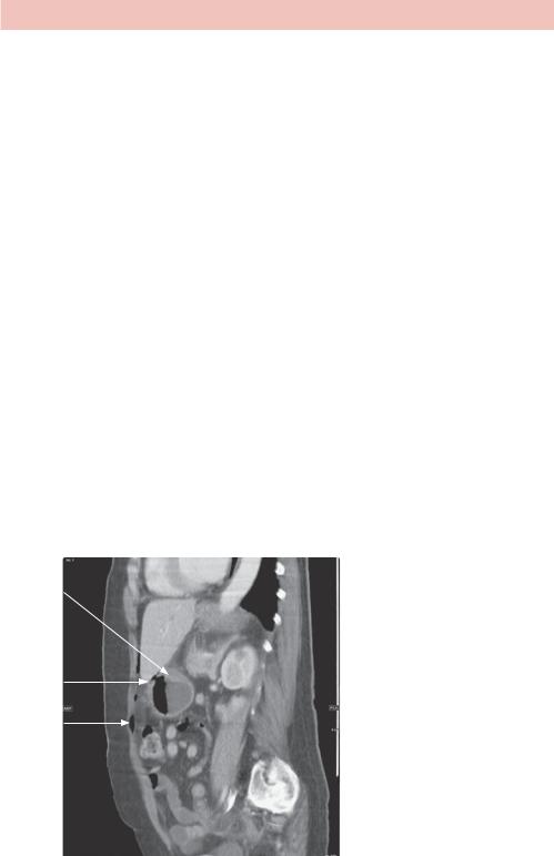
A contrast computed tomography (CT) is typically done provided the patient is stable, as the additional anatomical information may help locate the source of perforation (Figure 25.2).
Mass
Ulcer
Free gas
Figure 25.2 Sagittal reconstruction CT slice of the abdomen, showing free gas anterior to the stomach suggesting a perforated gastric ulcer and possible gastric mass. This is useful for the surgeon to know as tissue diagnosis is required and surgical planning may change.
80

Free gas can be seen after abdominal surgery although a significant quantity of gas 3 days after surgery is suspicious. If available, compare with any previous post-surgical images. CO2 insufflation used in laparoscopy absorbs rapidly and probably should be gone after 24 hours.
KEY POINTS
•Plain images of the patient can be done without moving the patient, although to allow free gas to appear under the diaphragm, the patient has to be upright before the chest X-ray.
•There are many causes of pneumoperitoneum and a combination of history and CT may determine the likely cause.
81