

Книги по МРТ КТ на английском языке / Thomas R., Connelly J., Burke C. - 100 cases in radiology - 2012
.pdfANSWER 60
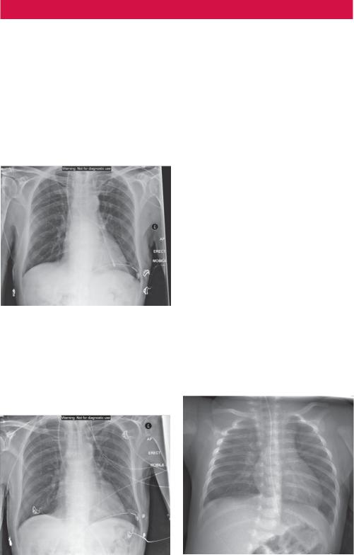
Figure 60.1 shows a normal heart size and mediastinal contour with no focal collapse, consolidation or active pulmonary lesion. The pleural spaces are clear. Incidental note is made of an azygos fissure.
The arrows in Figure 60.2 identify an azygos fissure (azygos lobe), which is a small accessory lobe sometimes found on the upper part of the right lung, separated from the rest of the upper lobe by a deep groove lodging the azygos vein, of little clinical significance.
The azygos lobe appears commencing in a ‘teardrop’ shape approximately at around the level of T5 to the right of the midline as a pale line curving outward and upward, then back in to meet the root of the neck. The line is the infolding of the pleura.
Abnormal fissures and lobes of the lungs are common and usually insignificant. A lobe related to the azygos vein appears in the right lung in about 1–2 per cent of people. It develops when the apical bronchus grows superiorly and medial to the arch of the azygos vein (instead of lateral to it). As a consequence, the azygos vein comes to lie at the bottom of a deep fissure in the superior lobe of the right lung.
In this patient, no focal active lung pathology was identified. Incidental note was made of the azygos lobe. No further investigation or treatment is required.
KEY POINTS
•An azygos lobe is frequently an incidental finding on chest radiograph and usually has no clinical significance.
162
CASE 61: A WORRIED NURSE IN INTENSIVE CARE
History
A 64-year-old man is admitted to intensive care following a coronary artery bypass graft and aortic valve replacement. The procedure was uncomplicated, but post-operatively he is slow to regain his appetite and a nasogastric tube is placed on intensive care for enteral feeding. The nurse uses litmus paper to test the acidity of the aspirates and is concerned. Simultaneously, the patient shortly afterwards begins to become dyspnoeic and is making gagging reflexes.
Examination
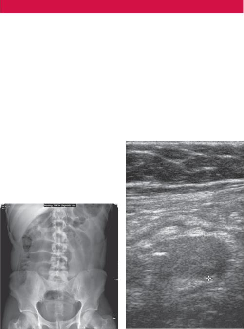
His saturations drop to 82–86 per cent on 8L of oxygen. His blood pressure and heart rate remain within normal limits. A chest radiograph is performed urgently (Figure 61.1).
Figure 61.1 Chest radiograph.
Questions
•What abnormality do you note on the chest radiograph in Figure 61.1?
•What other findings are there on the image?
•What has changed in Figure 61.2?
•What abnormality does Figure 61.3 show (taken from a different paediatric patient)?
Figure 61.2 Later chest radiograph.
Figure 61.3 Chest radiograph from a different (paediatric) patient.
163

ANSWER 61
The chest radiograph in Figure 61.1 shows that the nasogastric (NG) tube has been passed down the left main bronchus. This is unusual as the (less acute) angle of the right main bronchus is usually more favourable. There is a right internal jugular catheter which is appropriately placed with its tip projected over the superior vena cava/right atrium. A left intercostal chest drain is seen. Sternal wires are noted. Figure 61.2 demonstrates the appropriately repositioned NG tube extending below the diaphragm. Figure 61.3 demonstrates an NG tube in a different (paediatric) patient, which lies within the distal oesophagus and should be advanced into the stomach.
Chest radiographs for NG tube position are common. The NG tube is usually inserted either for providing enteral nutrition, administration of drugs or for gastric drainage. Nasogastric feeding is a common practice in all age groups. There is a risk that the NG feeding tube can be misplaced into the lungs during insertion (as in Figure 61.1) or may move out of the stomach at a later stage (Figure 61.3).
In the past, various methods have been used to determine the position of NG feeding tubes. These included:
•auscultation of air insufflated through the feeding tube (listening for a ‘whoosh’);
•testing acidity or alkalinity of the aspirate using litmus paper;
•looking for bubbling at the end of the NG tube;
•the appearance of the feeding tube aspirate.
Current recommendations, however, suggest that these measures are not reliable and therefore should not be used to detect the position of NG tubes.
The position of the tip of the NGT is assessed frequently in the first instance by drawing back gastric contents and testing with pH paper. If there is any concern then radiographic confirmation of NG tube position is performed.
Indeed current guidelines suggest:
•measuring the pH of the aspirate using pH indicator strips;
•use of radiography.
The most accurate method for confirming the correct position of an NG feeding tube is radiography. The aim of radiography is to positively confirm that the NG tube is within the gastrointestinal tract (within the stomach).
Sometimes longer nasojejunal (NJ) tubes are used. The objective is to place the tip of the tube past the pylorus, through the duodenum and beyond the duodenal–jejunal flexure into the jejunum. In doing so, this bypasses the regulatory function of the pylorus and delivers nutrition or therapeutic agents directly into the jejunum.
The importance of establishing the position of the NG tube cannot be understated. A patient who is fed or administered drugs via a malpositioned NG tube, such as that in Figure 61.1, can deteriorate significantly and it may even lead to iatrogenic death.
KEY POINTS
•Current guidelines for appropriate placement of NG tubes include measuring the pH of the aspirate using pH indicator strips and use of chest radiography to ensure the tip of the tube is sited appropriately.
164
CASE 62: YOUNG MAN WITH ABDOMINAL PAIN
History
A 24-year-old man presents to the accident and emergency department with vomiting and abdominal pain that has been worsening over the last 24 hours. He describes initially feeling unwell with mild fever and central abdominal discomfort that has slowly focused into the right iliac fossa. He has not been exposed to other people with similar symptoms and although he has bought and eaten freshly prepared food, none of his friends have has similar symptoms. He has no significant medical history.
Examination
His temperature is 37.8°C but his observations are otherwise normal. His chest and heart sounds are normal. On abdominal examination there is mild distension and tenderness over the right iliac fossa but no rebound tenderness. Bowel sounds are reduced.
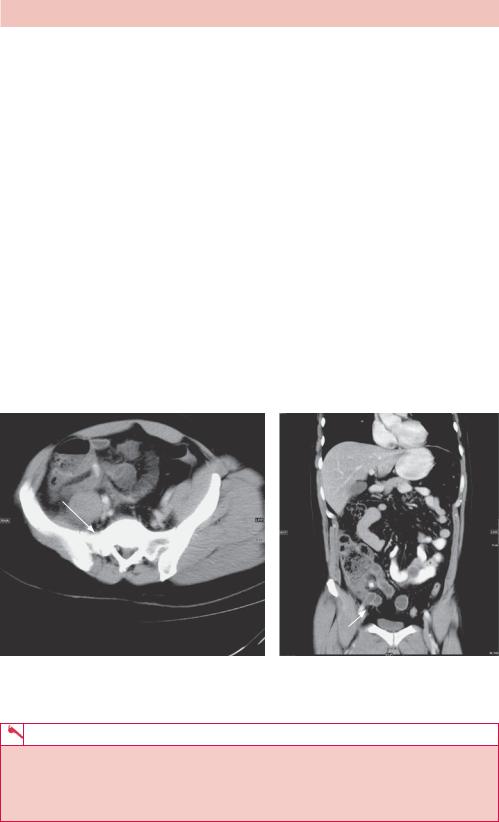
You arrange an abdominal radiograph (Figure 62.1) and an ultrasound (Figure 62.2), among other tests.
Figure 62.1 Abdominal radiograph. |
Figure 62.2 Ultrasound image of the right |
|
iliac fossa. |
Questions
•What do the investigations show?
•What differential diagnoses could give these appearances?
•What else would you like to know?
•Would you do more imaging?
165
ANSWER 62
The abdominal radiograph (Figure 62.1) shows dilated lucent loops of small bowel, likely to be partially gas filled in the upper abdomen. The distal small bowel is not seen, probably fluid filled and no comment about dilatation can be made. The large bowel gas pattern is not abnormal. The appearance could indicate a small bowel ileus or obstruction, although a transition point to indicate a mechanical obstruction is not seen.
The ultrasound of the right iliac fossa (Figure 62.2) shows a tubular structure measuring 1.3 cm containing a calcification that blocks the ultrasound beam and casts a shadow that may represent a dilated appendix (>6 mm diameter). The upper abdomen is normal. The bladder appears normal. No significant free fluid is seen.
The differential diagnosis includes appendicitis, mesenteric adenitis (usually children), gastroenteritis, Crohn’s disease, urinary tract infection and, in women, ovulation pain, ovarian cyst haemorrhage, torsion, ectopic pregnancy and pelvic inflammatory disease.
Appendicitis seems the most likely diagnosis, however tests should include urinalysis. In women, a pregnancy test should be done and it can be difficult to entirely rule out ovarian causes or pelvic inflammatory disease and a transvaginal ultrasound may be helpful although may not be acceptable in younger females. The diagnosis may end up being primarily clinical although a computed tomography (CT) may be done (as in this case due to the dilated small bowel) if it is considered that the additional information justifies the X-ray dose (Figure 62.3).
Appendicitis with small bowel dilatation is associated with a higher incidence of perforation of the appendix.
(a) |
(b) |
Figure 62.3 (a) Oblique axial and (b) coronal CT slices of the abdomen and pelvis showing a large appendicolith (present in 25 per cent on CT) within the dilated appendix (white arrows) with surrounding inflammatory change of the fat and caecal pole.
KEY POINTS
•Ultrasound may occasionally diagnose appendicitis but may struggle to rule out many of the differential diagnoses.
•Ultrasound may be useful where there is reasonably high suspicion for appendicitis in children and young women where a CT scan may be avoided.
166
CASE 63: SKIN PLAQUES AND ACHY HANDS
History
A 41-year-old woman is referred to the outpatient rheumatology clinic complaining of painful joints. The symptoms have been longstanding for 4 months and intermittently cause an achy pain in the finger joints of both hands. This is sometimes associated with joint swelling and causes stiffness, which is constant and not worse at any particular time of the day. She denies any stigmata of infection, weight loss or rash.
Approximately 5 years ago she was diagnosed with psoriasis for which she takes topical coal tar which controls her skin plaques well. She has no other relevant past medical history and does not take any other regular medication. She finds her job as a nursery school teacher very rewarding, but is finding her fine motor skill inhibited by joint stiffness when trying to help the children in arts and crafts lessons. She is a smoker of approximately 10 per week and drinks at weekends only. She is unmarried but lives with her boyfriend.
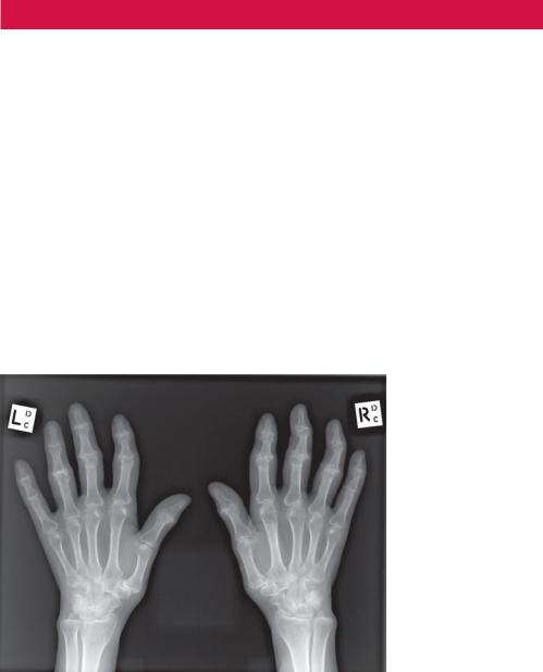
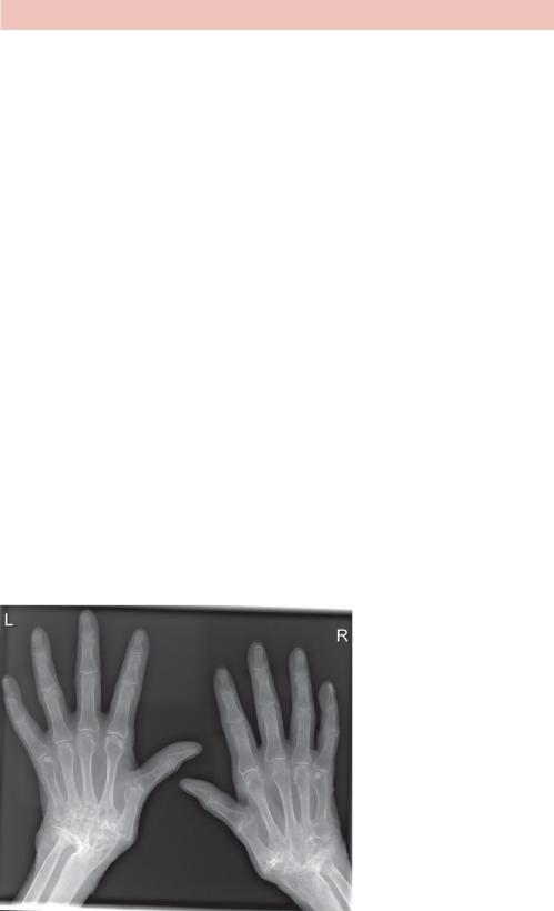
Examination reveals mildly swollen proximal interphalangeal joints bilaterally, which are tender and have slightly limited range of movement. Bloods are taken and a radiograph of both hands is ordered (Figure 63.1).
Figure 63.1 Anterior–posterior (AP) radiograph of both hands.
Questions
•What does the radiograph demonstrate?
•What is the main differential for these appearances?
•What are the extra-articular manifestations of rheumatoid arthritis?
167
ANSWER 63
Figure 63.1 is an AP radiograph of both hands in an adult female patient. Soft tissue swelling is seen involving the proximal interphalangeal joints (PIPJs). There is a symmetrical distal small joint polyarthropathy. This predominantly involves the second, third and fourth PIPJs of both hands with less marked involvement of the first interphalangeal joints and the distal interphalangeal joints (DIPJs). The involved joints demonstrate loss of normal cartilage and reduced joint space with marginal juxta-articular erosions. These are poorly demarcated and partially obscured with overlying new bone formation, suggesting periostitis, and are termed ‘proliferative erosions’. The PIPJ articular surfaces are irregular, but there is no subperiosteal osteoporosis or articular erosive changes seen to suggest a ‘pencil in cup’ appearance.
Bone density is preserved throughout and there is no bony ankylosis. The metacarpophalangeal joints are preserved, but there is mild radiocarpal and carpometacarpal joint space narrowing in keeping with degenerative change. These appearances are characteristic of a seronegative spondyloarthropathy most likely related to psoriasis, however a diagnosis of rheumatoid arthritis needs to be excluded.
Psoriasis is a chronic inflammatory skin condition, which can affect people at any age. It is characterized by the presence of red skin plaques that are raised from the skin and is often capped by silvery scales, associated with nail changes. It has a predilection for extensor sites and is most commonly identified at the elbow and knee joints. There are well-documented extracutaneous manifestations with arthropathy affecting 5 per cent of psoriatic suffers and sometimes preceding the skin plaques by many years. There five radiological subtypes of psoriatic arthropathy:1,2
•symmetrical polyarthritis of the interphalangeal joints;
•seronegative polyarthritis mimicking rheumatoid arthritis;
•monoarthritis or asymmetrical oligoarthritis;
•spondyloarthritis mimicking ankylosing spondylitis;
•arthritis mutilans – a severe form of arthritis with marked deformity and joint destruction.
In contrast, Figure 63.2 is a characteristic plain radiograph of both hands in a patient with longstanding rheumatoid arthritis. There is marked erosive change and fusion
Figure 63.2 Plain radiograph of both hands in a patient with longstanding rheumatoid arthritis.
168

related to the carpal bones bilaterally with erosive changes also seen related to the distal radius and ulna. Bone density is generally reduced and there is bony ankylosis of the carpometacarpal joints. The PIPJs and DIPJs are preserved throughout.
Rheumatoid arthritis is an immunologically mediated multisystem collagen vascular disease of unknown aetiology. Treatment in the acute setting of joint involvement is similar to that of psoriasis, but long-term treatment is complicated, often involving a delicate balance of disease modifying drugs (DMARDs) requiring regular clinical follow-up and blood tests. Being a multisystem disorder, rheumatoid arthritis can affect the patient in many ways outside of joint involvement. These include:
•systemic illness – fever, malaise and weight loss;
•vasculitis – predominantly affecting small vessels, the disease can mimic polyarteritis nodosa causing peripheral neuropathy;
•skin nodules – firm and tender skin nodules occur along tendon sheaths;
•respiratory disorders – lung nodularity with cavitation can be seen, with concomitant pulmonary fibrosis and pleural effusion;
•cardiovascular disorders – pericarditis, pericardial effusion and myositis are recognized;
•Felty’s syndrome – massive splenomegaly, often with neutropenia.
KEY POINTS
•Joint disease can precede the cutaneous manifestations of psoriasis by several years.
•Classically, psoriasis is associated with a symmetrical distal polyarthropathy of the interphalangeal joints.
•Rheumatoid arthritis predominantly affects the distal radius and proximal joints of the hand bilaterally.
References
1.Chapman, S. and Nakielny, R. Aids to Radiological Differential Diagnosis, 4th edn. Philadelphia: W.B. Saunders.
2.Jacobson, J.A., Girish, G., Jiang, Y. and Resnick, D. (2008) Radiographic evaluation of arthritis: inflammatory conditions. Radiology 248: 378–89.
169
This page intentionally left blank
CASE 64: TEACHING SPORT CAN BE A HEADACHE AT TIMES
History
A 38-year-old sports teacher was brought to the accident and emergency department by ambulance. He had been found unconscious in his office by another member of staff at the end of the afternoon. There was no sign of assault and the accompanying member of staff remembers seeing the sports teacher at lunch where they laughed together at something that had occurred that morning.
The sports teacher had taken his first class to the cricket nets for catching and batting practice. During the session a batsman had driven the ball hard, accidentally hitting the teacher on the left side of the head. Pupils near by heard a ‘crack’ but the sports teacher did not lose consciousness and carried on with the lesson. He did report at lunchtime feeling a little drowsy but had only administrative duties in the afternoon. On her way home, another teacher checked on the sports teacher to find him unrousable in his office and she raised the alarm.
Examination
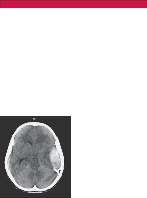
Ambulance paramedics urgently brought the sports teacher to the accident and emergency department. Glasgow Coma Scale score (GCS) on arrival was 7 (motor 4, eyes 2, speech 1). He had been intubated in the resuscitation room to protect his airway, and a cranial computed tomography (CT) scan was performed before admission to intensive care (Figure 64.1).
Figure 64.1 Axial cranial CT scan.
Questions
•What are the CT scan findings?
•What is the diagnosis?
171