

Книги по МРТ КТ на английском языке / Thomas R., Connelly J., Burke C. - 100 cases in radiology - 2012
.pdfANSWER 45
Triquetral fractures typically occur from a hyperextension injury with the wrist in ulnar deviation, however, can also occur with hyperflexion. Either the dorsal or volar radiotriquetral ligaments may avulse triquetral fragments at their attachments. Behind fractures of the scaphoid, triquetral fractures are the second most common carpal bone to fracture. They are frequently seen as dorsal chip fractures only on the lateral projection (Figure 45.2) since the pisiform usually overlies the triquetrum on the frontal projection of the wrist (Figure 45.1). In this case there is a small avulsion from the dorsum of the triquetrum seen only on the lateral projection (arrow, Figure 45.3).
Figure 45.3 Lateral view of the left wrist indicating the fracture.
Patients will usually complain of localized tenderness on the dorsum of the wrist.
The triquetrum may be identified by its pyramidal shape and by an oval isolated facet for articulation with the pisiform bone. It is situated just distal to the ulna and the triangular fibrocartilage complex, proximal to the base of the hamate.
The superior surface presents a medial, rough, non-articular portion, and a lateral convex articular portion, which articulates with the triangular articular disc of the wrist. The inferior surface, directed laterally, is concave, sinuously curved and smooth for articulation with the hamate. The dorsal surface is rough for the attachment of ligaments. The triquetrum articulates on its radial side with the lunate to which it is attached by the lunotriquetral ligament. On the volar (palmar) aspect there is an articulation with the pisiform.
Triquetral fractures may divided into two types:
•Chip fractures: A chip fracture, usually off the dorsal radial surface, typically occurs with a wrist hyperextension injury. Chip fractures can also occur with the hyperflexion.
•Mid-body fracture: Fractures through the mid-body of the triquetrum are less frequent than a chip fracture. This type of fracture is usually due to a direct blow, or may occur in conjunction with a perilunate dislocation. The dislocation may have been reduced, so a triquetral fracture from the proximal radial aspect of the bone may indicate the presence of a former dislocation.
KEY POINTS
•Triquetral fractures are the second most common fracture of the carpal bones.
•They are frequently seen as dorsal chip fractures only on the lateral projection as the pisiform usually overlies the triquetrum on the AP projection of the wrist.
132
CASE 46: SHORTNESS OF BREATH AND PLEURITIC CHEST PAIN
History
A 55-year-old man is admitted to the accident and emergency department complaining of gradual onset of shortness of breath over the course of several hours along with rightsided chest pain aggravated by deep inspiration. He is complaining of mild lightheadedness and feels the symptoms are getting worse. The pain is sharp and stabbing in nature. He was previously fit and well. One day earlier he returned on a long haul flight from a business trip to Asia. He denies any history of leg swelling.
Examination
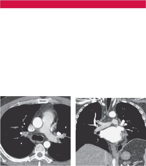
Upon admission to hospital his oxygen saturation on air was 94 per cent and his respiratory rate was 20/minute. A chest radiograph failed to demonstrate any focal lesion. Routine bloods were normal although D-dimer performed in the emergency department was elevated. Electrocardiogram (ECG) demonstrated a mild sinus tachycardia heart rate of only 102/minute. The accident and emergency team suspected a possible pulmonary embolism (PE) and so a computed tomography pulmonary angiogram (CTPA) was performed (Figures 46.1 and 46.2).
Axial CTPA image. |
Figure 46.2 Reformatted coronal CTPA |
|
image. |
Question
• What do the CT images demonstrate?
133

ANSWER 46
Figures 46.1 and 46.2 demonstrate axial and reformatted coronal CTPA images respectively, showing a large filling defect within the distal right main pulmonary artery extending into the upper lobe artery.
The initial chest radiograph in patients with pulmonary embolism is frequently normal. An initially normal chest radiograph may over time begin to show atelectasis, which can progress to cause a small pleural effusion and an elevated hemidiaphragm. After 24–72 hours, one third of patients with proven PE develop focal infiltrates that are indistinguishable from an infectious pneumonia. Occasionally, there may be evidence of Westermark sign, which is focal oligaemia (absence of blood vessel markings) beyond the location of the pulmonary embolism and dilatation of those vessels proximally. A rare late finding of pulmonary infarction is the Hampton hump, a triangular or rounded pleural-based infiltrate with the apex pointed toward the hilum, frequently located adjacent to the diaphragm.
CTPA is the most common study used for detection of pulmonary embolism and has become accepted both as the preferred primary diagnostic modality and as the standard for making or excluding the diagnosis of PE. In the majority of patients, multi-detector CT scans with intravenous contrast can resolve third-order pulmonary vessels.
It is important to note, however, that multi-detector CTPA carries a radiation dose and can miss lesions in a patient with pleuritic chest pain due to multiple small emboli that have lodged in distal vessels. In patients with a normal chest radiograph, nuclear scintigraphic ventilation–perfusion (V/Q) scanning of the lung is an alternative diagnostic modality for detecting PE with a lower radiation dose. This modality is recommended only for patients with a normal chest radiograph in order to prevent spurious perfusion mismatch from other lung processes.
KEY POINTS
•The initial chest radiograph in patients with PE is frequently normal.
•CTPA is the most common study used for detection of PE and has become accepted both as the preferred primary diagnostic modality and as the standard for making or excluding the diagnosis of PE.
•It is important to note, however, that in patients with a normal chest radiograph, ventilation–perfusion (V/Q) scanning is an alternative modality carrying a lower radiation dose.
134

CASE 47: A NEW ARRIVAL WITH COUGH AND A FEVER
History
A 40-year-old man arrives in the United Kingdom from Bangladesh to visit relatives. He has been complaining of feeling generally unwell with a cough and fever for around 3 weeks, which is worsening. He attends the accident and emergency department complaining of a productive cough and feels increasingly systemically unwell with fevers and sweating. He has has been well previously with no smoking history or history of exposure to dust.
Examination
His respiratory rate is 24 per minute and pulse 92 per minute. On respiratory examination there is limited expansion with resonance to percussion, rather quiet breath sounds but no bronchial breathing and no added sounds. He is febrile with a temperature of 40°C. Oxygen saturation is 92 per cent breathing air. Full blood count shows a white cell count of 20 × 103/mm3.
He is admitted to hospital under the care of the medical team but after a further 24 hours, the intensive care team are asked to review the patient as his breathing and oxygenation have deteriorated further. He is intubated and transferred to the intensive care unit where central lines and a nasogastric tube are inserted. A chest radiograph is performed (Figure 47.1) and, based on this, a computed tomography (CT) scan of the chest was requested (Figure 47.2).
Figure 47.1 Chest radiograph. |
Figure 47.2 Axial CT image. |
Questions
•Ignoring the bilateral internal jugular lines, what are the lung changes on the chest radiograph in Figure 47.1?
•How would you describe the pattern of disease on CT (Figure 47.2)?
135

ANSWER 47
The initial chest radiograph (Figure 47.1) shows diffuse patchy consolidation on a background of widespread tiny nodules distributed throughout all lobes bilaterally. Subsequent CT (Figure 47.2) confirms the presence of randomly distributed, diffuse tiny nodules and bilateral large pleural effusions. Findings are suggestive of miliary tuberculosis.
Miliary tuberculosis (TB) is the widespread dissemination of Mycobacterium tuberculosis via hematogenous spread. Classic miliary TB is defined as ‘millet like’ (approximately 2 mm) seeding of TB bacilli in the lung, as evidenced on chest radiography. This pattern is seen in 1–3 per cent of all TB cases.
Following exposure and inhalation of TB bacilli in the lung, a primary pulmonary complex is established, followed by development of pulmonary lymphangitis and hilar lymphadenopathy. Mycobacteraemia and haematogenous seeding occur after the primary infection. After initial inhalation of TB bacilli, miliary TB may occur as primary TB or may develop years after the initial infection. The disseminated nodules consist of central caseating necrosis and peripheral epithelioid and fibrous tissue.
Imaging findings may take weeks between the time of dissemination and the radiographic appearance of disease. Up to 30 per cent have a normal chest radiograph. When first visible, the nodules measure about 1 mm in size, but they can grow to 2–3 mm if left untreated. High-resolution CT scans are more sensitive at demonstrating small nodules. Nodules are either sharply or poorly defined and around 1–4 mm in size in a diffuse, random distribution. There may be associated intraand interlobular septal thickening.
Radiographically, nodules are not calcified, as opposed to the initial Ghon focus, which is often visible on chest radiographs as a small calcified nodule. Chest CT scanning is useful in the presence of suggestive and inconclusive chest radiography findings. When treated, clearing is frequently rapid. Under the age of 5 years, there is an increased risk of meningitis.
Risk factors include immunosuppression, cancer, transplantation, human immunodeficiency virus (HIV) infection, malnutrition, diabetes, silicosis and endstage renal disease.
KEY POINTS
•Miliary TB is caused by the widespread haematogenous dissemination of Mycobacterium tuberculosis. It is so named because the nodules are the size of millet seeds (1–5 mm with a mean of 2 mm).
•Miliary TB represents only approximately 1–3 per cent of all cases of TB.
•It is considered to be a manifestation of primary TB, although clinical appearance of miliary TB may not occur for many years after initial infection.
136
CASE 48: A SEATBELT INJURY
History
A 33-year-old man was a passenger of car involved in a head-on collision travelling at 60 mph (97 km/h). He is brought into the accident and emergency department as a trauma call. He was sitting in the rear of the car in the central passenger seat restrained by an old-style ‘lap belt’, unlike the other passengers who were wearing standard shoulder and lap belts. He was thrown forward into the seat in front. He was conscious throughout but is complaining of severe pain in the lower thoracic and lumbar spine.
Examination
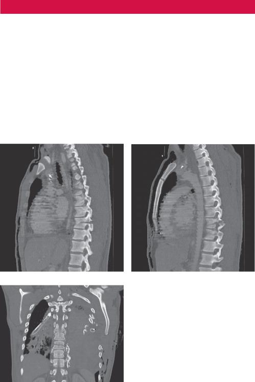
Primary survey is unremarkable and his observations are stable. There is marked tenderness over the lower thoracic and lumbar spine. On neurological examination of the lower limbs there are no motor or sensory abnormalities. Per rectal examination revealed normal tone. A computed tomography (CT) examination of the thorax and abdomen was obtained with sagittal (Figure 48.1a,b) and coronal (Figure 48.1c) reformats.
(a) |
(b) |
Question
• What abnormality do the CT images show?
(c)
Figure 48.1 CT scans: (a,b) sagittal and (c) coronal views.
137

ANSWER 48
The sagittal (Figure 48.1a,b) and coronal (Figure 48.1c) reformats of the spine show a ‘Chance’ fracture line extending through the spinous process, lamina, pedicles and vertebral body of T9 (a horizontal splitting of the vertebra beginning with the spinous process or lamina and extending anteriorly through the pedicles and vertebral body).
Chance fracture is caused by a flexion injury of the spine, first described by G.Q. Chance in 1948. It consists of a compression injury to the anterior portion of the vertebral body and a transverse fracture through the posterior elements of the vertebra and the posterior portion of the vertebral body. It is caused by violent forward flexion, causing distraction injury to the posterior elements.
Chance fractures later became known as ‘seatbelt’ fractures with the advent of lap seatbelts in cars. A head-on collision would cause the passenger wearing a lap belt to suddenly be flexed at the waist, hence creating significant stress on the posterior elements of the vertebra. From the 1980s when shoulder and lap belts became more common in cars Chance fractures have become less associated with road traffic accidents and are more commonly seen with falls or crush-type injuries where the thorax is acutely hyperflexed.
The most common site at which Chance fractures occur is the thoracolumbar junction and mid-lumbar region in paediatric populations.
An anterior–posterior (AP) view of the spine may reveal disruption of the pedicles and loss of vertebral body height. Frequently a transverse process fracture will be identified on AP projection. The lateral view will demonstrate the spinous process fracture and fractures through the laminae and pedicles. The vertebral body will usually look compressed and wedge shaped.
CT of the spine should be performed on all Chance fractures to assess the extent of the fracture and to evaluate the spinal canal. Sagittal reconstructions of the axial images provide a great deal of information about the fracture pattern. Posterior element fractures are better seen on lateral projection, although the AP view helps to demonstrate pedicle involvement.
Up to 50 per cent of Chance fractures have associated intra-abdominal injuries, including fractures of the pancreas, contusions or lacerations of the duodenum and mesenteric contusions or lacerations.
KEY POINTS
•The incidence of associated intra-abdominal injuries with a Chance fracture reaches 50 per cent. Therefore, when a Chance fracture is diagnosed a CT of the abdomen should be obtained.
•Injuries associated with Chance fractures include fractures of the pancreas, duodenum and mesentery contusions/rupture.
138
CASE 49: A DEVICE IN THE PELVIS
History
A 30-year-old woman is admitted via the accident and emergency department with diffuse cramping abdominal pain. She has not opened her bowels for 4 days. She has a history of painful endometriosis and has been using increasing amounts of opiate analgesia to control the pain.
Examination
Her observations are all within the normal range. Her haematology and biochemistry results are also normal.
The abdomen is soft with no guarding or rebound tenderness. Bowels sounds are present and normal. Per rectal exam demonstrates a rectum containing solid stool.
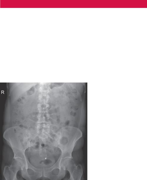
A plain radiograph of the abdomen was performed (Figure 49.1).
Figure 49.1 Plain radiograph of the abdomen.
Question
• What abnormalities can be seen in the abdomen radiograph?
139
ANSWER 49
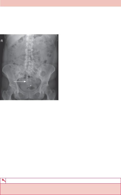
The radiograph of the abdomen demonstrates faecal loading of the large bowel but no significantly dilated loops (Figure 49.1) There is no evidence of free gas or pneumoperitoneum. Projected over the mid pelvis is an intrauterine contraceptive device (IUCD) (arrow, Figure 49.2). These are frequently seen as an incidental findings in radiographs of the abdomen and pelvis. A piercing is also present.
Figure 49.2
The IUCD is a form of birth control. It is an object, placed in the uterus, to prevent pregnancy. The aim of these devices is to confer long-term, reversible protection against unwanted pregnancy. They may, however, induce menstrual complications as well as an increased risk of pelvic inflammatory disease and ectopic pregnancy. They can also be spontaneously expelled from the uterus without being noticed by the patient.
Among modern IUCDs, the two types available are copper-containing devices and a hormone-containing device that releases a progestogen. Currently, there are several different kinds of copper IUCDs available in different parts of the world, and there is one hormonal device, called Mirena®.
An IUCD can increase the risk of spontaneous abortion unless removed in cases where intrauterine pregnancy occurs. Complications at the time of insertion include pain, syncope and uterine perforation.
In this case, however, the most likely cause of the patient’s abdominal pain is constipation due to opiate analgesia use and the IUCD is an incidental finding.
KEY POINTS
•Intrauterine contraceptive devices (IUCDs) are a commonly used form of birth control and are frequently seen as an incidental finding on radiographs.
140
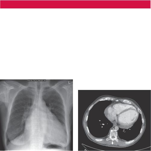
CASE 50: A CARDIAC ABNORMALITY
History
A 66-year-old woman is admitted to the accident and emergency department with sudden onset of chest pain 1 hour earlier. She had otherwise been fit and well apart from a history of hypertension for which she had been treated with amlodipine for the last 11 years.
Examination
The electrocardiogram (ECG) shows evidence of left ventricular hypertrophy. Troponin was not raised. D-dimer was moderately elevated. A chest radiograph was performed (Figure 50.1). The accident and emergency team were concerned about the possibility of a pulmonary embolism and therefore a computed tomography pulmonary angiogram (CTPA) was performed (one axial slice is shown in Figure 50.2). No pulmonary embolism or focal lung parenchymal abnormality was seen.
Figure 50.1 Chest radiograph. |
Figure 50.2 Axial CTPA image. |
Question
•What abnormalities do the chest radiograph in Figure 50.1 and axial enhanced CT image (at the level of the heart) seen in Figure 50.2 demonstrate?
141