

Книги по МРТ КТ на английском языке / Thomas R., Connelly J., Burke C. - 100 cases in radiology - 2012
.pdfANSWER 37
The radiographs show a well-defined sclerotic lesion that is eccentrically positioned in the distal femoral diaphysis. It appears to arise from the cortex and there is no associated bony expansion, soft tissue swelling or periosteal reaction. The ankle joint is normally positioned and regular in appearance. No fracture is seen. There is soft tissue swelling over the lateral malleolus.
When reviewing the ankle it is important to check the joint space between the talus and tibia/fibula which should be the same throughout. Small irregularities within the joint space might indicate an osteochondral defect. Check for avulsion below the fibula and posterior to the fifth metatarsal. Take care not to confuse fractures with ossicles commonly in this position and also posterior to the tibia.
There is quite a structured way of describing bony abnormalities that helps with deriving a differential diagnosis. Consider:
•age;
•number of lesions (may require more imaging);
•location;
•position within the bone (i.e. diaphysis, metaphysis, epiphysis, subarticular, cortex or medulla);
•density (sclerotic, lytic or mixed);
•borders – zone of transition well defined (narrow) or poorly defined;
•bony change – expansion, coarsened trabeculae, moth eaten (irregular holes also termed permeative lucency), osteopenia;
•periosteal reaction – lamellar, spiculated (tends to be associated with aggressive processes and can be hair on end, e.g. Ewing sarcoma, or sunray, e.g. metastases).
This patient’s lesion is a solitary sclerotic cortical lesion with benign appearance and, in this age group, a healing fibrous cortical defect is most likely. The differential includes osteoid osteoma (particularly with a central lucency or nidus). Less likely is a bone infarct (check for history of sickle cell disease, steroids), bone island or fibrous dysplasia. Unlikely is metastasis (age, appearance), primary bone sarcoma or osteomyelitis (typically aggressive periosteal reaction).
A fibrous cortical defect (or non-ossifying fibroma if greater than 2 cm) is the most frequent benign bony lesion in children and adolescents. These lesions are developmental abnormalities and usually an incidental finding on radiographs. The lesion develops in the distal metaphysis of a long bone as a radiolucent and eccentrically located lesion, with thin cortex and sclerotic or scalloped margins while the growth plate is open. There is typically spontaneous healing with sclerosis once the growth plate has ossified. Surgery is considered if there is risk of a fracture (occupying greater than 50 per cent of the transverse diameter), symptoms or enlargement.
KEY POINTS
•A good description of the lesion helps with deriving a differential diagnosis.
•Age is one of the main discriminators in deciding differentials.
112

CASE 38: PAINFUL SHOULDER
History
A 45-year-old man slipped and fell backwards onto his outstretched right arm. He presents complaining of severe shoulder pain and loss of movement. The shoulder is also swollen.
Examination
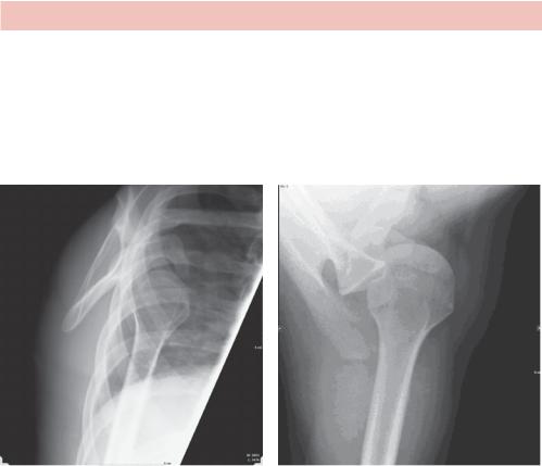
The patient holds his arm in slight abduction and external rotation. The shoulder is ‘squared off’ (i.e. box-like) with loss of deltoid contour compared with opposite side. The humeral head is just palpable anteriorly below the clavicle in the subcoracoid area. A careful assessment is made to check his radial pulses for a vascular injury and the axillary nerve function for regimental badge sensory loss over the deltoid and deltoid contraction on attempted abduction. Plain images are taken of the shoulder (Figure 38.1).
Figure 38.1 Posterior–anterior (PA) view of the right shoulder.
Questions
•What differential diagnoses should you consider?
•What complications can arise?
•What further imaging would be helpful?
113
ANSWER 38
The history and examination all suggest an anterior shoulder dislocation. Often in the case of trauma, shoulder injuries have to be assessed initially on the basis of a PA projection, which in this case confirms displacement of the humeral head inferiorly relative to the glenoid. Axial and Y views are nearly perpendicular to the PA projection and show the position of the humeral head. Figure 38.2 shows the humeral head anterior to the glenoid.
(a) |
(b) |
Figure 38.2 (a) Y view facing the glenoid fossa with the scapula describing a Y; (b) axial view of the right shoulder.
The imaging helps to distinguish the differential diagnosis of glenohumeral dislocation, of which anterior dislocation (96 per cent) is much more frequent than posterior (3 per cent) and inferior (1 per cent). Pseudo dislocation, where the humeral head is subluxed but not consistently dislocated, is associated with chronic shoulder joint instability, brachial plexus injury or haemarthrosis. Acromioclavicular dislocation and sternoclavicular dislocation should also be considered although much less common.
Posterior dislocations characteristically occur in trauma or seizure and can be harder to recognize, particularly if an axial or Y view has not been obtained. Suspicious features include fixed internal rotation (lightbulb sign), widening of the glenohumeral joint space (rim sign) with loss of overlap of the humerus over the glenoid.
Anterior dislocation can be associated with fracture of the greater tuberosity, anterior tear of the glenoid labrum (Bankart lesion), fracture of the anterior rim of the glenoid and an impaction fracture of the posterolateral surface of the humeral head (Hill–Sachs lesion) where it impacts on the glenoid rim. If not identified, recurrent dislocations are likely. The rotator cuff muscles may also be injured by traction.
Treatment requires adequate analgesia and sedation before relocating the joint. Imaging and neurovascular examination is performed before and after reduction.
114

Magnetic resonance (MR) is the investigation of choice to follow up a shoulder dislocation, particularly in younger patients who are more at risk of recurrent dislocations. Contrast injection into the joint (an arthrogram) may be required to see labral injuries reliably.
KEY POINTS
•Anterior dislocations are much the commonest type of shoulder dislocation.
•Two views are required to identify dislocation, particularly posterior dislocations.
•The risk of recurrent dislocations increases with younger age and further imaging with MR may be required.
115
This page intentionally left blank
CASE 39: CHEST PAIN AFTER FALLING
History
A 75-year-old man is brought to his general practice by his daughter. He complains of left-sided chest pain on inspiration. On further questioning it becomes apparent that he has had a recent fall. He lives alone and independently with frequent visits from his daughter who is concerned that there have been several falls. The patient is on blood pressure medication and takes a statin for previously high cholesterol. Reviewing his notes you see a previous attendance 2 years earlier for a fall and several episodes of treatment for pneumonia. There is a past history of smoking but no other significant medical history.
Examination
The observations are normal. He is thin. There is some bruising over the left side of the thorax and a notable low thoracic kyphosis. There are bilateral fine inspiratory crackles. The heart sounds are normal. The abdomen is unremarkable and no neurological or significant cognitive abnormality is noted.
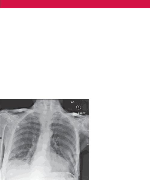
A chest radiograph (Figure 39.1), electrocardiogram (ECG) and blood tests are arranged.
Figure 39.1 Chest radiograph.
Questions
•What does the radiograph show?
•What is the differential diagnosis?
•What further imaging would you consider?
•What other investigations should you consider?
117

ANSWER 39
Although the history and examination make rib fractures very likely, radiology is rarely indicated simply to look for low energy traumatic rib fractures. In this patient’s case there are questions as to the cause of the fall. A chest radiograph is indicated for a possible chest infection or lesion and a thoracolumbar spine radiograph for the possible cause of the kyphosis.
The chest radiograph shows recent fractures of the left sixth to ninth ribs posterolaterally. There are also old fractures of the right ribs posterolaterally. Features that are common in trauma are fractures in a line and a posterolateral position, although this will depend on the type of trauma. Old fractures have healed and the bone cortex is continuous but the rib may be distorted. Recent and new fractures have discontinuous cortex but recent fractures show signs of healing with callus and bone remodelling that is absent in new fractures.
Do not stop looking once you have seen the fractures! Also look for other fractures (e.g. spine), bone lesions and examine the lungs, pleura and heart. On this radiograph there is consolidation in the right lower zone and a probable hiatus hernia gas bubble behind the heart.
The spine radiograph reported wedge compression fractures of the T11 and T12 vertebral bodies together with some small sclerotic and lytic lesions in the lumbar vertebrae. The initial blood tests show iron deficiency anaemia and raised erythrocyte sedimentation rate (ESR). This suggests an underlying systemic disease that affects bone and includes bowel or chest malignancy with bone metastases, myeloma or prostate carcinoma. The patient should be referred to the older person team for further investigation. From an imaging point of view this probably includes a computed tomography (CT) scan of the chest, abdomen and pelvis, although a bone scan could be helpful if sclerotic bone metastases from prostate or bowel carcinoma are suspected.
If blood tests suggest myeloma, a skeletal survey for lytic lesions could be done. Bone scans have lower sensitivity for lytic bone lesions which appear as photopenic regions of reduced uptake. The tracer used in bone scans, 99Tc-MDP (technetium-99 conjugated with methylene diphosphonate), is taken up primarily by osteoblastic activity typical in sclerotic bone lesions.
The National Service Framework for older person care recommends a formal falls assessment in patients at risk and regular medication review.
KEY POINTS
•Radiology is rarely indicated simply to look for rib fractures.
•In older patients the ‘Occam’s razor’ approach of assuming only one underlying pathology has to be modified to allow for several interacting pathologies.
•Avoid ‘satisfaction of search’ and keep looking after spotting the first abnormality.
118
CASE 40: SWELLING OF THE BIG TOE
History
A 57-year-old man presents to his general practitioner (GP) with pain and swelling of his left big toe. He reported that this had been a longstanding problem over the last 7 years but had just put it down to ‘old age’ and did not want to trouble his GP with it. The pain and swelling would come and go in waves. He noted, however, that these episodes were now more frequent and he would struggle to weight bear on that side at times. On this occasion the pain had suddenly started at night and had been getting worse in flares ever since. In his past medical history he had a hernia operation 14 years ago. He was on treatment for hypertension and hyperlipidaemia with bendroflumethiazide and simvastatin, respectively. He is an ex-smoker and drinks 14 units of alcohol per week. He is not aware of any history of trauma.
Examination
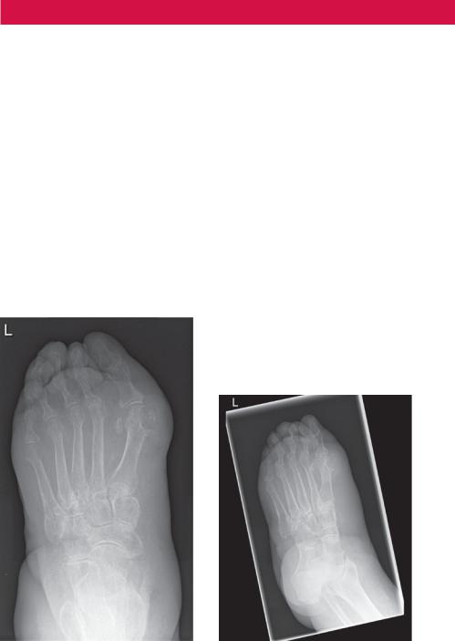
Examination revealed a tender, erythematous right big toe with a markedly reduced range of motion at the first metatarsophalangeal joint. He had a moderately raised white cell count of 12 and his erythrocyte sedimentation rate (ESR) was also raised. Serum urate levels were elevated. He was afebrile and observations were otherwise normal. Radiographs of the foot were taken (Figure 40.1).
(a) |
(b) |
Figure 40.1 (a) Anterior–posterior and (b) oblique radiographs of the foot.
Questions
• What do the radiographs of the foot demonstrate?
119

ANSWER 40
Figure 40.1 demonstrates ‘punched-out’ lytic bone lesions with overhanging edges involving the first metatarsophalangeal joint with marginal sclerosis. There is the appearance of ‘rat bites’ with an overlying calcified soft-tissue swelling. These classical findings are consistent with longstanding gout.
Gout is defined as a peripheral arthritis that results from the deposition of sodium urate crystals in one or more joints. It has many contributing factors, but two central processes are involved in its development: the overproduction of uric acid and the underexcretion of uric acid. A multitude of conditions, including renal disease, have been implicated, but the vast majority of cases are idiopathic. Podagra (another name for pain in the first metatarsophalangeal joint) is the classic presentation of gout. In general, the symptoms of gout appear suddenly at night and occur in men with hyperuricaemia who are aged 30–60 years.
Gout is also associated with hyperlipidaemia, hypertension, kidney failure, obesity and insulin resistance. Other ‘social’ factors such as alcohol intake also increase the risk of gout. An increase in uric acid levels and resulting precipitation of gout is a side effect of thiazide diuretics.
Patients with new onset of acute gout usually have no radiographic findings and the classic features seen in this case are usually not seen until 6–12 years after initial attack. Early radiologic findings in gout are limited to the soft tissues and involve asymmetric swelling in the affected joints. There is preservation of joint space initially. In the intermediate stage of disease, gout causes subtle changes in the bony structures on plain film radiographs. In the periphery of affected joints, small ‘punched-out’ lytic bone lesions arise, often with sclerosis of margin. Obtaining two views is important to appreciate these subtle findings. Definitive diagnosis is made by finding the negatively birefringent crystals on polarized microscopy of synovial fluid aspirate.
The hallmark sign of late-phase gout is the appearance of large and numerous interosseous tophi on plain film radiographs. Calcific deposits in gouty tophi are seen in approximately 50 per cent of cases (only calcium urate crystals are opaque) and ‘mouse/ rat bite’ erosions develop due to longstanding soft tissue tophi. Joint space narrowing and cartilage destruction is seen late in the course of the disease.
KEY POINTS
•Classical plain radiographic features of longstanding gout include ‘punched-out’ lytic bone lesions with overhanging edges, marginal sclerosis and the appearance of ‘rat bites’ with an overlying calcified soft tissue swelling (tophus).
•These features usually are not usually seen until several years after initial attack and are present in around 50 per cent of patients.
120
CASE 41: A YOUNG MAN WITH PROGRESSIVE DYSPNOEA ON
EXERTION
History
A 38-year-old man presents to his general practitioner (GP) with progressive dyspnoea on exertion developing steadily over the past 2 months. He says that he is now unable to walk 100 m before becoming severely short of breath and having to stop. He also noted the development of a non-productive cough over a similar time scale and more recently developing a fever. He had never experienced anything like this before and was previously fit and well with no past medical history of note. In his social history he reported having unprotected sex with both men and women.
Examination
On examination his respiratory rate was 22 per minute, fever (with a temperature of 37.9°C), a few crackles and wheezes over both lung fields. On the basis of the examination the GP sent the patient to the accident and emergency department.
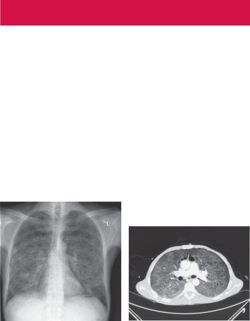
In casualty, oxygen saturation was 90 per cent breathing air. Haematology results showed a normal white cell count. Biochemistry and liver function tests were normal. A retroviral screen showed he was human immunodeficiency virus (HIV) positive with a CD4 count below 200 cells/μL, consistent with acquired immune deficiency syndrome (AIDS). He was referred for a chest radiograph (Figure 41.1), on the basis of which a computed tomography (CT) was requested and performed (Figure 41.2).
Figure 41.1 Chest radiograph. |
Figure 41.2 CT scan. |
Questions
•What is the likely cause of his increasing shortness of breath?
•What do the chest radiograph (Figure 41.1) and subsequent CT scan (Figure 41.2) show?
121