

Книги по МРТ КТ на английском языке / Thomas R., Connelly J., Burke C. - 100 cases in radiology - 2012
.pdfANSWER 29
Given the history of inversion, a common injury, the lateral side of the foot, particularly the base of the fifth metatarsal should be checked. Forced dorsiflexion suggests the shafts of the metatarsal and phalanges could also be injured.
The radiographs show a complex lucency through the base of the right fifth metatarsal, in keeping with a fracture (Figure 29.2). There is associated soft tissue swelling. No other fractures are seen.
The appearance of the fracture is complex. Reviewing the whole foot, multiple epiphyses are noted consistent with a 13 year old with active growth plates which may cause some confusion in interpretation of the radiographs. An unfused apophysis (secondary ossification centre) lies parallel to the lateral edge of the base of the fifth metatarsal. The transverse fracture line crosses the apophysis and base of the metatarsal. Fractures at the base of the fifth metatarsal are common and reflect an avulsion injury of the peroneus brevis tendon, typically on inverting the foot. The fracture edge is typically at right angles to the metatarsal lateral cortex and should not be confused with the apophysis, if present. There may also be an ossicle close the fifth metatarsal base. Ossicles should have a smooth outline with a regular or corticated edge.
Other common foot injuries to be considered that can be subtle in appearance are:
•Lisfranc fractures (see Case 87), in which the Lisfranc ligament at the base of the first to fourth metatarsals is injured; there may be avulsion fragments between the first and second metatarsal bases and the alignment of the metatarsal shafts and cuneiforms is lost;
•avulsions, appearing as small flakes of bone around the interphalangeal joints where the flexor, extensor tendons insert;
•stress fractures of the shafts of predominantly second and third metatarsals in long distance runners or people with a walking injury; these may be difficult to see and appear initially only as a periosteal reaction;
•fracture through the first metatarsal ossicles.
KEY POINTS
•The mechanism of injury and site of symptoms may help to find subtle injuries.
•It is important to look around the edge of every bone, as small avulsions and stress fracture periosteal reactions are easy to miss.
•Review the whole foot and beware of ‘satisfaction of search’ – where you stop looking after finding an injury. Multiple injuries are common in trauma.
92
CASE 30: LEFT-SIDED LOIN PAIN
History
A 24-year-old man presents with sudden onset left upper quadrant pain radiating to the groin with mild haematuria. He has no history of previous episodes or past renal problems. There is no history of lower urinary tract symptoms. He is otherwise fit and well with no medical problems or relevant family history. He smokes 10 cigarettes per day and drinks around 10 units of alcohol per week.
Examination
He is well with a pulse of 94 per minute but otherwise normal observations. The chest examination is normal with normal heart sounds. The abdomen is soft but tender on the left side, most notably over the left renal angle and left inguinal fossa. Urinalysis shows blood 4+ but no protein or nitrites.
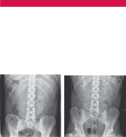
You arrange an urgent intravenous urogram (IVU) (Figure 30.1).
(a) |
(b) |
Figure 30.1 (a) Control and (b) 20-minute images from an intravenous urogram. Pelvic views were normal.
Questions
•What is a urogram?
•What are you looking for on the control image and do you see any abnormality?
•What does the 20-minute image show?
•What is the differential diagnosis?
93
ANSWER 30
An IVU consists of a control image without contrast to look for calcification. Contrast is then given intravenously and images are taken while the contrast passes through the kidneys (nephrogram phase) and then as it drains through the collecting system and ureters into the bladder. The IVU is becoming a rather old-fashioned test because it provides only limited anatomical information and is being replaced by pre, post and delayed contrast phase computed tomography (CT).
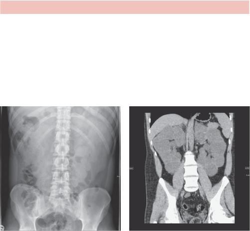
The control image (Figure 30.2a) is reviewed for calculi (none seen) and the renal outline, which in this case appears enlarged and lobulated bilaterally (Figure 30.2b).
(b)
Figure 30.2 (a) Control and (b) corresponding coronal CT slice demonstrating the renal outline and appearance.
The 20-minute IVU radiograph shows drainage of contrast through the collecting system on the right that has a slightly distorted appearance. No drainage is seen on the left, suggesting obstruction, confirmed on later images with a delayed nephrogram and slow accumulation of contrast in a dilated collecting system and due to a small stone, not seen on the plain images, at about the level of the pelviureteric junction (outflow of the left kidney) seen on CT.
There is an underlying bilateral kidney disorder with lobulated increase in size due to cyst formation. The differential for cystic diseases of the kidney includes acquired simple cysts (most common with increasing age and few in number), developmental disorders (e.g. multicystic dysplastic kidney), genetic causes (e.g. autosomal recessive (ARPKD) and dominant (ADPKD) polycystic kidney disease), systemic diseases (e.g. Von Hippel–Lindau syndrome and tuberous sclerosis) or malignancy in the form of cystic renal cell carcinoma. This patient has newly diagnosed ADPKD.
Unlike ARPKD that presents in childhood with renal failure and may be diagnosed prenatally, ADPKD is often clinically silent until it presents in adulthood, either with complications such as stones, haematuria, hypertension or renal failure (typically mean age
94

for endstage renal failure is over 50). However, as an autosomal dominant disease, the patient may be aware of a family history of renal disease and may present for ultrasound screening. Cysts may be seen in other organs and there is an association with cardiac and vascular anomalies, such as intracranial berry aneurysms.
KEY POINTS
•There are many causes of renal cysts; sporadic simple cysts are the most common.
•ADPKD typically becomes symptomatic later in life and is a significant cause of endstage renal failure.
95
This page intentionally left blank
CASE 31: UNABLE TO WEIGHT BEAR AFTER A CYCLING ACCIDENT
History
A 45-year-old woman presents to the accident and emergency department after collision with a car while on her bicycle. She complains that the car hit her left knee from the side and she is unable to bend the knee or support her weight. Previously she was fit and well.
Examination
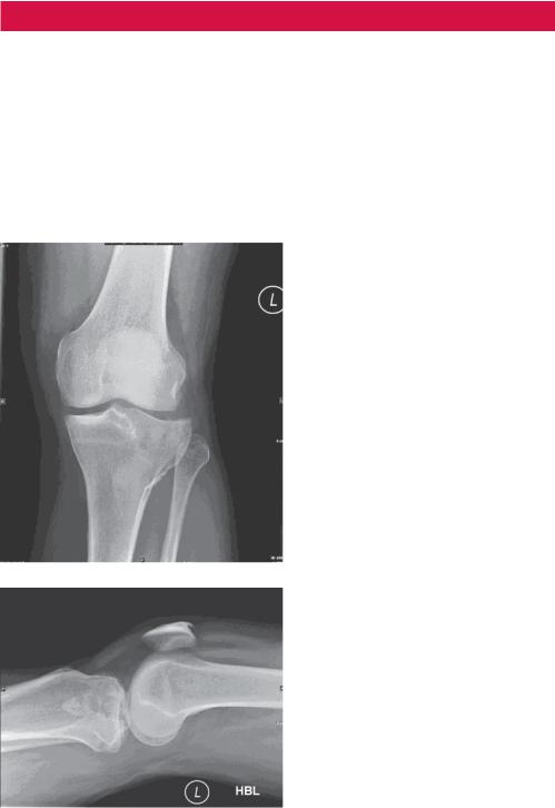
She is initially immobilized with a hard collar. Her neck, chest and abdomen examination is unremarkable and plain images of the neck, chest and pelvis are normal. The left knee appears swollen and bruised but there is no penetrating injury. Plain anterior–posterior (AP) and lateral radiographs of the knee are taken (Figure 31.1a,b).
(a)
Figure 31.1 (a) AP and (b) lateral radiographs of the left knee.
Questions
•What abnormality is seen?
•What would you do next?
97
ANSWER 31
There is a horizontal fat–fluid line, a lipohaemarthrosis, in the suprapatella pouch seen on the cross-table horizontal lateral view. There is also a fracture of the lateral tibial plateau, seen on the AP projection, with minimal displacement and loss of height.
A lipohaemarthrosis results from an intra-articular fracture with escape of fat and blood from the bone marrow into the joint. Ideally the patient has been lying supine for 5 minutes to allow the fat and blood to separate. The fat rises and is less radio-opaque. If seen, a lipohaemarthrosis indicates a tibial plateau or distal femoral fracture even if the fracture is not apparent. Conversely, in a significant proportion of tibial fractures a lipohaemarthrosis is not seen, although a haemarthrosis or effusion that appears as soft tissue without a fluid level in the suprapatella bursa is likely to be present.
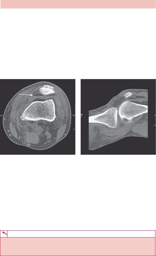
A computed tomography (CT) scan is done to image the extent of injury and plan surgery (Figure 31.2).
Fat on fluid level
(b)
Figure 31.2 (a) Axial and (b) sagittal CT slices of the right knee demonstrating the suprapatellar lipohaemarthrosis.
There are a number of classification systems, such as the Schatzker classification, that recognize patterns of fragmentation and displacement for tibial fractures. It is important to recognize that a significant proportion of fractures will have associated meniscal, collateral and cruciate ligamentous injury. These are better assessed by magnetic resonance (MR) which can also identify occult fractures.
Tibial plateau fractures may be either low or high energy. The majority of tibial plateau fractures are in patients over 50 years. Osteoporosis in older women is a contributing factor in low energy fractures and typically results in a depressed fracture. Tibial plateau fractures in younger patients are commonly the result of high energy injuries. The most common mechanism is a valgus force at the knee while weight bearing or with axial loading, typically either road traffic accidents or sports-related injuries.
KEY POINTS
•A fat–fluid level in the suprapatella bursa indicates a lipohaemarthrosis and is likely to indicate a tibial plateau fracture even if not seen on plain radiographs.
•Increased size of the suprapatella bursa most likely indicates an effusion or a haemarthrosis.
98
CASE 32: STRANGE BONE APPEARANCE AFTER FALLING
History
A 77-year-old woman presents to the accident and emergency department after slipping on ice and falling, hurting her left hip. She is unable to weight bear on her left leg. There is no history of significant past joint pain or swelling. Before falling she had mildly limited mobility but was otherwise active and well. Other than bendroflumethiazide for hypertension there is no significant medical history.
Examination
The left leg is shortened with deformity around the hip. There is bruising over the lateral aspect but no neurological or vascular abnormality is noted in the leg. Her observations and the rest of the examination are normal.
You organize a pelvic and left hip radiograph and review a previous image of the left hip (Figure 32.1).
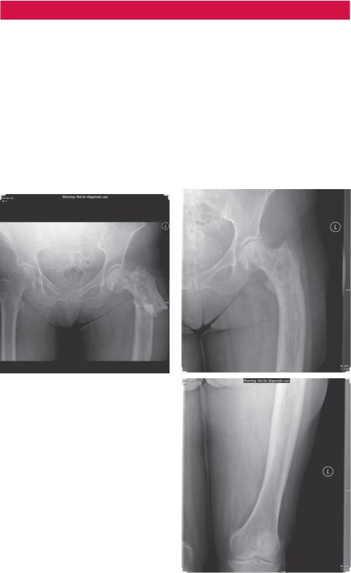
Figure 32.1 Current AP pelvic radiographs (left) and AP hip radiograph (right) from
1 year earlier.
Questions
•What abnormality is seen on the pelvic radiograph.
•What is the differential diagnosis for this appearance?
•What would you do next?
99
ANSWER 32
The pelvic radiograph shows a fracture through the proximal femoral shaft with displacement of the distal femur and angulation of the proximal fragment. The radiographs also show a longstanding abnormal bony appearance of most of the left femur with expansion (compared with the right), cortical thickening and trabecular coarsening, most prominent around the femoral head and neck. There is also bowing of the femoral shaft.
The differential diagnosis includes Paget’s disease, osteomyelitis, metastatic carcinoma and myelofibrosis. Trabecular coarsening and cortical thickening is quite characteristic of Paget’s disease. There is no history or other findings to suggest metastatic cancer (typically multiple sclerotic lesions in breast or prostatic cancer) or osteomyelitis, and the asymmetry makes myelofibrosis less likely.
Paget’s disease is a disorder of bone remodelling which may occur in single or multiple bones and typically affects spine, pelvis, femur and skull. The aetiology is not known, however, the disease progresses through a resorption, lytic phase with excessive osteoclastic activity to a bone formation osteoblastic sclerotic phase with a mixed phase in between. Paget’s disease occurs predominantly in older patients, affecting less than 3 per cent of patients around 50, rising to as much as 10 per cent in the over 80s. There is a slightly higher incidence in Europeans and males.
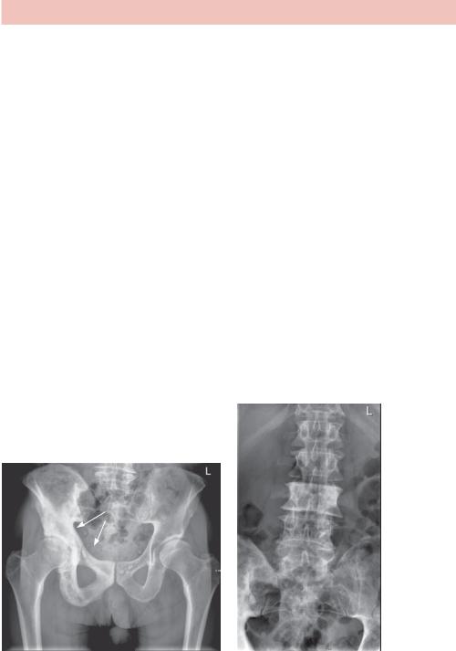
Figure 32.2 demonstrates Paget’s disease in other bones. In the spine, the vertebral bodies typically become enlarged with a prominent cortical margin (picture frame vertebrae) or become sclerotic, mimicking lymphoma or metastatic disease. In the pelvis, typical findings include thickening of the iliopectineal line (see arrows, Figure 32.2a) in early stages, progressing to patchy sclerosis and lucency in later stages.
(a) |
(b) |
Figure 32.2 Paget’s disease in (a) the right side of the pelvis and (b) an L3 vertebral body of different patients.
100

Complications of Paget disease depend on the bone and stage of disease. The majority of people with Paget’s disease are asymptomatic, but those with symptoms may experience bone pain (most common symptom), osteoarthritis of adjacent joints, insufficiency fractures, bowing of affected long bones, excessive warmth (due to hypervascularity) and neurologic complications such as deafness and cranial nerve involvement, particularly when the spine or skull is involved. Beware of sarcomatous change in 1 per cent of patients (rising to 5–10 per cent if more than one bone is affected).
KEY POINTS
•Paget’s disease is usually asymptomatic and a not infrequent incidental finding in older people.
•The disease typically affects spine, pelvis, femur and skull, and characteristically demonstrates cortical thickening, trabecular coarsening late in the disease, although lucency is a feature of early Paget’s disease.
101