

Книги по МРТ КТ на английском языке / Thomas R., Connelly J., Burke C. - 100 cases in radiology - 2012
.pdfThis page intentionally left blank
CASE 76: SHOOTING LEG PAIN FOLLOWING LIFTING
History
A 63-year-old man is lifting boxes at home when he experiences the sudden onset of lower back pain. The pain is so severe that he is unable to move and consequently calls an ambulance. Upon admission to accident and emergency he describes the pain as sharp and radiating down his legs, more severe on the left.
Examination
On examination there are reduced ankle reflexes and weakness of plantar flexion at the ankle on both sides. He is unable to straight leg raise on the left. He is admitted under the orthopaedic team for management of the pain and a magnetic resonance imaging (MRI) scan is performed (Figures 76.1 and 76.2).
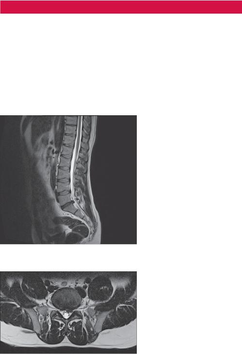
Figure 76.1 Sagittal T2-weighted MR image of the lumbar-sacral spine.
Figure 76.2 Axial T2-weighted MR image.
Questions
•What do the MRI images show?
•What is the reason for these appearances?
213

ANSWER 76
Figure 76.1 is a sagittal T2-weighted MR image of the lumbar-sacral spine, which demonstrates a disc prolapse at the level of L5/S1. Figure 76.2 is an axial T2-weighted MR image through the level of the disc herniation, which demonstrates the disc effacing the left L5 nerve root as it exits the spinal canal.
A spinal disc herniation is a medical condition affecting the spine, in which a tear in the outer, fibrous ring (annulus fibrosus) of an intervertebral disc allows the soft, central portion (nucleus pulposus) to bulge out. Tears are frequently posterolateral owing to the presence of the posterior longitudinal ligament in the spinal canal. Nuclear material that is displaced into the spinal canal is associated with an inflammatory response, and the tear in the disc ring can result in the release of inflammatory chemical mediators which may directly cause pain, even in the absence of nerve root compression. This is the rationale for using anti-inflam- matory medication for pain associated with disc herniation, protrusion, bulge or disc tear.
A weakened annulus is a necessary condition for herniation to occur. Many cases involve trivial trauma, sometimes in the presence of repetitive stress. Traumatic injury to lumbar discs commonly occurs when lifting while bent at the waist, rather than lifting with the legs while the back is straight.
Lumbar disc herniations occur most often between the fourth and fifth lumbar vertebral bodies or between L5 and the S1 (as in this example). Symptoms can affect the lower back, buttocks, thigh, anal or genital region (via the perineal nerve) and may radiate into the foot. The sciatic nerve is the most commonly affected nerve, causing symptoms of sciatica. The femoral nerve may also be affected.
Plain radiographs do not demonstrate disc herniation but are useful in the diagnosis of other conditions, particularly fracture, bone metastases or infection, and should be the primary imaging modality when trauma, malignancy or infection are suspected.
MR imaging is the modality of choice for delineating herniated nucleus pulposus and its relationship with adjacent soft tissues. On MRI, disc prolapses appear as focal, asymmetric protrusions of disc material beyond the confines of the annulus. Herniation of the nucleus pulposus are usually hypo-intense, however, because disc herniations are often associated with a radial annular tear, high signal intensity in the posterior annulus is often seen on sagittal T2-weighted images.
On sagittal MR images the relationship of herniation of the nucleus pulposus and degenerated facets to exiting nerve roots within the neural foramina is well delineated. In addition, free fragments of the disc are easily detected on MRI.
In cases of disc bulging, early MR findings include loss of the normal posterior disc concavity. Moderate bulges appear as non-focal protrusions of disc material beyond the borders of the vertebrae. Bulges are typically broad based, circumferential and symmetric.
A radial tear of the annulus fibrosus is considered a sign of early disc degeneration. It is accompanied by other signs of disc degeneration, such as a bulging annulus, loss of disc height, herniation of the nucleus pulposus and changes in the adjacent endplates.
KEY POINTS
•Herniation of the nucleus pulposus through an annular defect causes focal protrusion of the disc material beyond the margins of the adjacent vertebral endplate.
•MR imaging is the modality of choice for demonstrating the relationship of the herniated nucleus pulposus and degenerated facets to exiting nerve roots within the neural foramina.
214
CASE 77: A CHRONIC PRODUCTIVE COUGH
History
A 30-year-old woman is admitted to hospital with a fever and a cough. She has a history of repeated chest infections for several years, two of which have required hospital admission. She has been coughing up yellow-green purulent sputum for two days. She has had some streaky haemoptysis but is not worried as this has occurred on previous occasions.
Examination
Her observations were stable with a heart rate of 80 per minute and a blood pressure of 124/82. Her respiratory rate was 22 per minute, with reduced air entry bilaterally and diffuse coarse crackles most pronounced over the lower zones posteriorly. Her blood haematology showed a leucocytosis and raised C-reactive protein. Her biochemistry and liver function tests were normal. Chest radiograph and computed tomography (CT) scans were taken (Figures 77.1 and 77.2).
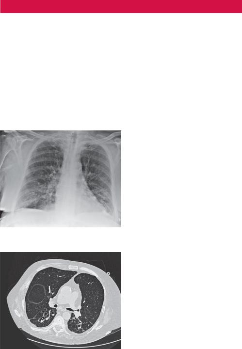
Figure 77.1 Chest radiograph.
Figure 77.2 Axial CT image taken at the level of the lower lobes.
Question
• What do the chest radiograph and axial CT image demonstrate?
215

ANSWER 77
The chest radiograph (Figure 77.1) shows ring opacities (cystic changes) due to the dilatation of bronchi with crowding of the vascular markings and prominent parallel lines (tram tracks) presenting thickened walls of the dilated bronchi.
On the axial CT scan through the lower lobes the bronchi are dilated and larger than the accompanying vessels. This is abnormal and sometimes referred to as the ‘signet ring sign’. The signet ring sign consists of a small circle of soft tissue attenuation that abuts a ring of soft tissue attenuation surrounding a larger low attenuating circle of air. The ring of soft tissue attenuation represents the wall of the dilated bronchus seen on an axial CT scan, whereas the low attenuating circle of air represents air within the dilated bronchus. The circle of soft tissue attenuation abutting the ring represents the pulmonary artery that lies adjacent to the dilated bronchus seen in cross-section. In Figure 77.2 dilated, thickwalled, slightly ectatic bronchi are demonstrated within both lower lobes.
Bronchiectasis is the irreversible dilatation of part of the bronchial tree. The bronchi involved are dilated, inflamed and collapsible. This results in airflow obstruction and impaired clearance of secretions. Bronchiectasis is associated with a wide range of disorders, some inherited, such as cystic fibrosis or agammaglobulinaemia, and some acquired, such as necrotizing bacterial infections caused by Staphylococcus or Klebsiella or early childhood infections caused by measles or Bordetella pertussis.
Haemoptysis, as experienced by the patient in this case, is common and may occur in up to 50 per cent of patients. Episodic haemoptysis with little sputum production or ‘dry’ bronchiectasis is a sequela of tuberculosis. When massive haemoptysis occurs, the bleeding usually originates in dilated bronchial arteries, which contain blood at systemic as opposed to pulmonary pressures.
The diagnosis of bronchiectasis is based on a clinical history of frequent viscid sputum production and characteristic CT scan findings.
Chest radiography is usually the first imaging examination, but the findings are often non-specific and the images may appear normal in patients with minor to moderate disease. Abnormal radiographic findings may be non-specific and confirmation using high-resolution CT (HRCT) scanning may be required.
Although subtle, thick-walled and dilated bronchi are present on the radiograph in Figure 77.1. Potential abnormal plain radiographic findings in bronchiectasis include: parallel line ‘tram track’ opacities caused by thickened dilated bronchi; ring opacities or cystic spaces as large as 2 cm in diameter resulting from cystic bronchiectasis, sometimes with air–fluid levels; tubular opacities caused by dilated fluid-filled bronchi; increased size and loss of definition of the pulmonary vessels in the affected areas as a result of peribronchial fibrosis; crowding of pulmonary vascular markings from the associated loss of volume, usually caused by mucous obstruction of the peripheral bronchi; oligaemia as a result of reduction in pulmonary artery perfusion in severe disease; signs of compensatory hyperinflation of the unaffected lung.
CT, in particular HRCT, scanning has become the imaging modality of choice for demonstrating and defining the extent of bronchiectasis. HRCT allows evaluation of the surrounding lung tissue and assessment for other lesions.
On HRCT scans in patients with bronchiectasis, the internal bronchial diameter may be greater than that of the adjacent artery and there may be a lack of bronchial tapering. The bronchi may be within 1 cm of costal pleura or abut the mediastinal pleura and bronchial
216

wall thickening may be seen. A cystic cluster of thin-walled cystic spaces may be present, often with air–fluid levels.
KEY POINTS
•Plain chest radiographs are usually the first imaging examination, but the findings are often non-specific and the images may appear normal in patients with minor to moderate disease.
•HRCT scanning is the diagnostic modality of choice and has few limitations.
217
This page intentionally left blank
CASE 78: GENERAL FATIGUE AND WEAKNESS
History
A 65-year-old man presents to his general practitioner (GP) with breathing difficulty over the past month, along with problems chewing, talking and walking up the stairs in his house. Of particular concern, he had noticed his eyelids drooping and he had difficulty maintaining a steady gaze. He noted that he has felt persistently fatigued and his symptoms worsened with activity and improved with rest.
Examination
He was afebrile. His pulse was 74/minute and blood pressure 126/78. The chest was clear, breath sounds vesicular although there was reduced expansion and a respiratory rate of 22/minute.
Neurological examination demonstrated bilateral ptosis along with weakness of the arms, legs and swallowing muscles. The jaw was slack and the voice had a nasal quality. Muscle weakness was worsened by repetitive or sustained use of the muscles involved. Recovery of strength was seen after a period of rest.
Full blood count and biochemistry were normal. Due to the difficulty in breathing, the patient was referred for a chest radiograph (Figure 78.1) and computed tomography (CT) scan (Figure 78.2).
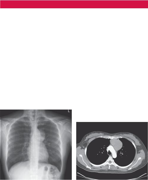
Figure 78.1 Chest radiograph. |
Figure 78.2 CT scan. |
Questions
•What is the abnormality on the chest radiograph?
•Where is the lesion anatomically located?
•Does Figure 78.2 confirm your suspicion?
•What is the differential diagnosis?
219

ANSWER 78
Figure 78.1 shows a smooth, well-defined mass arising from the left side of the mediastinum. By virtue of the fact that the margin of the descending thoracic aorta can be clearly seen, this mass does not lie within the posterior mediastinum. Furthermore, the hilar structures can also be seen making the middle mediastinum an unlikely location. Indeed the axial CT scan seen in Figure 78.2 demonstrates that the mass lies within the anterior mediastinum.
Anatomically the mediastinum lies between the right and left pleura, in and near the median sagittal plane of the chest. It extends from the sternum in front to the vertebral column behind, and contains all the thoracic viscera except the lungs.
It may be divided into superior and inferior parts:
•The superior mediastinum lies above the upper level of the pericardium at the plane drawn from the sternal angle to the disc of T4–T5 (angle of Louis).
•The inferior mediastinum lies below the superior margin of the pericardium and is further subdivided into three parts: anterior – in front of the pericardium; middle – containing the pericardium and its contents; posterior – behind the pericardium.
The mediastinum is surrounded by the chest wall anteriorly, the lungs laterally and the spine posteriorly. It is continuous with the loose connective tissue of the neck, and extends inferiorly onto the diaphragm.
The location of a mass within the mediastinum can be deduced radiologically on the plain chest radiograph by assessing a number of radiological landmarks. In Figure 78.1 the margin of the descending aorta and thoracic vertebrae/ribs, which are posterior mediastinal structures, can be seen crisply and clearly. This suggests that the lesion is not within the posterior mediastinum (where the presence of a mass located in opposition to these structures would cause a silhouette sign). The vascular structures of the hilum are also preserved. Therefore, the mass most likely lies within the anterior compartment. This is confirmed on CT (Figure 78.2).
The anterior mediastinum contains the following structures: thymus, lymph nodes, pulmonary artery, phrenic nerves and thyroid. The most common lesions encountered in the anterior mediastinum arise from thyroid, thymic (thymoma) or lymph node (lymphoma) origin. Germ cell tumours (teratoma) arise from pluripotent cells of the thymus.
This case was later proven to be a thymoma, in the context of myasthenia gravis. Indeed as many as 30–40 per cent of patients who have a thymoma experience symptoms suggestive of myasthenia gravis. The thymus is a lymphoid organ located in the anterior mediastinum. In early life, the thymus is responsible for the development and maturation of cell-mediated immunological functions.
The thymus gland is located behind the sternum in front of the great vessels. It reaches its maximum weight at puberty and undergoes involution thereafter. Peak incidence of thymoma occurs in the fourth to fifth decade of life. No sexual predilection exists.
Of patients with a thymoma, one-third to one-half are asymptomatic. Others often present with local symptoms related to the tumour encroaching on surrounding structures. These patients may present with cough, chest pain, superior vena cava syndrome, dysphagia and hoarseness (if the recurrent laryngeal nerve is involved). One-third of thymoma cases are found on radiographic examinations during a work-up for myasthenia gravis, as in this case.
KEY POINTS
•The anatomical location of a mediastinal mass may be deduced from the plain radiograph by assessing for anatomical landmarks.
220
CASE 79: A CERVICAL SPINE INJURY FOLLOWING A HORSE-
RIDING ACCIDENT
History
A 14-year-old girl is thrown from her horse while making a jump. She landed on her head with her neck in the hyperflexed position. She denies any loss of consciousness, although she is taken into accident and emergency with her neck immobilized, complaining of severe pain in her lower cervical spine. She had sustained no other obvious injury and has no significant medical history.
Examination
She is maintaining her airway. Examination of the chest, abdomen and pelvis is unremarkable. Neurological assessment fails to demonstrate a focal deficit. Sensation, power and reflexes are normal in all four limbs. Per rectal examination reveals normal tone.
There is concern regarding her cervical spine injury, therefore a lateral radiograph is performed (Figure 79.1). A computed tomography (CT) scan is subsequently performed with a sagittal reformat image shown in Figure 79.2.
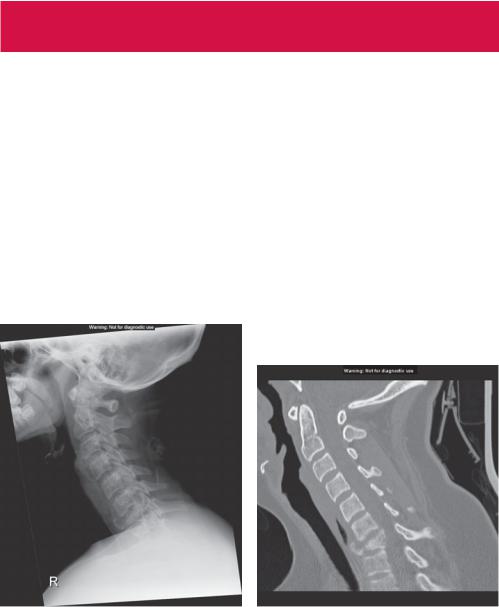
Figure 79.1 Lateral radiograph. |
Figure 79.2 Sagittal reformat CT scan. |
Questions
•What does the lateral cervical spine radiograph show?
•What does the CT scan demonstrate?
221