

Книги по МРТ КТ на английском языке / Thomas R., Connelly J., Burke C. - 100 cases in radiology - 2012
.pdfANSWER 41
The most likely diagnosis based on the clinical history and imaging findings is pneumocystis pneumonia (PCP) or pneumocystosis, which is a form of pneumonia, caused by the yeast-like fungus Pneumocystis jirovecii. The older name Pneumocystis carinii (which now only applies to the Pneumocystis variant that occurs in animals) is still in common usage.
Plain chest radiograph (Figure 41.1) demonstrates perihilar interstitial reticular shadowing with cyst formation. There is relative sparing of the apices and both bases. The axial enhanced CT image at the level of the pulmonary trunk (Figure 41.2) demonstrates perihilar ground-glass change and cystic changes, indicating the development of pneumatoceles.
Pneumocystis organisms are commonly found in the lungs of healthy individuals. It is believed most children have been exposed to the organism by the age of 4 years, and its occurrence is worldwide. The organism is a rare cause of infection in the general population, however it is a frequent cause of morbidity and mortality in persons who are immunocompromised, especially patients with AIDS.
Patients who do not have AIDS but are immunocompromised and at risk for PCP include individuals with haematologic malignancies, organ transplant recipients and those receiving long-term steroid or cytotoxic therapy, including patients with systemic vasculitis or other autoimmune deficiency. Other patients with immune deficiency disorders who are at particular risk for PCP include those with thymic dysplasia, those with severe combined immunodeficiency, and those with hypogammaglobulinaemia. Severe malnutrition may also predispose individuals to PCP.
The risk of pneumonia due to PCP increases when CD4 levels are less than 200 cells/μL.
The symptoms of PCP are non-specific. PCP in patients with HIV infection tends to run a more subacute, indolent course and tends to present much later, often after several weeks of symptoms, compared with PCP associated with other immunocompromising conditions.
Chest radiographs should be included in the initial evaluation for PCP. Frequently, these are the only images required. Characteristically, the distribution is central in location with bilateral diffuse symmetric finely granular, reticular interstitial/airspace infiltrates with perihilar and basilar distribution. Chest radiograph is normal in 10–39 per cent of patients with PCP. Hilar lymphadenopathy and pleural effusions are uncommon (seen in less than 5 per cent).
CT (in particular, high-resolution CT) scanning is more sensitive than chest radiography for the detection and exclusion of PCP pneumonia, and the results may be positive when chest radiograph findings are normal. CT findings include patchwork pattern bilateral asymmetric patchy mosaic appearance with sparing of segments/subsegments of pulmonary lobe or a ‘ground-glass’ pattern as in this case with bilateral diffuse airspace disease (fluid + inflammatory cells in alveolar space) in symmetric distribution. Cysts are visible on chest radiographs in 10 per cent of patients, although these are appreciated far more commonly on HRCT scans (33 per cent). Findings of cysts or pneumatoceles are not infrequent in patients with PCP.
KEY POINTS
•PCP is the most common cause of interstitial pneumonia in immunocompromised patients, which quickly leads to airspace disease.
•Chest radiograph findings may be normal in a significant number of patients with PCP.
•The classical chest radiograph features of PCP pneumonia, when present, are bilateral, diffuse, often perihilar, fine, reticular interstitial opacification, which appears to be granular.
122

CASE 42: PAIN ON DEEP INSPIRATION
History
A 67-year-old man presents to the accident and emergency department as a referral from his general practitioner (GP). Over the course of the preceding 12 hours he has been experiencing pain in the left side of his chest, worst on deep inspiration. This is the first ever such episode and he describes the pain as sharp and stabbing. He is an ex-smoker with a 30 pack-year history. Emphysematous changes have been noted on a previous chest radiograph and he takes a salbutamol inhaler as required along with an inhaled corticosteroid regularly. Aside from a history of mild to moderate chronic obstructive pulmonary disease he was otherwise well. He denies any history of trauma.
Examination
He is tachypnoeic with a respiratory rate of 33/minute and tachycardic with a heart rate of 104/minute which electrocardiogram (ECG) confirms as sinus rhythm with no acute changes. On examination of the respiratory system there is reduced expansion, a slightly hyperresonant percussion note and reduced air entry on the left. Vocal resonance is also reduced on the left. No added sounds are identified, however. Full blood count (FBC), biochemistry and liver function tests are all normal. Blood gas analysis demonstrates a PaO2 of 9 kPa with a PaCO2 of 4.5 kPa. The patient is referred for a chest radiograph (Figure 42.1).
Figure 42.1 Posterior–anterior (PA) chest radiograph.
Questions
•What abnormality do you see on the chest radiograph?
•What concerning features would you look for?
•How would you manage this patient?
123

ANSWER 42
The PA chest radiograph (Figure 42.1) shows a large left pneumothorax, which appears tethered to the left costal pleura. There is a background of emphysematous change but no evidence of mediastinal shift to suggest that the pneumothorax is under tension, which would be an alarming feature requiring immediate intervention.
Pneumothorax refers to the presence of air within the pleural space. Diagnosis is established on the plain chest radiograph by demonstrating an outer margin of the visceral pleura known as the pleural line (delineating collapsed lung), separated from the parietal pleura (chest wall) by a lucent space occupied by gas and devoid of any pulmonary markings.
The pleural line demonstrating the margin of collapsed lung can sometimes be difficult to detect in cases of a small pneumothorax. It is important to note that a skin fold may mimic the pleural line. When a pneumothorax is suspected but not confirmed on inspiratory radiograph, an expiratory image may confirm the diagnosis. This is because at the end of expiration, the constant volume of the gas within the pneumothorax is accentuated by the reduction of the hemithorax.
Chest radiography is the first investigation performed to assess pneumothorax, because it is straightforward, rapid, cheap and non-invasive. CT may be used in more complex cases, for example in planning pleurodesis (usually in recurrent pneumothoraces) and it is more sensitive than plain radiographs in detecting blebs or bullae or a small pneumothorax.
Pneumothorax is classified as spontaneous (atraumatic), traumatic, or iatrogenic:
•Spontaneous pneumothorax may be either primary (occurring in persons without clinically or radiologically apparent lung disease) or secondary (in which lung disease is present and apparent, as in this example). Most individuals with primary spontaneous pneumothorax have unrecognized lung disease; it is often thought to occur due to rupture of a subpleural bleb.
•Traumatic pneumothorax is caused by penetrating or blunt trauma to the chest. Gas enters the pleural space directly through the chest wall through visceral pleural penetration or alveolar rupture resulting from sudden compression of the chest.
•Iatrogenic pneumothorax results from a complication of a diagnostic or therapeutic intervention. With the increasing use of invasive diagnostic procedures, iatrogenic pneumothorax has become more common.
In this large secondary pneumothorax with significant symptoms drainage is required to remove the air from the pleural space. Needle aspiration is not usually adequate in such cases and insertion of an intercostal drain is required. This has been done in Figure 42.2 with some resulting but as yet incomplete re-expansion of the collapsed left lung.
124

Figure 42.2 Chest radiograph post drainage.
KEY POINTS
•In the erect position, pleural gas collects over the apex where the space between the lung and the chest wall is most notable.
•It is important to assess for radiographic manifestations of tension pneumothorax are mediastinal shift, diaphragmatic depression and rib cage expansion.
•Tension pneumothorax is an emergency requiring immediate intervention.
125
This page intentionally left blank

CASE 43: AN ELBOW INJURY
History
A 45-year-old woman attends the accident and emergency department following a fall off a stepladder from a height of 1 metre onto her outstretched right arm while decorating at home. She immediately noted marked pain and is unable to flex or extend the elbow joint. She is previously fit and well, with no previous illnesses. She is right hand dominant.
Examination
She is unable to pronate or supinate the forearm and unable to flex or extend the elbow. She demonstrates bony tenderness maximal over the radial head and there is swelling of the elbow joint. Her arm pulses are intact distally, capillary refill is less than 2 seconds, and sensation and power in the hand and wrist are normal. Plain radiographs are taken (Figure 43.1a,b).
(a) |
(b) |
Figure 43.1 (a) Lateral and (b) magnified anterior–posterior (AP) images.
Question
•What is the likely injury?
•What do the plain radiographs show?
127
ANSWER 43
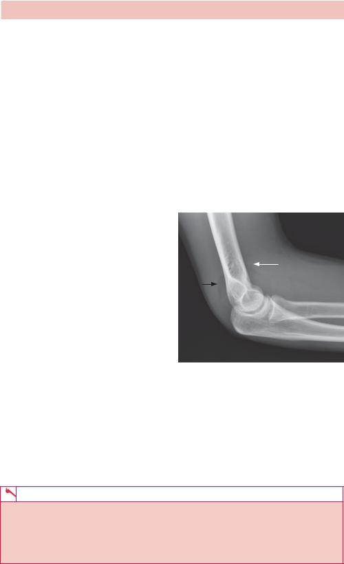
The lateral radiograph (Figure 43.1a) demonstrates elevated anterior and posterior elbow fat pads, suggesting a joint effusion and is suspicious for an occult fracture. The magnified AP image (Figure 43.1b) confirms a fracture of the radial head.
The preferred study for the evaluation of elbow trauma is conventional radiography and the radiographic examination requires the acquisition of two views: the lateral view, ideally in 90 degree flexion, and AP view in full extension.
The classic elbow ‘fat pad’ sign seen on lateral elbow radiograph is an invaluable soft tissue finding in cases of intra-articular injury of the elbow. Fat is normally present within the joint capsule of the elbow, but outside the synovium. As it is usually ‘hidden’ in the concavity of the olecranon and coronoid fossae, fat is usually not visible on the lateral radiograph.
Injuries causing intra-articular haemorrhage/effusion, however, cause distension of the synovium which forces the fat out of the fossa, producing triangular radiolucent shadows anterior and posterior to the distal end of the humerus – the radiographic sail sign (Figure 43.2).
The posterior fat pad (black arrow – Figure 43.2) is not normally seen on radiographs and its presence is always an abnormal finding that requires further investigation for an occult fracture.
The anterior fat pad may be normally visualized on lateral radiographs as a triangular radiolucency, although in the presence of joint effusion it is displaced anteriorly, becoming more pronounced and the anterior margin becomes convex. Fat pad signs may not be evident if the fracture is extracapsular.
Fracture of the radial head is the most common type of elbow fracture in adults, whereas fractures of the radial neck are more common in children. Fractures of the
radial head and neck of the radius generally result from a hard fall on an outstretched hand with the impact of fall driving the head of radius axially onto the capitulum of the humerus.
Treatment depends on the degree of displacement, angulation and articular involvement. Minor degrees such as that shown are often treated by early mobilization in a brace to minimize later elbow stiffness.
KEY POINTS
•Radiographic examination requires two views: AP view in full extension and lateral view, ideally in 90 degrees of flexion.
•The posterior fat pad is not normally seen on radiographs, and its presence is always an abnormal finding that should prompt further investigation for occult fractures.
•Fat pad signs may not be evident if the fracture is extracapsular.
128
CASE 44: PAIN IN THE HAND FOLLOWING A PUNCHING INJURY
History
A 22-year-old man attends the accident and emergency department following a fight. He remembers punching another man and, although intoxicated, is complaining of pain in the knuckles. He is otherwise fit and well.
Examination
On examination his knuckles are swollen and tender to palpation. There is maximal tenderness in the region of the fifth metacarpal head (base of the little finger) with virtually no range of movement at the fifth metacarpophalangeal joint. Neurovascular function is intact distally. Plain radiographs are taken (Figure 44.1).
(a) |
(b) |
Figure 44.1 (a) Anterior–posterior (AP) and (b) oblique plain radiographs.
Questions
•What is the likely injury?
•Describe the injury seen in the plain radiographs of the hand.
129

ANSWER 44
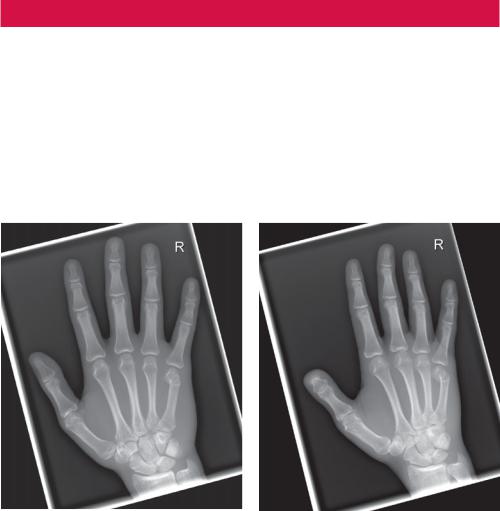
The AP and oblique images of the hand demonstrate a metacarpal neck fracture, with volar angulation and displacement of the distal fragment. Note also prominent soft tissue swelling of the hand.
A ‘boxer’s fracture’ is the common name for a fracture involving the neck of the fifth metacarpal, which forms the knuckle of the little finger (but the same name may also be used for a fracture at the neck of any of the metacarpals). It is usually caused by the impact of a clenched fist with a skull or a hard, immovable object.
Only the collateral ligaments remain attached to the proximal phalanx and therefore the metacarpal head is freed from any proximal stabilizing influence. The metacarpal head tilts volarly, causing the joint to lie in hyperextension and the collateral ligaments become slack. If the joint is allowed to remain in hyperextension, collateral ligaments will shorten, leading to limited metacarpophalangeal flexion.
The little finger carpometacarpal articulation allows a flexion–extension arc of 20–30 degrees in addition to a rotatory motion, facilitating little finger opposition to thumb.
A true lateral radiograph is necessary with these fractures in order to measure the angle of displacement of the distal fragment. The normal metacarpal neck angle is about 15 degrees and therefore a measured angle on film of 30 degrees is actually approximately 15 degrees.
These fractures are often angulated, and if severely so, require pins to be put in place and realignment as well as the usual splinting. However, the prognosis on these fractures is generally good.
KEY POINTS
•A boxer’s fracture is the common name for a break in the end of the little finger metacarpal bone, also known as a fifth metacarpal fracture.
•The injury is commonly caused by punching something harder than the hand, for example, a wall. The end of the metacarpal bone takes the brunt of the impact, which usually breaks through the neck (which is the narrowest area near the end) and bends down toward the palm.
130
CASE 45: A FALL ON THE HAND AND PAIN IN THE WRIST
History
A 20-year-old man attends the accident and emergency department following a fall onto his flexed left wrist while playing football.
Examination
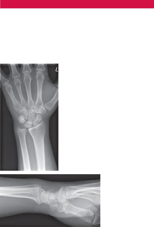
He demonstrates a markedly reduced range of movement and is complaining of point tenderness on the dorsum and ulnar aspect of the left wrist. There is no pain on palpation of the anatomical snuffbox or upon axial loading of the thumb. Radiographs are taken (Figures 45.1 and 45.2).
Figure 45.1 (a) Anterior–posterior (AP) view of the left wrist.
Figure 45.2 Lateral view of the left wrist.
Questions
•What injury do you suspect he may have sustained?
•What do the wrist radiographs demonstrate?
131