

Книги по МРТ КТ на английском языке / Thomas R., Connelly J., Burke C. - 100 cases in radiology - 2012
.pdfANSWER 14
Figure 14.1 is a single ultrasound image of the left lobe of the liver obtained with a curvilinear transducer (C5–2) in a longitudinal orientation. The liver appears of normal echogenicity and echo texture with a smooth capsular contour. No focal lesion is seen on this image. There are anechoic linear structures seen which extend to the periphery with a maximal diameter of 4 mm, and colour Doppler assessment in Figure 14.2 demonstrates no flow within them. This is in keeping with intrahepatic biliary duct dilatation, and the remainder of the study did not demonstrate an obstructing lesion although the common bile duct (CBD) was 7 mm in diameter, which is within normal limits for a patient post cholecystectomy. The patient was referred for a liver magnetic resonance imaging (MRI) scan for further characterization. The liver function tests of raised bilirubin and alkaline phosphatase with normal transaminases and albumin suggest obstruction with normal cellular and synthetic function.
Figure 14.3 is a coronal maximal intensity projection (MIP) image of the same patient acquired from heavily T2-weighted sequences. It confirms moderate intrahepatic biliary duct dilatation and also demonstrates a focal tapered stenosis at the level of the common hepatic duct. There is no intraductal filling defect or stenosing soft tissue mass lesion, and the CBD and pancreatic duct are of normal calibre. The appearances suggest a stricture of the common hepatic duct which may be ischaemic, post inflammatory or neoplastic in nature. Endoscopic retrograde cholangiopancreatography (ERCP) was advised to obtain cytology brushings, attempt to correct the obstruction and decompress the intrahepatic ducts, but a stent could not be inserted. The patient was therefore referred to the interventional radiology department for a percutaneous transhepatic cholangiogram (PTC).
The single image of the PTC (Figure 14.4) was acquired by opacifying the intrahepatic ducts with radiographic contrast during a fluoroscopy procedure. The dilated ducts of the left system were punctured under ultrasound guidance, and a guidewire was then manipulated across the hilar stricture and down into the duodenum. A sheath (8F) was inserted to stabilize the position, and contrast was injected through this to perform the cholangiogram. A stent can be passed over the wire and placed across the stenosis to relieve the obstruction and improve patient symptoms. This procedure is performed by the interventional radiology department.
Interventional radiology (IR) is an expanding subspeciality of radiology that uses imaging guidance and minimally invasive techniques to diagnose and treat a patient. A trained radiologist uses their experience in ultrasound, computed tomography (CT) and fluoroscopy to guide the passage of a needle or catheter to a site of interest and perform a task that would otherwise be surgically difficult and involve significant morbidity in the form of an open operation. Interventional radiology consultants can use the veins, arteries and biliary ducts to access deep or distal lesions, vessels or organs, often leaving only a pinhole size scar at the site of puncture (often the groin) as a sign of recent treatment. This allows tissue conservation, reduced morbidity and faster recovery for patients. The scope of the speciality is too broad to be effectively covered in this answer, but the procedures used include the following:
•Angiography: A vein or artery can be punctured with ultrasound guidance, and contrast is injected mapping the vessel anatomy under fluoroscopy. Stenoses and occlusions can be characterized and an expandable balloon is used to improve blood flow. This is termed ‘angioplasty’.
42
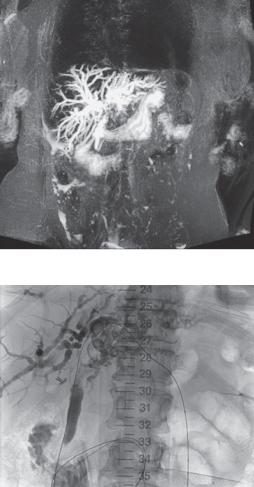
Figure 14.3 Coronal maximal intensity projection image.
Figure 14.4 Percutaneous transhepatic cholangiogram.
•Biopsy: Ultrasound (superficial lesions) and CT (complicated or deep lesions) can help to guide a needle accurately to a lesion of interest for core biopsy and histological characterization.
•Drainage: Inserting a drain can offer a conduit for decompression of infected collections or uncomfortable ascites. Optimal and accurate placement is obtained by imaging guidance.
•Stenting: Expandable stents can be inserted into a vessel or duct to act as ‘scaffolding’ and can exert radial force to maintain patency in atherosclerosis or tumour stenosis.
•Line insertion: Patients on long-term therapy (e.g. dialysis, antibiotics, chemotherapy) require definitive vascular access (e.g. Hickman line, portacath) to avoid the discomfort of recurrent peripheral cannulation and thrombophlebitis.
•Embolization: Instilling an embolic agent (coils, particles or glue) into a selectively cannulated vessel can control active haemorrhage, prevent aneurismal rupture or infarct a tumour (e.g. uterine fibroid). An adjunct to this is chemoembolization, where a chemotherapy agent is instilled directly to a tumour and then the blood vessel is embolized to cause tumour infarction.
43

•Radiofrequency ablation: Small malignant lesions can be cauterized via a specialized electrical probe that is inserted under image guidance for accurate placement.
•Vertebroplasty: Guiding the infusion of inert cement into a collapsed spinal vertebra can provide stability in cases of osteoporotic or metastatic collapse.
KEY POINTS
•Ultrasound is excellent for the assessment of the liver and has a high sensitivity for detecting intrahepatic biliary duct dilatation.
•Biliary obstruction can be circumvented by stent insertion either via ERCP or PTC.
•Interventional radiology is a subspeciality of radiology that uses image guidance to perform minimally invasive techniques.
44
CASE 15: INFANT WITH CLICKING HIPS
History
A 6-week-old female infant is brought by her mother as part of the 6-week check in general practice. The baby was delivered at term by caesarian section due to a breech presentation. Her maternal grandmother and aunt are known to have chronic hip problems.
Examination
On examination she moves both lower limbs normally. The hip creases appear a little asymmetric. On Barlow’s manoeuvre (adduction of the hip with light pressure on the knee) the right hip appears to pop posteriorly and then anteriorly on Ortolani’s manoeuvre (the hips and knees are flexed to 90 degrees and anterior pressure is applied to the greater trochanters while using the thumbs to abduct the legs).
You expedite the hip ultrasound that has already been arranged due to the risk factors (Figure 15.1).
 a angle
(a) |
(b) |
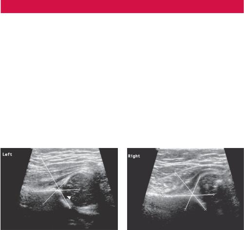
Figure 15.1 Left and right hip ultrasound images (coronal flexion view).
Questions
•Who gets hip ultrasound?
•What do the images in Figure 15.1 show?
•Is a radiograph helpful?
•What happens next?
45
ANSWER 15
Hip ultrasound is done on neonates and young infants (<6 months) to screen or check for developmental dysplasia of the hip. Screening is done on infants with risk factors that include family history, breech presentation, foot deformities or neuromuscular disorders. Examination features that raise suspicion for hip abnormality include asymmetric groin creases, a click on movement and a click or subluxation on provocation tests.
As with all ultrasound, gel is used to couple the ultrasound beam into the soft tissue and allow movement of the probe without loss of image. The baby is placed in the lateral position with the hip flexed and the probe is placed parallel to the ilium (bright line on the left of the image) and the orientation optimized to produce a horizontal image like a golf ball (stippled cartilage of the femoral head) on a tee (cup is the acetebulum, stalk is the ilium). If the golf ball appears to be falling off the tee (upwards on the image), then the femoral head is subluxed and the acetabular cup is shallow. This is all formalized by measuring angles (indicated on the image). The alpha angle measures the acetabular roof angle with the ilium and normally measures over 60 degrees. The beta angle assesses the prominence of the labrum (cartilaginous flange around the acetabulum).
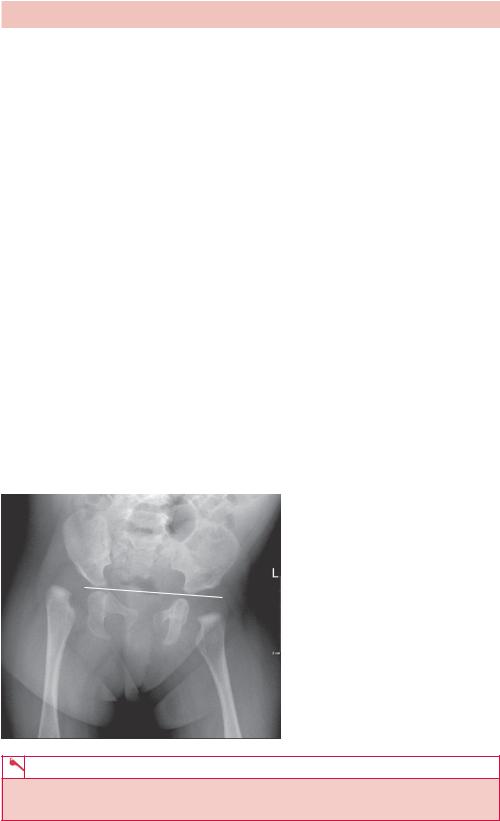
Figure 15.1 shows a normal left hip but a low alpha angle (53.5 degrees) on the right, corresponding with a shallow acetabulum and hip instability. The ultrasound shows a lot of soft tissue detail, including the unossified cartilage as well as the bone, although the anterior bone edge blocks the beam and no deeper structure is seen. Plain radiographs complement this view by demonstrating the bone structure but with very poor soft tissue detail. Ultrasound is the investigation of choice in infants with unossified femoral heads but as the ossification centre develops and blocks the ultrasound from about 6 months onwards, radiographs are more useful. Figure 15.2 shows the hip radiograph at 4 months.
The aim is to diagnose a hip abnormality as soon as possible to minimize the degree of intervention required to fix the problem. Treatment ranges from observing (if very mild and picked up on a neonatal scan) to braces or surgical intervention.
Figure 15.2 Radiograph at 4 months with right hip subluxation and a shallow right acetabulum (relative to Hilgenreiner’s line drawn on the radiograph).
KEY POINTS
•Ultrasound is the method of choice for examining infant hips.
•Hip screening in infants with family history or breech presentation occurs at 4–6 weeks.
46
CASE 16: PAINFUL WRIST AFTER FALLING
History
A 13-year-old girl presents to the accident and emergency department with a painful right wrist after falling while roller skating. She fell backwards with her arm outstretched. Her wrist appears deformed, but she tells you that although it hurts, the wrist has had that appearance for a long time and that it was investigated at another hospital many years ago. She gives a family history of bone problems but is otherwise well with no medical problems.
Examination
On the volar aspect of the right forearm just proximal to the wrist there is a firm swelling that extends laterally. There is some associated tenderness but no malalignment of the wrist or hand. There is normal movement, pulses and sensation. The chest and abdomen are normal. Given the history you also briefly examine the arms and spine that appear normal and the legs. The bone around the knees is prominent, with an asymptomatic bony nodule over the lateral aspect of the proximal right tibia.
You arrange radiographs of the arm (Figure 16.1a,b).
(a) |
(b) |
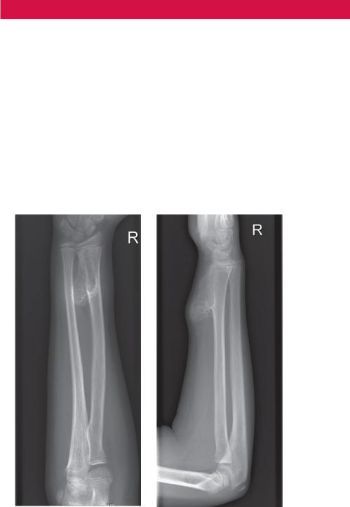
Figure 16.1 Anterior–posterior (AP) and lateral radiographs.
Questions
•What do the radiographs show?
•Can you name some differential diagnoses?
•What other imaging should be obtained?
47
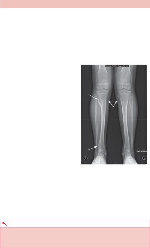
ANSWER 16
The radiographs show a lobulated expansile lesion arising from the cortical bone of the radial metaphysis and extending proximally. The bone is well delineated with a narrow zone of transition, an appearance suggesting the lesion is benign. No fracture is seen.
The appearance is in keeping with an exostosis, also known as an osteochondroma, that results from dysplasia of the growth plate. This may be solitary or multiple in a condition known as multiple hereditary exostosis (or diaphyseal aclasis), or rarely as part of various syndromes such as Turner’s syndrome or tuberous sclerosis.
Rather than take more images of the arm, the best course of action would be to obtain previous images. Picture archiving and communication system (PACS) is the digital system developed to manage the images prima-
rily produced in hospital radiology departments. In the last decade it has almost entirely replaced film-based archives with the advantages of rapid search and comparison, remote and web-based access and integration into other hospital information
systems. In this patient, a previous skeletal image on PACS (Figure 16.2) suggests the
diagnosis of multiple hereditary exostosis.
Multiple hereditary exostosis is inherited as an autosomal dominant trait, with an incidence of about 1 in 50000. Children are diagnosed on average around 3 years old. As seen in this case, bony growths (exostoses) arise from the metaphyses, point away from epiphysis, and extend down the diaphysis during growth. They increase in size and number with age, arising in several characteristic sites. Over 90 per cent of cases are at the distal and proximal tibia, proximal femur and proximal humerus. Ribs, scapula, radius, ulna, ilium and phalanges are also common sites.
Clinical complications include trauma and fractures, particularly at exposed positions
at the wrist and knees. The exostosis may exert pressure on surrounding soft tissue and cause neurovascular compromise. There can be inequality in limb length or short stature. In a small proportion of patients (1–2 per cent) at a later age (>21) an exostosis undergoes sarcomatous transformation into a chondrosarcoma. This is more likely in exostoses of the pelvis, scapula, proximal humerus, proximal femur and spine, and may be associated with a change in size or onset of pain.
KEY POINTS
•Exostoses can be solitary or multiple and develop from the metaphysis while the growth plate is open.
•Exostoses can cause symptoms. In older symptomatic patients, malignant transformation should be considered although it is uncommon.
48
CASE 17: CONSTIPATION IN WOMAN WITH OVARIAN TUMOUR
History
A 78-year-old woman presents to the oncology clinic with a 1 week history of right upper quadrant and epigastric tenderness and constipation for 3 days. She has a history of metastatic ovarian cancer treated with abdominal hysterectomy, oophorectomy and omentectomy for peritoneal disease 15 months ago. She has also had chemotherapy. On her last scan she was noted to have lung nodules, mediastinal and mesenteric lymph node enlargement that appeared stable and a new caecal metastatic deposit.
Examination
On examination, she is not acutely unwell. Her observations are normal. The chest and heart sound normal. The upper abdomen is tender and there is a palpable fullness or mass below the right costophrenic margin.
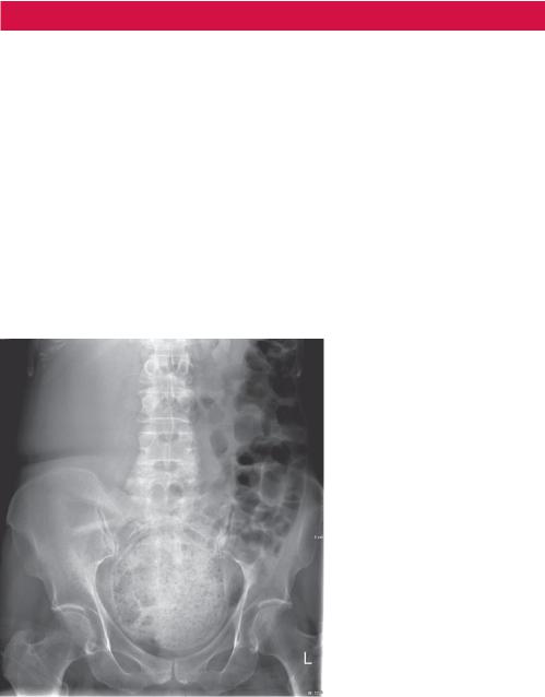
You assess an abdominal radiograph (Figure 17.1) during clinic and decide to admit the patient for assessment and a computed tomography (CT) scan.
Figure 17.1 Abdominal radiograph.
Questions
•What is the likely cause of her pain?
•What does the radiograph show?
•What appearance do bowel contents have?
49
ANSWER 17
Based on the history of cancer and surgery, the patient is at risk of bowel obstruction secondary to adhesions associated with surgery or tumour infiltration. Metastatic ovarian carcinoma frequently metastasizes through the peritoneal space, seeding deposits onto the omentum (removed in this patient’s case) and mesentery. Liver metastases are also possible, causing pain from stretching the liver capsule, biliary obstruction or ascites. Other common causes that are less likely in this case are cholecystitis and appendicitis.
The radiograph shows a large volume of faeces in the pelvis within large bowel or rectum and gas-filled empty bowel loops on the left side of the abdomen. There is a paucity of bowel gas on the right together with impression of a large soft tissue region extending throughout the right side of the abdomen. Small bowel can be distinguished from large by its central position and a fold pattern likened to a coiled spring (valvulae conniventes) less than 3 cm in normal diameter. Large bowel tends to frame the abdomen with haustral folds that are widely spaced and not circumferential (i.e. they do not cross the whole diameter of the visible bowel).
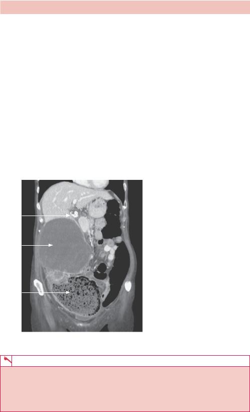
The patient’s CT (Figure 17.2) shows a large right upper quadrant mass, probably a metastatic deposit, displacing empty transverse colon to the left and partially obstructing the caecum that is distended by faeces. Gallstones are also seen; these are not apparent on the plain radiograph.
Gallstone
Mass
Caecum
Figure 17.2 Reconstructed coronal CT slice of the abdomen and pelvis.
The colon receives the sterile dilute product of digestion and then proceeds to remove a large proportion of the fluid content as well as employing microbial digestion (fermentation) to extract various products not produced in small bowel digestion, such as vitamin K. As a result, faeces take on a more solid appearance as they move round the colon. In addition, the microbial activity produces gas and the stool has a characteristic foamy appearance. Stomach contents also have a similar appearance with food mixed with fluid and air. Small bowel tends to be predominantly homogeneously fluid filled in appearance although with a small amount of gas that increases if there is obstruction.
KEY POINTS
•The distribution of stool tends to be of more significance than its appearance.
•Constipation may be indicated by a large volume of stool throughout the bowel, a rather solid appearance to the stool in the rectum or impacted faeces above an obstruction.
•Radiographs are rarely indicated for constipation unless obstruction is suspected.
50
CASE 18: THIRTY-YEAR-OLD MAN WITH HEADACHE
History
A 30-year-old man presents to you with a chronic history of headache that has worsened significantly in the last week. Investigations for the same problem 9 months ago ruled out sinus disease. He is concerned that this may be due to a brain tumour. Several relatives have died of various types of cancer. There is no history of definite head trauma, although he has had various sporting injuries. There is no other history of significant illness. On careful questioning the headache is reported to be present on waking and worsens on coughing.
Examination
On examination he is well. His observations are normal. There is papilloedema on examination of the eyes but no focal neurology is demonstrated. Examination is otherwise normal.
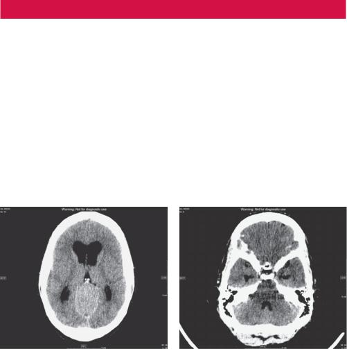
You arrange a computed tomography (CT) scan of the head.
(a) |
(b) |
Figure 18.1 Axial CT image of the brain at the level of (a) the third ventricle and (b) the fourth ventricle.
Questions
•Headache is a common problem. What red flag symptoms help to decide on investigations?
•What does the CT show?
•What would you do next?
51