

Книги по МРТ КТ на английском языке / Thomas R., Connelly J., Burke C. - 100 cases in radiology - 2012
.pdfThis page intentionally left blank
CASE 26: MAN WITH ATYPICAL CHEST PAIN
History
A 50-year-old man presented with sudden onset moderate central chest pain. He smokes 15 cigarettes a day and drinks around 10 pints of beer over each weekend. He has had no previous illnesses.
Examination
On examination he looks well. His blood pressure is 164/90 but observations are otherwise normal. Nothing abnormal is found on examination of respiratory, cardiovascular, abdominal and nervous systems. The electrocardiogram (ECG) is normal.
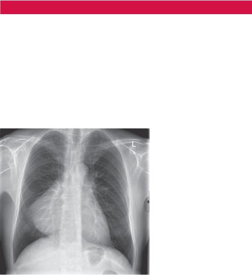
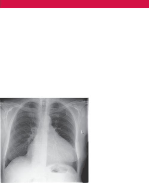
A posterior–anterior (PA) chest radiograph is requested as part of the work up for possible acute coronary syndrome (Figure 26.1).
Figure 26.1 PA chest radiograph.
Questions
•What abnormality is present and where is it?
•List some possible differential diagnoses.
83
ANSWER 26
The radiograph shows an abnormal but well-defined right heart border. This may reflect cardiac enlargement or a separate soft tissue mass. In general, the position of a soft tissue mass may be indicated by the loss of definition of the edge of adjacent structures – the silhouette sign. In this case the right heart border is abnormal, suggesting a mass adjacent to the right atrium of the heart obscuring the normal air–tissue interface. The differential diagnosis therefore includes anterior and middle mediastinal or lung masses such as pericardial cyst, lipoma, fat pad, bronchogenic cyst, sequestration, massive lymphadenopathy, diaphragmatic hernia, ventricular aneurysm or right atrial enlargement.
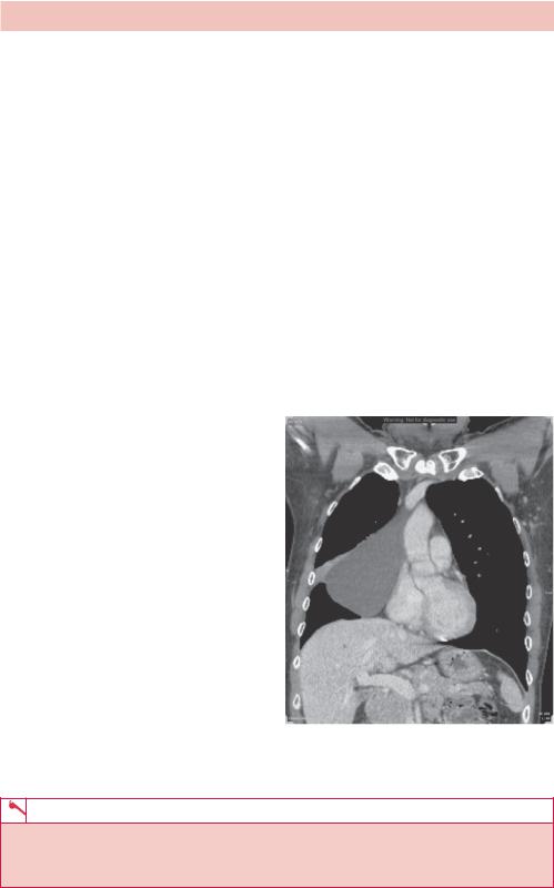
A computed tomography (CT) scan is the next step in imaging (Figure 26.2). This not only localizes the abnormality but also displays the soft tissue attenuation (measured in Hounsfield units, HU), which allows distinction between fat (e.g. in a fat pad or lipoma – typically negative HU), water-like fluid (HU <10) in a cyst, soft tissue and contrast (>100 HU) in, for example, a ventricular aneurysm. In this case there is a fluid attenuation mass adjacent to the heart, which otherwise appears normal, consistent with a pericardial cyst. There is also a small area of adjacent right upper lung lobe collapse (atelectasis).
Pericardial cysts are congenital malformations that are attached to the parietal pericardium, but do not communicate with the pericardial space. If there is communication with the pericardial space, the structure is termed a pericardial diverticulum. They are usually found incidentally in asymptomatic
patients. The most frequent location is in the right cardiophrenic angle, but they can be found in the left cardiophrenic angle, anterior mediastinum or middle mediastinum. The vast majority are unilocular and they usually range in size between 3 and 8 cm. Because they are soft, they may change shape with position. Rarely, they may cause symptoms due to compression of surrounding structures and require surgical removal.
Bronchogenic cysts arise out of the tracheobronchial tree as a congenital malformation, typically pericarinal but also paratracheal, oesophageal, retrocardiac and pulmonary in location. They are usually asymptomatic but may cause stridor, compression or become infected.
Figure 26.2 Reconstructed coronal CT slice through the thorax.
KEY POINTS
•Pericardial cysts are occasionally incidental, asymptomatic findings on chest X-ray.
•They abut the heart, resulting in a silhouette sign, and may be diagnosed on CT if they have classic cystic appearance.
84
CASE 27: YOUNG WOMAN WITH SHORTNESS OF BREATH
AND CHEST PAIN
History
This 37-year-old woman presents to the accident and emergency department after referral from her general practitioner (GP) complaining of increasing breathlessness and intermittent right-sided chest pain. The symptoms have come on over 2–3 weeks without associated fever or significant cough. There is a medical history of pelvic pain that is undergoing investigation for suspected endometriosis. She is otherwise well, a nonsmoker and taking no medication.
Examination
The right hemithorax is dull to percussion with reduced breath sounds throughout. The left lung and heart sound normal. The abdomen is soft and there is tenderness to palpation diffusely over both iliac fossae.
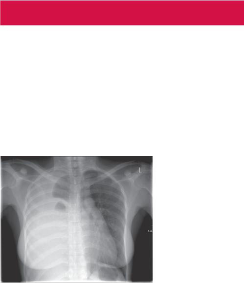
You arrange some tests including a chest radiograph (Figure 27.1).
Figure 27.1 Chest radiograph.
Questions
•What is the main abnormality and what effect is it having on the normal anatomy?
•What is the differential diagnosis?
•What is the appropriate management?
85
ANSWER 27
There is uniform opacification of most of the right hemithorax. The right hemidiaphragm and heart border are not seen. There is displacement of the heart and trachea to the left. Some residual aerated right lung is noted, probably the apices of the right upper and lower lobes. The left lung and heart are normal in appearance. No bone abnormality is seen.
We are looking for causes of opacification of a hemithorax. This can be within the lung, mediastinum or pleural space and may involve consolidation, soft tissue or fluid. The position of the mediastinum gives a clue as it is displaced away from the opacity even though the lung only appears partially aerated. This suggests a differential of pleural fluid or soft tissue, such as from a diaphragmatic hernia. Other causes such as consolidation, lung collapse, tumours (such as mesothelioma) or congenital agenesis or hypoplasia tend either not to displace or to displace the mediastinum
towards the opacity. The appearance with smooth edges and some thickening of the horizontal fissure suggests pleural effusion.
Effusions can be transudates (protein |
|
||
<30 g/L; e.g. in cardiac failure), exudates |
|
||
(>30 g/L; e.g. in infection, malignancy, |
|
||
pulmonary infarction), haemorrhagic (e.g. |
|
||
trauma, carcinoma) or chylous (e.g. due |
|
||
to obstructed thoracic duct caused by |
|
||
trauma, malignancy or parasites). Systemic |
|
||
diseases often cause bilateral effusions but |
|
||
can be unilateral. |
(a) |
||
To investigate this further, cross-sectional |
|
||
imaging to determine the underlying cause |
|
||
may be done. Computed tomography (CT) |
|
||
is not typically sensitive to the type of |
|
||
effusion. The effusion may also be drained, |
|
||
particularly if the patient is symptomatic, |
|
||
and a sample of fluid may help to decide |
|
||
on the cause. This is typically done with |
|
||
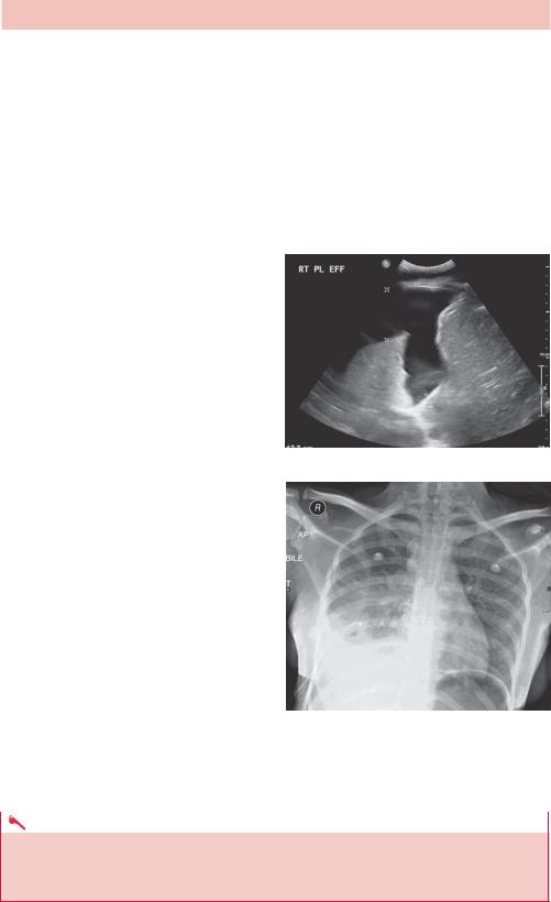
ultrasound guidance (Figure 27.2a), often |
|
||
using a Seldinger technique with a needle |
|
||
to insert a guidewire over which a drain |
|
||
tube is inserted. Thoracoscopy may be per- |
|
||
formed to look into the pleural space and |
|
||
allows biopsy of any abnormal pleural tis- |
(b) |
||
sue. Figure 27.2b shows the result of drain- |
|||
|
|||
age. The effusion in this case was stained |
Figure 27.2 (a) Ultrasound of the right lung |
||
with old blood and eventually shown to be |
base; (b) anterior–posterior (AP) radiograph |
||
caused by an endometriosis deposit. |
showing a right basal chest drain and partial |
||
|
|
drainage of the effusion. |
|
|
|
|
|
|
KEY POINTS |
|
|
|
|
|
|
•There are multiple causes of thoracic opacification and noting the effect on the mediastinum and anatomy helps to narrow down the possible causes.
•Consider pleural effusions in terms of transudate, exudates, blood and chyle.
86
CASE 28: CHEST DISCOMFORT AND DYSPNOEA
History
A 68-year-old man on treatment for non-Hodgkin’s lymphoma presents to the accident and emergency department with mild chest discomfort, worse on lying flat and eased by leaning forward. It has been getting slowly worse over the last few weeks. Now he gets dizzy on standing, rapidly breathless on exertion and has noticed some bilateral ankle swelling over the last week. He does not have a significant past cardiac or respiratory history.
Examination
On examination his blood pressure is 144/88, pulse 94/minute and respiratory rate 22/minute. The JVP is a little raised and fine crackles are heard at both lung bases. The heart sounds are difficult to hear but otherwise regular. The abdomen is soft and there is moderate left flank tenderness to deep palpation. The electrocardiogram (ECG) shows small QRS complexes and T wave inversion.
You organize a chest radiograph as well as blood tests (Figure 28.1).
Figure 28.1 Posterior–anterior (PA) chest radiograph.
Questions
•What abnormalities are seen?
•What others symptoms or signs might you find?
•What other tests would you do?
87
ANSWER 28
The heart is enlarged. The hilar vessels are not enlarged. There is some blunting of the costophrenic angles but no lung abnormality is seen.
The heart size is usually estimated by measuring the cardiothoracic ratio (CTR, maximum cardiac width/maximum inner thoracic width) on a PA projection radiograph with adequate inspiration (6 ribs seen anteriorly, 10 posteriorly). Beware anterior–posterior (AP) projections and poor inspiration as this will artificially increase the size of the heart. Typically in adults a CTR ratio >0.5 suggests cardiomegaly.
In addition to the CTR, the heart shape may indicate an underlying cause such as valve disease or a shunt. Increase of the right atrium size shifts the right heart border laterally; the left ventricle shifts the left heart border. The right ventricle lifts the heart, moving the apex superolaterally. The left atrium is behind the heart and on enlarging may project a second border over the right side of the heart. The left atrial appendage may enlarge and may produce a bump at the upper left heart border. Often the heart just appears generally enlarged and correlates with heart failure; occasionally this may be due to a pericardial effusion and is termed the ‘water bottle sign’.
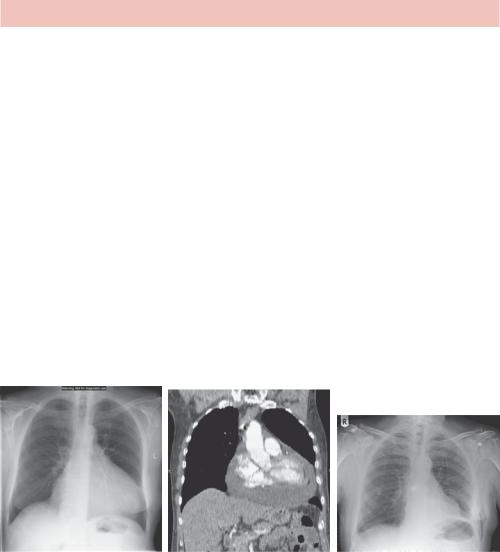
The differential diagnosis for cardiomegaly includes ischaemic heart disease, valve disease, pericardial effusion/cardiac tamponade, dilated cardiomyopathy and pulmonary embolism. In this patient, the presentation is suspicious for a pericardial effusion, probably malignant in origin and is confirmed on computed tomography (CT) (Figure 28.2).
(a) |
(b) |
(c) |
Figure 28.2 (a) The presenting radiograph, (b) the coronal CT shows a rim of lower attenuation fluid around the heart, (c) radiograph after paracentesis.
The pericardium covers the heart and great vessels, with the exception of only partially covering the left atrium and normally contains less than 50 mL of transudate fluid. To be distinctive on chest X-ray more than 250 mL needs to be present. The type of excess fluid depends on the cause:
•transudate – congestive heart failure, hypoalbuminaemia;
•exudate – infection, autoimmune disease (e.g. rheumatoid arthritis, systemic lupus erythematosus, hypersensitivity);
•blood – trauma, surgery, rupture, myocardial infarction, neoplasm;
•lymph – neoplasm, surgery.
An echocardiogram is the next investigation of choice and may be used to guide insertion of a pericardial drain.
88

KEY POINTS
•A cardiothoracic ratio greater than 0.5 on a PA projection radiograph with good inspiration indicates cardiomegaly.
•Pericardial effusions are difficult to see on chest X-ray unless large, although a rapid change in heart size on successive X-rays is suspicious.
89
This page intentionally left blank
CASE 29: SKATEBOARDER WITH A PAINFUL FOOT
History
A 13-year-old boy was skateboarding when he fell forwards onto steps and twisted his right foot. His description of the injury sounds as if it involved inversion of the foot. He is taking no medication and there is no significant medical history.
Examination
The boy is a fit 13 year old who appears well but in discomfort. The right mid foot is swollen and tender, more over the lateral aspect. The ankle joint is not painful but there is pain in the foot on moving the ankle joint. There are no other injuries.
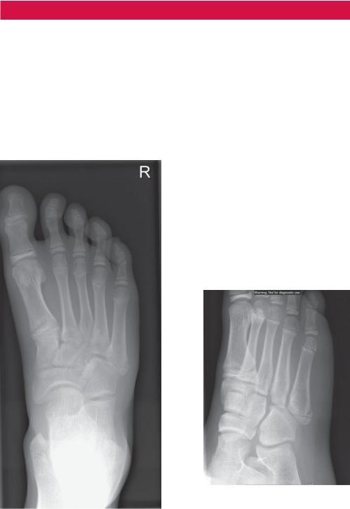
You arrange plain radiographs of the right foot (Figure 29.1).
Figure 29.1 Anterior–posterior radiograph of the right foot.
Figure 29.2 Magnified oblique radiograph of the right foot.
Questions
•Given the history, where would you look for an injury?
•What do the radiographs show?
•What might be confusing about the appearance of this injury?
•What other foot fractures should you consider?
91