

Книги по МРТ КТ на английском языке / Thomas R., Connelly J., Burke C. - 100 cases in radiology - 2012
.pdfANSWER 70
The cranial–caudal (CC) and medio-lateral-oblique (MLO) views shown in Figures 70.1b and 70.2b, respectively, demonstrate a large irregular, spiculate tumour containing microcalcifications. The lesion is located lateral (see CC view) and slightly superior (see MLO view) to the nipple in approximately the 2 o’clock position in the left breast. This is the typical appearance of a malignant breast carcinoma. For comparison the corresponding mammograms of the normal right breast are included in Figures 70.1a and 70.2a. The mass is causing parenchymal distortion and extends posteriorly, hence explaining some fixation seen on clinical examination.
Mammography can be performed both in the context of screening (in the asymptomatic woman) or diagnosis in the symptomatic patient. Until recently, women in the United Kingdom were invited to attend breast screening from age 50 with a 3-year cycle and continued to age 70. This has now been extended from 47 to 73 with self or GP referral for older women. In younger, typically pre-menopausal women the breast tissue is denser, making mammography less sensitive as a screening tool.
If an abnormality is found on screening it is graded, which determines the type of followup. Suspicious lesions are referred to a breast clinic. The standard breast mammogram views are bilateral CC and MLO views, which are obtained with the breast compressed, as seen in Figures 70.1 and 70.2.
Mammography is a form of low-dose radiograph used to create detailed images of the breast and can demonstrate microcalcifications smaller than 100 μm. It may reveal a lesion before it is palpable by examination, hence its use in screening. Mammographic features suggestive of malignancy include microcalcifications, soft tissue mass, asymmetry or architectural distortion, all of which are demonstrated in Figure 70.1b.
In the breast clinic the patient undergoes a triple assessment of clinical review, mammography and ultrasound. The ultrasound shown in Figure 70.3 confirms a 3.1 cm mass with ill-defined margins, casting an acoustic shadow. Ultrasound is a useful adjunct to mammography and has the benefit of not using ionizing radiation. Ultrasound can help to determine if a breast lesion is likely to be benign (e.g. a cyst or fibroadenoma) or malignant. Malignant lesions are characteristically poorly defined with irregular margins and posterior acoustic shadowing, as seen in Figure 70.3. Ultrasound is also used to guide biopsy and therapeutic procedures. In this patient’s case, the lesion was an invasive ductal carcinoma.
In certain settings magnetic resonance (MR) imaging of the breast may be indicated. This is used for:
•further characterization of an indeterminate lesion (despite full assessment with examination, mammography and ultrasound);
•detection of occult breast carcinoma in a patient with carcinoma in an axillary lymph node;
•multifocal or bilateral tumours;
•invasive lobular carcinoma, which has a high incidence of multifocality;
•evaluation of suspected, extensive, high-grade intraductal carcinoma;
•detection of recurrent breast cancer.
KEY POINTS
•Breast clinic assessment typically consists of the triple assessment of clinical review, mammography and ultrasound.
•Mammography can be used both in screening and in symptomatic presentations such as a breast lump or discharge.
•Ultrasound can be used to characterize breast lesions and guide biopsy.
192
CASE 71: A RISING CREATININE
History
A 57-year-old man was admitted overnight complaining of haematuria and lethargy. His symptoms have been intermittent over the last month, with occasional macroscopic blood seen when passing urine. He complains of an increase in frequency and nocturia but no dysuria. There is no history to suggest any stigmata of infection.
He has no relevant past medical history and lives at home with his wife and two children. As an ex-smoker he has a tobacco history of 20 pack-years with only occasional alcohol usage.
Examination
Examination reveals that he is afebrile and in no obvious discomfort. Cardiovascular and respiratory examinations are normal, but on abdominal examination there is fullness at both renal angles and mild discomfort on deep palpation.
Blood results reveal a creatinine of 515 μmol/L with normal urea and potassium. A routine blood result from 3 months previously recorded his creatinine as 72 μmol/L. His inflammatory markers are not elevated and a blood gas does not demonstrate any acidosis. Ultrasound images were taken (Figures 71.1 and 71.2).
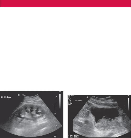
Figure 71.1 Ultrasound of left kidney. |
Figure 71.2 Ultrasound of bladder. |
Questions
•What does the ultrasound show?
•What options are available to resolve the situation?
193
ANSWER 71
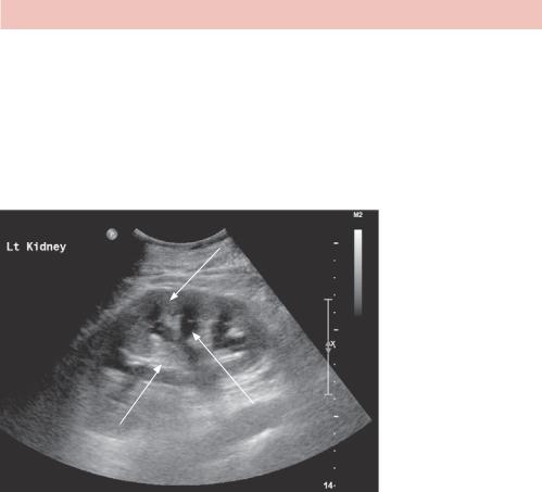
These are two static ultrasound images of the left kidney and the bladder. The left kidney image is acquired in a longitudinal orientation and demonstrates normal echogenicity and shape with preserved corticomedullary differentiation and cortical thickness. There are serpiginous areas of anechoic echogenicity associated with the renal medulla with cortical extension in keeping with moderate pelivocalyceal dilatation (Figure 71.3). Although not accurately demonstrated on this image, there is the suggestion of proximal ureteric dilatation distal to the pelvico-ureteric junction (PUJ). Imaging of the right kidney revealed identical appearances.
Renal cortex
Calyceal separation
Renal medulla
Figure 71.3 Annotated ultrasound of left kidney.
The second image (Figure 71.2) is acquired in a transverse orientation within the pelvis and demonstrates an adequately distended bladder with an area of irregular echogenicity seen posteriorly at the expected level of the bladder trigone. This mass lesion measures approximately 4.3 × 2.9 cm contiguous with the bladder wall. The vesico-ureteric junction (VUJ) and distal ureters are not demonstrated on this image. The features are likely to represent a transitional cell carcinoma (TCC) of the bladder with obstruction of both the left and right kidneys. A staging computed tomography (CT) examination of the chest, abdomen and bladder is recommended to characterize this further, with referral to a urologist for continued management.
The CT scan confirmed a surgically resectable TCC with bilateral renal obstruction and no evidence of local or distant spread. The patient ultimately requires a definitive surgical procedure to remove the cancer, but at present, the ureteric obstruction and deterioration in renal function makes the patient biochemically unstable. Surgery carries significant risk, and in a non-life threatening situation, the patient should be physically and biochemically optimized to encourage a safe transition through the operation, reduce the risk of intra-operative mortality and improve post-operative recovery. This patient is currently a poor candidate for general anaesthesia, and as a bridge to surgery there are two options available:
194
• Cystoscopy and retrograde ureteric stent insertion: Trained urologists can pass a camera through the patient’s urethra and visualize the bladder TCC directly. Not only does this allow for biopsy and tissue confirmation of the TCC, but if the VUJ is adequately visualized, a guidewire can be passed into the ureter and a ‘double-J’ stent inserted to relieve the VUJ obstruction. Cystoscopy can be performed under conscious sedation but general anaesthesia is preferred. In this situation, it was felt that the tumour mass would make VUJ cannulation very difficult, making this option inappropriate.
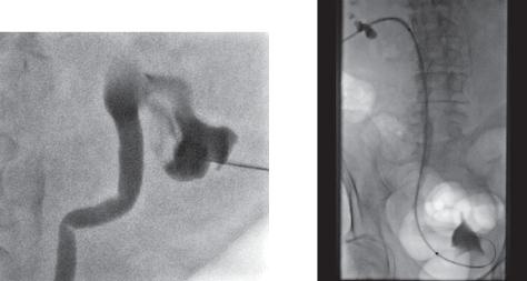
•Nephrostomy and antegrade ureteric stent insertion: This minimally invasive procedure is performed by a trained interventional radiologist and can be performed under local anaesthesia only, although light sedation is sometimes required. The patient lies on a fluoroscopy table in the prone position, and a micropuncture needle is passed percutaneously under direct ultrasound imaging into a dilated lower pole calyx. Ideally this should transgress Brodel’s bloodless line, an avascular area between the anterior and posterior renal segments to reduce the risk of renal haemorrhage. Instilling contrast into the collecting system under fluoroscopy can confirm satisfactory positioning (Figure 71.4).
Adopting the Seldinger technique, a guidewire can then be manipulated into the proximal ureter over which a sheath is passed to stabilize the position. A hydrophilic wire and catheter combination is then used to transgress the VUJ and obstructing lesion for safe passage of a stiff guidewire from the renal pelvis into the bladder. This allows careful manipulation of an appropriately sized renal stent to be passed over the wire so that one end lies within the bladder, and the other lies more proximally within the renal pelvis. This acts as a conduit through which urine can drain for renal decompression (Figure 71.5).
Figure 71.4 Contrast fluoroscopy image |
Figure 71.5 Fluoroscopy study |
confirming position of lower pole colyx. |
positioning ureteric stent. |
Stent positioning requires a high level of accuracy and it may also become obstructed both by tumour overgrowth and stent migration. As a precautionary measure, an additional pigtail catheter is often left within the renal pelvis, running along the line of the original percutaneous puncture to act as a urinary diversion both for initial decompression and in case of stent failure. This is termed a covering nephrostomy and remains in position for the short term (Figure 71.6).
195
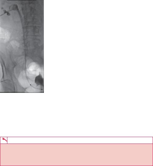
Figure 71.6 Fluoroscopy image confirming ureteric stent placement with covering nephrostomy.
Renal decompression will allow time for the renal function to return to normal and means that the operation can be performed in a less urgent and more controlled environment. Holistically, it also allows for the patient to make appropriate decisions about their own management. The nephrostomy and stent can be removed intra-operatively, leaving only a small cutaneous scar.
KEY POINTS
•Nephrostomy allows for biochemical and physical optimization before surgery.
•Nephrostomy insertion is ideally along Brodel’s bloodless line.
•Ultrasound guidance and a Seldinger technique are employed for nephrostomy insertion.
196
CASE 72: NECK PAIN AFTER FALLING
History
A 21-year-old woman is brought to the accident and emergency department with head and neck trauma after falling from a horse. She is immobilized on a board with a hard neck collar and complains of diffuse neck pain and mild intermittent tingling in the arms and legs. There is no history of loss of consciousness, no evidence of head injury (she had a riding helmet on) and no complaint of injury elsewhere. There is no significant past medical history although the patient has had intermittent neck pain in the past that has not been investigated.
Examination
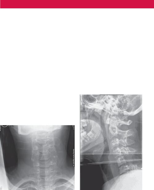
Routine observations are stable and she is maintaining her airway, breathing and circulation. You perform a primary survey which reveals diffuse tenderness in the mid cervical spine. There is abnormal sensation and hyperreflexia in lower and upper limbs. The chest, abdomen and pelvis are normal. No limb fractures or dislocations are suspected. You arrange trauma series radiographs of the neck (Figure 72.1).
(a) |
(b) |
Figure 72.1 (a) Anterior–posterior (AP) and (b) lateral cervical spine views from the trauma series (normal AP peg view not shown). There is overlying artefact as the images are obtained with immobilization blocks.
Questions
•Which lines do you review on cervical spine radiographs?
•Is there an abnormality on the radiograph?
•Do you have a differential diagnosis?
•What would you do next?
197
ANSWER 72
A trauma series of cervical spine radiographs includes a peg view of the dens through the open mouth and various other views to ensure the C7/T1 junction is seen. If clear views are not obtained or an acute abnormality is noted then a computed tomography (CT) scan is the next step.
When assessing cervical spine radiographs, there are a number of lines that should be viewed to help pick up abnormalities. On the lateral projection, the anterior and posterior vertebral edges describe lines. The line through the junction of the lamina and the anterior edge of the spinous processes (spinolaminar line) and the curve through the posterior tips of the spinous processes should be reviewed. These lines should all be smooth and continuous with no steps, although the natural curvature of the neck may be altered due to pain or immobilization. Any small fragments of bone should be considered for fractures although may be ossification centres in younger patients or degenerative changes in older patients. On the AP view look at the lines through and spacing between the spinous processes and the pedicles.
The radiographs show a gap in the spinolaminar line at C5 with absence of a normal spinous process and an expanded appearance of both pedicles and lamina, best seen on the AP projection. The anterior and posterior vertebral lines appear normal. No fracture is identified, however, the appearance suggests a chronic expansile bone lesion involving the pedicles and spinous process of C5 that may be narrowing the spinal canal. No significant soft tissue swelling or periosteal reaction is seen to suggest an aggressive process.
In the absence of a fracture, the patient’s acute symptoms are likely to be due to cord trauma secondary to spinal stenosis (narrowing) that may be due to the underlying bone lesion or a new haematoma. Spinal stenosis can be congenital or acquired and most often affects the cervical or lumbar spine. Degenerative change or metastatic bone lesions are common causes in patients over 50. Congenital or acquired primary bone lesions as well as trauma are more common in younger patients.
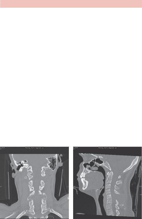
A CT is required to characterize the bone changes and ensure there is no traumatic injury (Figure 72.2).
(a) |
(b) |
Figure 72.2 (a) Coronal and (b) sagittal images of the patient’s cervical spine showing the expansile lesion in the posterior elements of the C5 vertebra. No fracture is seen.
198
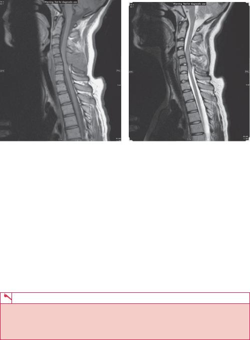
Given the patient’s neurology, an emergency magnetic resonance (MR) scan is required to examine the spinal cord, nerve roots and soft tissue associated with the bone lesion (Figure 72.3).
(a) |
(b) |
Figure 72.3 Sagittal MR (a) T1and (b) T2-weighted images of the patient’s cervical spine.
The MR shows an expansile lesion of the C5 spinous process with associated narrowing of the spinal canal and increased T2 signal (altered water content) within the spinal canal at this point. The high signal is most likely to be inflammatory due to trauma and cord swelling rather than a tumour or haemorrhage.
Given the patient’s age, possible differential diagnoses for expansile lesions in the cervical spine include benign tumours from the bone such as an aneurysmal bone cyst, as in this case, or from the spinal cord or nerve roots such as a neurofibroma. Malignant bone tumours are less likely given the age and history but could include lymphoma. Osteomyelitis should also be ruled out although unlikely in this case.
The patient requires urgent referral to a neurosurgical/spinal orthopaedic centre for possible removal of the lesion to decompress the cord. Steroids may help to reduce cord swelling.
KEY POINTS
•Acute or symptomatic spinal stenosis is an emergency and one of the few situations requiring an emergency MR to view the cord.
•Degenerative changes and disc prolapse are the most common causes of spinal stenosis, but the differential includes tumours and haematomas.
199
CASE 73: A YOUNG MAN WITH BACK PAIN
History
This 25-year-old man presents to you in the accident and emergency department with lower back pain that is so bad he is unable to walk. He gives a history of gradually worsening back pain over the last few months. He also complains of night sweats and bowel disturbance. He does not give a history of other medical problems and says that he is not working currently. He smokes 15 cigarettes a day and drinks at least 30 units a week. He takes cannabis regularly but denies use of intravenous drugs.
Examination
On examination he is thin and appears to neglect his appearance. His blood pressure, pulse and temperature are a little elevated. His chest and heart sounds are normal. The abdomen is soft. There is evidence of injection sites in the forearms and antecubital fossae. He is extremely tender over the lower spine. Neurologically, he has reduced power (4/5) on leg raising bilaterally.
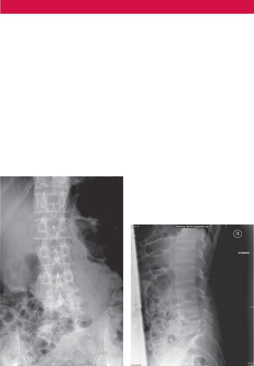
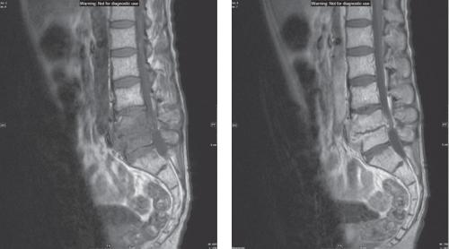
You arrange investigations including a chest and lumbar spine radiograph (Figure 73.1) and, on the basis of that, a magnetic resonance (MR) scan of the spine is arranged (Figure 73.2).
(a) |
(b) |
Figure 73.1 (a) Anterior–posterior (AP) and (b) lateral lumbar spine plain radiographs.
200
(a) |
(b) |
Figure 73.2 T1-weighted sagittal MR image of the lumbar spine, pre (a) and post (b) gadolinium contrast.
Just to give a brief summary of MR, hydrogen is a ubiquitous atom present in water and most biological molecules. In a magnetic field, the magnetic moment of the hydrogen nucleus becomes polarized and can be switched between aligned parallel and antiparallel with the magnetic field by radiofrequency pulses. Add in a magnetic field gradient to change the frequency at which the protons resonate according to position and you have the beginnings of spatial resolution and an MR image.
The protons relax from an ordered polarized state once the radiofrequency pulse has stopped. The relaxation of polarization is measured with the T1 rate constant, and the rate at which they become disorganized by T2. T1 and T2 are used as contrast parameters.
Fat and proteinaceous fluid have high T1 and T2 signals. Water has low T1and high T2 signal. Soft tissues are somewhere inbetween. Abnormalities often have high T2 signal. T1 images tend to be used for anatomy, comparison with T2 images or used with gadolinium contrast (high T1 signal).
Questions
•What do the figures demonstrate?
•Why was an MR scan ordered?
•What is the differential diagnosis.
•What would you do next and how?
•What is the appropriate treatment?
201