

Книги по МРТ КТ на английском языке / Thomas R., Connelly J., Burke C. - 100 cases in radiology - 2012
.pdfANSWER 1
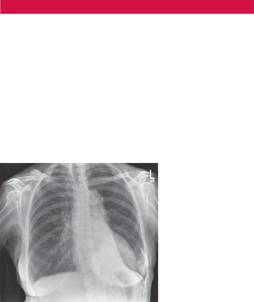
This is a posterior–anterior (PA) chest radiograph of an adult male. The lungs are hyperexpanded as evidenced by visualizing more than six anterior ribs above the diaphragm. The distance between the apex of the hemidiaphragm and a line drawn from the costophrenic to the cardiophrenic angle is less than 1.5 cm, in keeping with diaphragmatic flattening. The lung parenchyma demonstrates bullous emphysematous disease, most marked in the upper zones. There is no evidence of consolidation, collapse or pneumothorax. The cardiomediastinal borders are within normal limits, and both hila are of normal morphology. This chest radiograph suggests a diagnosis of chronic obstructive pulmonary disease (COPD).
COPD is a combination of increased mucus production, small airway obstruction and emphysematous change, with a slow and progressive history of increasing shortness of breath, usually in association with significant tobacco usage. Most commonly, the emphysematous component is ‘centrilobular’, with irreversible destruction of normal lung most in the apical segments of the upper lobes. On computed tomography (CT) this is clearly seen as central black holes of destroyed lung ‘punched-out’ from normal parenchymal architecture (Figure 1.2), although a CT is not a necessary investigation in most cases of COPD. Sometimes the clinical symptoms of COPD are confused with asthma, which usually starts in childhood and shows greater reversibility of airflow obstruction. Some patients develop asthma later in life, and in practice both conditions may coexist or be difficult to differentiate.
The most important investigation in a |
|
|
patient with COPD is lung function test- |
|
|
ing. Spirometry shows the reduced forced |
|
|
expiratory volume in 1 second : forced |
|
|
vital capacity (FEV1 : FVC) ratio charac- |
|
|
teristic of obstructive conditions. There is |
|
|
an increase in the total lung capacity (TLC) |
|
|
and residual volume (RV) in COPD as a |
|
|
result of air trapping. |
|
|
Reversibility to bronchodilators is limited |
|
|
in COPD. Assessment of functional capac- |
|
|
ity is an important part of the evaluation |
|
|
in chronic COPD. In acute exacerbations |
|
|
such as that described, it is important to |
|
|
assess blood gases to look for significant |
Figure 1.2 CT scan. |
|
hypoxia and/or carbon dioxide retention. |
||
|
KEY POINTS
•Flattening of the diaphragms and lung hyperexpansion are characteristic chest radiograph features of COPD.
•COPD is a combination of increased mucus production, small airways obstruction and emphysematous change.
•Lung function tests are the most important investigation in a patient with COPD.
2
CASE 2: THE BREATHLESS ASTHMATIC
History
A 36-year-old woman presented to the accident and emergency department complaining of progressively increasing breathlessness over the last 2 weeks. This was accompanied by a wheeze and cough productive of white sputum. Her exercise tolerance had reduced and she denied any orthopnoea or chest pain. She had a history of asthma which was usually well controlled with inhalers and had never previously required a hospital attendance. There was no other history of note and she denied ever being a smoker. She lived at home with her husband and two children.
Examination
On examination, her respiratory rate was 22 breaths per minute. She was afebrile and normotensive with a regular pulse rate of 88 per minute. Her cardiovascular and abdominal examinations were normal, but on auscultation of her lungs there was a prolonged expiratory wheeze with reduced air entry at the left base.
A chest radiograph was performed as part of her initial investigations (Figure 2.1).
Figure 2.1 PA chest radiograph.
Questions
•What radiological abnormality is present?
•What is the most likely cause considering her history?
3

ANSWER 2
This patient has left lower lobe collapse. Depending on the airway obstructed, each lobe collapses in a characteristic way. This was originally described by Benjamin Felson, a professor of radiology in the United States in 1973. In the case of the left lower lobe, when there is proximal occlusion, the lobe collapses posteriorly and medially towards the spine. Lying behind the heart, it assumes a triangular shape with a straight lateral border being classically described as a ‘sail sign’ on posterior–anterior (PA) chest radiograph as shown in Figure 2.2.
It usually overlies the cardiac shadow and can be easily missed on poorly windowed or under-penetrated films. The collapsed lobe obscures the left medial hemidiaphragm and the horizontal fissure swings downwards with the hilar displaced inferiorly. Other features to help confirm the diagnosis would include mediastinal and tracheal shift towards the side of the collapse, and possible herniation of the contralateral lung across the midline from compensatory hyperinflation. The degree of hilar depression and compensatory hyperaeration is variable depending on the degree of collapse. Less commonly, a stenosing bronchogenic tumour may be seen as a soft tissue density overlying the left hilar point.
Figure 2.2 PA chest radiograph with ‘sail sign’ indicated.
The causes of lobar collapse are numerous; incidence varies with age and clinical history. Overall, the commonest cause of collapse is related to a proximal stenosing bronchogenic carcinoma, and although the majority of lung cancer is seen in men, the incidence in women is rising. Lung cancer is rarely diagnosed in people younger than 40, but the incidence rises steeply thereafter with most cases (85 per cent) occurring in people over the age of 60 with a past medical history of smoking. In ventilated patients, including neonates, malpositioning of the endotracheal tube can aerate one lung and occlude the contralateral side, while in infants, collapse related to an inhaled foreign body (e.g. a peanut) should always be considered. In older children and young adults, the commonest cause of lobar collapse is as a complication of asthma.
Asthma is a chronic inflammatory disease characterized by reversible airflow limitation and airway hyperresponsiveness. In response to immunological stimuli, mucus hypersecretion from goblet cell hyperplasia can cause airway plugging. Proximal occlusion of a bronchus causes loss of aeration, and as the residual air is gradually absorbed, the lung volume reduces with eventual collapse. Considering the patient’s age and clinical history, this is the most likely cause of her left lower lobe collapse.
KEY POINTS
•Depending on the airway affected, each lobe collapses in a characteristic way.
•The ‘sail sign’ on a PA chest radiograph is indicative of left lower lobe collapse.
•In paediatric cases, always consider inhaled foreign body as a possible cause of lobar collapse.
4
CASE 3: AN ICY FALL
History
A 39-year-old woman is sent for an X-ray following a fall. She slipped on some ice while out shopping and raised her right hand to break the fall but her little finger was hyperflexed in the palm of her hand. She felt an instant sharp and stabbing pain in her little finger, which was centred over the distal and interphalangeal joints. Over the next few hours, her finger began to swell and was increasingly uncomfortable. No other injury was sustained and she attended her local general practitioner (GP) practice for further advice.
Examination
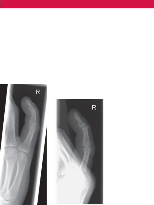
On examination there was soft tissue swelling and a partially flexed little finger that the patient was unable to completely straighten. There was no evidence of skin breach and the patient was otherwise fit and healthy. Concerned that a fracture had been sustained, the GP referred her to hospital for an X-ray and definitive treatment (Figure 3.1a,b).
(a) |
(b) |
Figure 3.1 (a) Oblique and (b) lateral radiographs of right little finger.
Questions
•What does this radiograph show?
•What other sites are commonly involved in this form of injury?
•How are X-rays made?
5
ANSWER 3
Figure 3.1a is a single oblique radiograph of the right little finger of adequate quality and penetration. There is partial flexion of the distal interphalangeal joint (DIPJ) with a small bony fragment seen in the dorsal aspect of the distal phalynx that is separated from the parent bone. Reduced cortication of the separated surfaces in association with generalized soft tissue swelling is in keeping with an acute fracture, and the bony fragment has been retracted proximally. These appearances are most likely related to a hyperflexion injury, with a fragment of bone avulsed by the extensor tendon at its insertion into the distal phalynx. In summary, there is an avulsion fracture to the distal phalynx of the right little finger.
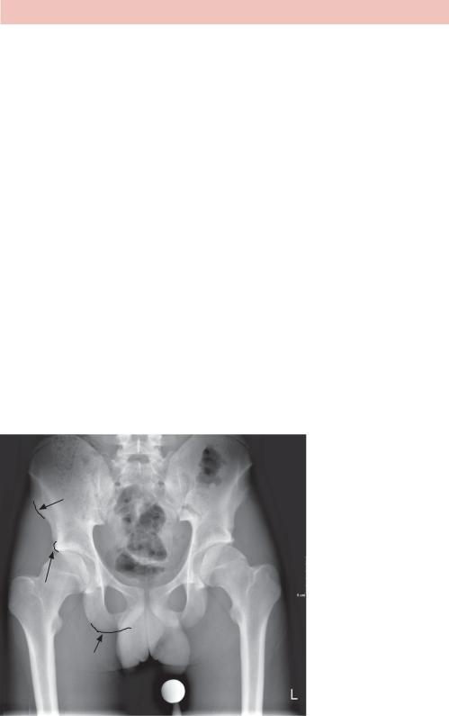
The term ‘avulsion’ is used medically to describe one part of the body forcibly detached from another in response to trauma. Commonly seen in the accident and emergency department related to skin degloving from road traffic accidents and nail bed trauma from a crush injury, radiological avulsion fractures occur when a bony fragment is separated from the parent bone in response to forcible contraction of a ligament or tendon. During puberty, secondary ossification centres lay down advancing margins of new bone for continued growth and development, with muscle insertions at this site forming the ‘apophysis’. The newly ossified bone is a site of weakness and is vulnerable to separation under extreme force. Any bone subjected to a forceful and usually unbalanced muscle contraction is subject to potential injury, however avulsion fractures are most often seen in active adolescent children commonly at muscle insertions into the pelvis. Sprinters, footballers and tennis players are at greatest risk of such injury. The three commonest sites of pelvis apophyseal avulsion, as shown in Figure 3.2, are:
•ischial tuberosity at the insertion of the adductor magnus muscle of the hamstring;
•anterior inferior iliac iliac spine at the insertion of the rectus femoris muscle; and
•anterior superior iliac spine at the sartorius muscle insertion.
Anterior Superior
Iliac Spine
Anterior Inferior
Iliac Spine
Ischial Tuberosity
Figure 3.2 Annotated plain film of pelvis.
6

First discovered by the German physicist W.C. Roentgen in 1895, the discovery of X-rays changed the landscape of medicine forever. To train as a radiologist, the basic physics of X-ray production need to be understood. Every atom is made up of a positively charged nucleus with numerous negatively charged electrons of different energy levels electrostatically held in place around it. Superheating a metal filament (e.g. tungsten) allows a negatively charged electron to free itself from the atom, and this can be accelerated along an X-ray tube attracted by a positively charged ‘anode’ target plate (also commonly made of tungsten). The fast-moving electron strikes the target plate with such force that it can eject a static electron from a target plate atom out of its normal stable trajectory around its nucleus. This makes the target atom unstable, and to protect itself, another static electron encircling the same nucleus will demote itself from a higher energy band to plug the hole left by the ejected electron. In doing so, it releases energy in the form of a single photon called an ‘X-ray’.
Rapidly repeating this procedure can generate an X-ray beam, which when passed through a human body can generate an image on X-ray sensitive material, as the X-rays interact with tissues of differing densities (e.g. bone versus fat).
KEY POINTS
•Avulsion describes the forcible detachment of one part of the body from another in response to trauma.
•A high index of suspicion of a pelvic apophyseal avulsion in athletic but skeletally immature adolescents is advisable.
•X-Rays were discovered in 1895 by W.C. Roentgen.
7
CASE 4: DIFFICULTY SWALLOWING
History
A 75-year-old man presents complaining of difficulty swallowing together with intermittent regurgitation of undigested food, often some time after eating. This has been slowly worsening. There is occasionally choking and coughing at night. There is no associated pain or heart burn and no history of weight loss or chest symptoms. He has a 30 packyear smoking history.
Examination
He looks well. The neck and chest examination is normal. No oropharyngeal abnormality is seen on visual examination. The abdomen is soft and non-tender.
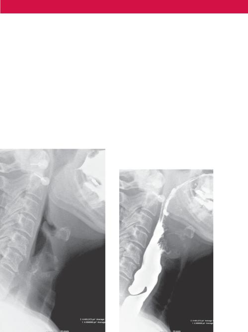
A recent chest X-ray is unremarkable. You organize a contrast swallow test (Figure 4.1).
(a) |
(b) |
Figure 4.1 (a–c) Three sequential lateral projections and (d) an anterior–posterior (AP) projection contrast swallow images. (continued overleaf)
8
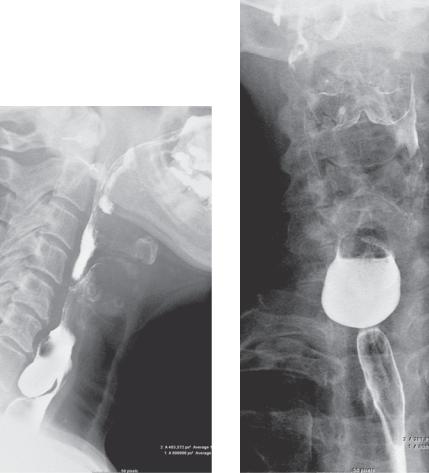
(c) |
(d) |
Figure 4.1 (a–c) Three sequential lateral projections and (d) an anterior–posterior (AP) projection contrast swallow images.
Questions
•What differential diagnosis should be considered?
•What do the images demonstrate?
•What other investigations can be used and what are their relative benefits?
9

ANSWER 4
The differential diagnosis for dysphagia is usefully split up into anatomical regions corresponding with the phases of swallowing (i.e. oral, pharyngeal and oesophageal). The type of symptom and the most appropriate investigations depend on whether the problem is primarily oropharyngeal or oesophageal.
Oropharyngeal dysphagia can be caused by:
•central neurological disorders such as stroke, brainstem tumours or degenerative diseases (e.g. Parkinson’s disease, multiple sclerosis and Huntington’s disease);
•peripheral neurological disorders including peripheral neuropathy, poliomyelitis and syphilis;
•systemic disorders such as myasthenia gravis, polymyositis, dermatomyositis or muscular dystrophy;
•oropharyngeal lesions including cricopharyngeal achalasia, tumours, inflammatory masses, Zenker’s diverticulum, oesophageal webs, extrinsic structural lesions, anterior mediastinal masses and cervical spondylosis; see Case 66.
Oesophageal dysphagia can be caused by:
•achalasia;
•spastic motor disorders, such as diffuse oesophageal spasm, hypertensive lower oesophageal sphincter and nutcracker oesophagus;
•scleroderma;
•obstructive lesions, such as tumours, strictures, lower oesophageal rings (Schatzki rings), oesophageal webs, foreign bodies, vascular compression and mediastinal masses.
Endoscopy is the investigation of choice for both oropharyngeal dysphagia, which is typically investigated in the ear, nose and throat department, and oesophageal dysphagia, which is investigated in the upper GI gastroenterology department. If endoscopy does not provide the answer, or the patient refuses the test, then a contrast swallow test can be done to visualize swallowing function. Videofluoroscopy is a low X-ray dose film of the very fast swallowing action in the oropharynx and is useful if there is a motor problem or unsafe swallow. A barium swallow is a series of images taken of the oesophagus while swallowing barium contrast.
This patient’s symptoms are suggestive of a pharyngeal or oesophageal problem. On the fluoroscopy images there is barium pooling in an oesophageal diverticulum arising from the posterior midline of the upper oesophagus, the typical position for a pharyngeal pouch (Zenker’s diverticulum). This is thought to be caused by spasm or uncoordinated peristalsis of the upper oesophageal sphincter and is located in Killian’s triangle, formed by the overlap of the oblique muscles of the inferior constrictor muscle and the transverse muscle fibres of the cricopharyngeus muscle.
The patient’s symptoms probably reflect progressive increase in size and compressive effect of the pouch. There is also increased risk of aspiration. The treatment is usually surgical excision or endoscopic stapling. The cricopharyngeal muscle may be separated to prevent recurrence. Complications of pouches include aspiration and, rarely, a carcinoma within the pouch.
Diverticula in other positions are possible. A Killian–Jamieson diverticulum is a lateral cervical oesophageal diverticulum just a little lower in position. Pulsion diverticula asso-
10

ciated with abnormal oesophageal contractions sometimes form in the lower third of the oesophagus. Pseudodiverticula are rare dilated glandular pouches in the mucosa of the mid oesophagus associated with reflux.
KEY POINTS
•Although endoscopy is the investigation of choice, contrast swallow tests provide evidence of functional problems that may not be seen on endoscopy and often underlie dysphagia.
•Common symptoms of a pouch are dysphagia, regurgitation and cough.
11