

Книги по МРТ КТ на английском языке / The Embryonic Human Brain An Atlas of Developmental Stages. Third Edition. 2006. By Ronan O'Rahilly
.pdf
THE
EMBRYONIC HUMAN BRAIN
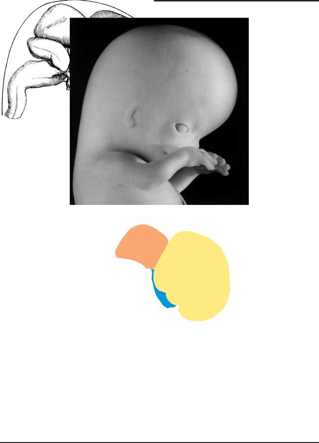
The brain at the end of the embryonic period (stage 23, 8 postfertilizational weeks). The color scheme used here (telencephalon in yellow, diencephalon in blue, mesencephalon in brown) will be repeated in a number of illustrations throughout the book.

THE
EMBRYONIC HUMAN BRAIN
AN ATLAS OF
DEVELOPMENTAL STAGES
THIRD EDITION
RONAN O’RAHILLY,
¨
FABIOLA MULLER,
M.D., D.Sc., Dr.h.c.
Dr.habil.rer.nat.
School of Medicine
University of California at Davis
Davis, California and
Institut d’Embryologie Sp´eciale Universit´ de Fribourg, Fribourg, Switzerland
A JOHN WILEY & SONS, INC., PUBLICATION
Illustrations on the cover:
Ultrasonic in vivo image at 6 weeks showing the fourth ventricle. cf. Fig. 23-35. Courtesy of Dr. Harm-Gerd Blaas.
Perspektomat reconstruction at 8 weeks, stage 23.
See Fig. 2-1.
Oblique section at 7 weeks, stage 20.
See Fig. 20-4.
Copyright C 2006 by John Wiley & Sons, Inc., Hoboken, NJ. All rights reserved.
Published simultaneously in Canada
No part of this publication may be reproduced, stored in a retrieval system, or transmitted in any form or by any means, electronic, mechanical, photocopying, recording, scanning, or otherwise, except as permitted under Section 107 or 108 of the 1976 United States Copyright Act, without either the prior written permission of the Publisher, or authorization through payment of the appropriate per-copy fee to the Copyright Clearance Center, Inc., 222 Rosewood Drive, Danvers, MA 01923, (978) 750-8400, fax (978) 750-4470, or on the web at www.copyright.com. Requests to the Publisher for permission should be addressed to the Permissions Department, John Wiley & Sons, Inc., 111 River Street, Hoboken, NJ 07030, (201) 748-6011, fax (201) 748-6008, or online at http://www.wiley.com/go/permission.
Limit of Liability/Disclaimer of Warranty: While the publisher and author have used their best efforts in preparing this book, they make no representations or warranties with respect to the accuracy or completeness of the contents of this book and specifically disclaim any implied warranties of merchantability or fitness for a particular purpose. No warranty may be created or extended by sales representatives or written sales materials. The advice and strategies contained herein may not be suitable for your situation. You should consult with a professional where appropriate. Neither the publisher nor author shall be liable for any loss of profit or any other commercial damages, including but not limited to special, incidental, consequential, or other damages.
For general information on our other products and services or for technical support, please contact our Customer Care Department within the United States at (800) 762-2974, outside the United States at
(317) 572-3993 or fax (317) 572-4002.
Wiley also publishes its books in a variety of electronic formats. Some content that appears in print may not be available in electronic formats. For more information about Wiley products, visit our web site at www.wiley.com.
Library of Congress Cataloging-in-Publication Data:
ISBN-13: 978-0-471-69462-5
ISBN-10: 0-471-69462-2
Printed in the United States of America
10 |
9 |
8 |
7 |
6 |
5 |
4 |
3 |
2 |
1 |
In memory of
Ernest Gardner, M.D., neuroscientist, colleague, and friend
. . . ce feroit vn grand bon-heur pour le genre humain, fi cette partie, qui eft la plus delicate de toutes, & qui eft fujette ´a des maladies tres-frequentes, & tres-dangereufes, eftoit auffi bien connu¨e, que beaucoup de Philofophes & d’Anatomiftes fe l’imaginent.
Niels Stensen, Discovrs svr 1’Anatomie du cerveau, 1669.

CONTENTS
Preface to the Third Edition |
ix |
|
1. |
Historical Aspects |
1 |
2. |
Techniques |
5 |
3. |
Prenatal Measurements |
9 |
4. |
Embryonic Staging |
11 |
5. |
Prenatal Age |
15 |
6. |
Terminology |
17 |
7. |
Early Stages |
21 |
8. |
Stage 8: The First Appearance of the Nervous System |
25 |
9. |
Stage 9: The Major Divisions of the Brain |
31 |
10. |
Stage 10: The Neural Tube and the Optic Primordium |
39 |
11. |
Stage 11: Closure of the Rostral Neuropore |
51 |
12. |
Stage 12: Closure of the Caudal Neuropore and the Beginning of |
|
|
Secondary Neurulation |
61 |
13. |
Stage 13: The Closed Neural Tube and the First Appearance of |
|
|
the Cerebellum |
75 |
14. |
Stage 14: The Future Cerebral Hemispheres |
84 |
15. |
Stage 15: Longitudinal Zoning in the Diencephalon |
93 |
16. |
Stage 16: Evagination of the Neurohypophysis |
103 |
17. |
Stage 17: The Future Olfactory Bulbs and the First Amygdaloid Nuclei |
115 |
18. |
Stage 18: The Future Corpus Striatum, the Inferior Cerebellar Peduncles, |
|
|
and the Dentate Nucleus |
133 |
19. |
Stage 19: The Choroid Plexus of the Fourth Ventricle and the Medial |
|
|
Accessory Olivary Nucleus |
149 |
20. |
Stage 20: The Choroid Plexus of the Lateral Ventricles, the Optic and |
|
|
Habenular Commissures, and the Interpeduncular and Septal Nuclei |
169 |
21. |
Stage 21: The First Appearance of the Cortical Plate in the Cerebral |
|
|
Hemispheres |
187 |
22. |
Stage 22: The Internal Capsule and the Olfactory Bulbs |
205 |
23. |
Stage 23: The Brain at the End of the Embryonic Period |
219 |
24. |
Trimester 1, Postembryonic Phase |
263 |
|
Early Postembryonic Phase |
265 |
|
Later Postembryonic Phase |
285 |
25. |
Trimester 2 |
297 |
vii

viii CONTENTS
26. Trimester 3 and the Newborn |
|
|
305 |
Supplement: Early Postnatal Life |
|
|
319 |
Bibliography |
|
|
323 |
Glossary |
|
|
331 |
Appendix 1: Changing Lengths of the Brain and its Subdivisions from 3 |
1 |
to 5 |
1 |
/2 |
/2 |
||
Weeks (Stages 9–16) |
|
|
341 |
Appendix 2: Computer Ranking of the Sequence of Appearance of Features |
|
||
of the Brain |
|
|
343 |
Appendix 3: Sequence and Stage of Appearance of Median Features of the Brain 349 |
|||
Appendix 4: Sequence and Stage of Appearance of Tracts of the Brain |
|
|
351 |
Appendix 5: Sequence and Stage of Appearance of Features Associated |
|
|
|
with the Rhombencephalon |
|
|
353 |
Index |
|
|
354 |

PREFACE TO THE THIRD EDITION
The main objectives of this monograph are to provide and to interpret drawings, photographs, and photomi-
crographs of the human embryonic brain. The drawings include at least a lateral view and a median reconstruction of the brain at each stage, as well as a clear indication of the plane of section of further illustrations, either drawings or photomicrographs. Summarizing statements of the morphological status of the brain at each stage are supplied, and brief statements concerning relevant anomalous conditions are included.
The embryonic period proper (the first eight postfertilizational weeks) is particularly important because (1) the embryonic brain is extremely difficult to comprehend and to visualize, (2) serial sections of first-class quality that show the human embryonic brain are rarely accessible, and (3) most major congenital anomalies appear during that time. A briefer account of the fetal period has been included and emphasizes the continuity of development.
Despite a general, superficial similarity in the development of the brains of various vertebrates, significant, subtle differences exist so that, undoubtedly important as are the findings in other species, contributions specifically to the prenatal human brain are as essential as they are fascinating.
The external and internal structure of the developing brain, the primary concern of this book, is an essential prolegomenon for subsequent investigations ranging from immunocytochemistry to imaging in vivo. Structural representation, however, needs to be completed by interpretation. In the present context two necessary components of precise interpretation are (1) morphological staging (as distinct from mere seriation, such as length or supposed age), without which individual events cannot be arranged in correct sequence, and (2) accurate three-dimensional reconstructions, without which erroneous assumptions of interpretation can be (and frequently are) made. These desiderata, which have been used by the authors in their research over many years, are a unifying feature of this treatise: (1) the Carnegie system of staging originated by Streeter and named and developed by the authors is fundamental to the presentation, and (2) most of the drawings here are based on extremely precise graphic reconstructions prepared by the authors. No similar work is available.
The authors’ scheme of the neuromeres, which differs from others in being based on the human, is detailed here. Other topics emphasized include segmentation, not only of the neural axis both rostrally and caudally, but also that of occipitocervical differentiation. Stem and crest cells in the olfactory region are elaborated, and the cerebellum, the amygdaloid complex, the hippocampal region, and the corpus striatum are given special attention.
This edition has been enhanced by the use of color, as well as by the inclusion of more that 100 new illustrations and 26 new tables. Moreover, some representative examples of ultrasonic images have been interspersed. The definitions formerly given in Chapter 6 have been expanded considerably and collected as an important Glossary near the end of the book.
The Bibliography has been enlarged and brought up to date. It comprises more than 350 references to the chief studies of the prenatal human brain. The profuse literature relating to other species, however, is beyond the scope of this book.
A set of standardized abbreviations, mostly selfevident, is used throughout for the illustrations, and a list of them is placed immediately inside the front cover.
The number assigned to chapters 8–23 and the first number given to each illustration indicate the developmental stage.
The authors wish to acknowledge the valuable support for their research provided by the U.S. National Institutes of Health over many years. Except where otherwise indicated, the photomicrographs as well as many of the photographs are from the Carnegie Collection. The scanning images are included through the kindness of Dr. med. Harm-Gerd Blaas and other colleagues who are credited in the appropriate places. It is a pleasure to thank all at WileyLiss for their encouragement and assistance.
RONAN O’RAHILLY
FABIOLA MULLER¨
Villars-sur Glane,ˆ Switzerland
July 2006
ix