

Книги по МРТ КТ на английском языке / The Embryonic Human Brain An Atlas of Developmental Stages. Third Edition. 2006. By Ronan O'Rahilly
.pdfTABLE 6–1. Examples of Improvements in Terminology
Undesirable Terms |
Comment |
|
|
Preferable Terms |
|
|
|
||
Anterior and posterior |
Best avoided for the early embryo |
Rostral and caudal |
||
Basal ganglia |
Ganglia are in the peripheral nervous system |
Basal nuclei |
||
Central fissure |
In forebrain, fissure (see Glossary) has restricted usage |
Central sulcus |
||
Cerebral vesicles |
Based on avian species |
|
Forebrain, midbrain, and hindbrain |
|
Cerebrum |
When considered to be the telencephalon only, it is in |
Telencephalon |
||
|
contradiction with the essential and comprehensive |
|
||
|
adjective cerebral |
|
|
|
Chorda dorsalis |
Acceptable but clumsy term |
|
Notochord |
|
Ganglionic eminences |
Ganglia are in the peripheral nervous system |
Ventricular eminences |
||
Gestational age |
Scientifically useless term |
|
Postfertilizational age |
|
Lateral fissure |
In forebrain, fissure (see Glossary) has a restricted usage |
Lateral sulcus |
||
Medullary folds and groove |
Medulla has other implications |
Neural folds and groove |
||
Menstrual age |
Menstural weeks acceptable, but they are not age |
(Post)menstrual weeks |
||
Midline; midsagittal |
Unofficial and unnecessary terms |
Median |
||
Parasagittal |
Misinterpretation of term sagittal |
Sagittal |
||
Yolk sac |
Yolk is not involved |
|
Umbilical vesicle |
|
|
|
|
|
|
A |
|
|
The |
|
|
|
A sagittal |
median |
|
|
|
plane |
|
|
|
GL |
plane |
|
|
|
|
|
||
|
|
|
|
|
A |
|
|
|
|
coronal |
|
|
|
|
plane |
|
|
|
|
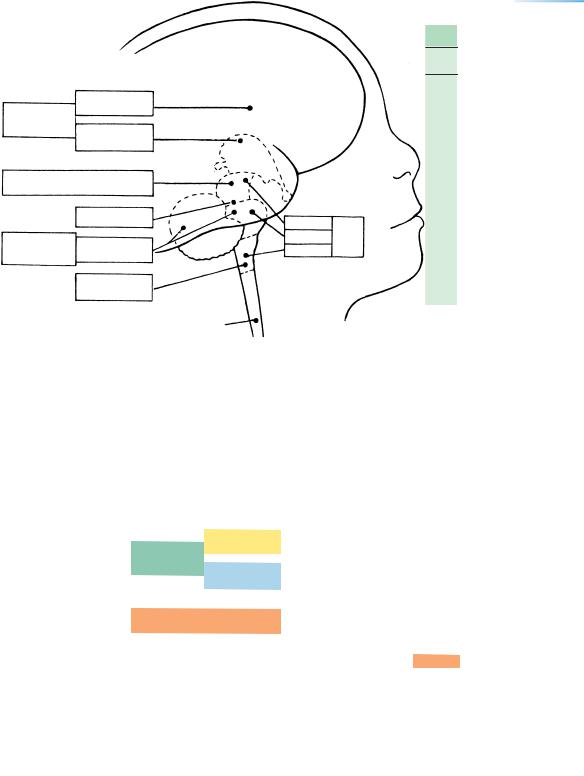
A transverse plane
B |
|
Axis of mesencephalic flexure |
|
A transverse |
A coronal |
|
plane |
|
|
plane |
|
|
|
The orbitomeatal plane
Figure 6–1. The main planes of reference and the chief terms of position shown in an embryo of stage 23 (8 weeks). (A) The body as a whole. (B) The head, including the brain.
TERMINOLOGY |
19 |
TABLE 6–2. The Main Developmental Subdivisions of the Brain as Given by His
Divisions |
Subdivisions |
Cavities |
EProsencephalon
n |
Telencephalon |
Lateral ventricles |
c |
Diencephalon |
Third ventricle |
e |
|
|
p |
|
|
hMesencephalon
a |
Mesencephalon |
Aqueduct |
l |
|
|
oRhombencephalon
n |
Isthmus rhombencephali |
|
|
Metencephalon |
|
|
Cerebellum |
|
|
Pons |
Fourth ventricle |
|
Myelencephalon |
|
|
Medulla oblongata |
Central canal |
|
|
|
3 |
6 |
DIVISIONS |
SUBDIVISIONS |
Telencephalon
Prosencephalon
Diencephalon
Mesencephalon
Isthmus
Rhomb-
encephalon Metencephalon
Myelencephalon
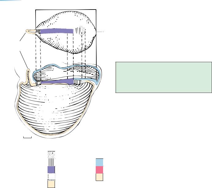
Figure 6–2. The location of the three divisions and six subdivisions of the brain as seen in the newborn.
Spinal
cord
Cerebral hemisphere
Midbrain
Pons Brain
stem
Medulla

C H A P T E R 7
EARLY STAGES
Because the first morphological indication of the nervous system appears at stage 8 (approximately 3 postfertilizational weeks), the following brief
statement concerning stages 1 to 7 is provided. Full details of these early stages have been published in a monograph by O’Rahilly and Muller¨ (1987a).
Some evidence exists in mammals (and probably including the human) that a labile axis of bilateral symmetry is determined by the site of penetration of a spermatozoon (reviewed by Denker, 2004).
Stage 1 is the unicellular embryo that is formed at fertilization, normally in the lateral end (ampulla) of the uterine tube. A new, genetically distinct human organism is thereby formed, although the embryonic genome does not become activated until the next stage.
Stage 2 (2–3 days) is the cleaving embryo that proceeds along the uterine tube.
Stage 3 (4–5 days) is characterized by the appearance of a cavity within the cellular mass, at which time the embryo is termed a blastocyst and lies in the uterine cavity. The embryonic disc now presents dorsal and ventral surfaces. The inner cell mass, mainly the epiblast, has been used for the production of embryonic stem cells that retain the ability to develop into all types of cells, including germ cells.
Stage 4 (6 days) is the attachment of the blastocyst to the uterine lining (endometrium), an event that heralds the beginning of implantation within the uterine mucosa.
Stage 5 (7–12 days), the continuation of implantation, is complicated and is subdivided into three substages,
according to whether (5a) the trophoblast is solid, (5b) the trophoblast contains lacunae, or (5c) the lacunae form a vascular circle. The amniotic cavity and umbilical vesicle (so-called yolk sac) appear during stage 5. At stage 5c a thickening of the hypoblast at one end of the embryonic disc may possibly indicate the rostral end of the body even before the appearance of the primitive streak (Luckett, discussed by O’Rahilly, 1973).
Stage 6 (about 17 days) is marked by the appearance of chorionic villi. During this stage, at approximately 2 12 postfertilizational weeks, axial features (particularly a cellular proliferation termed the primitive streak) develop, and hence the embryo acquires right and left sides, as well as rostral and caudal ends. The embryonic disc is now about 0.2 mm in length. Twins (other than certain conjoined examples) arise before the appearance of axial features, i.e., during approximately the first fortnight. Retinoic acid is believed to be implicated in the patterning of the rostrocaudal axis and the induction of the expression of Hox genes.
Stage 7 (Fig. 7–1, about 19 days). An axial structure known as the notochordal process is characteristic of this stage, when the embryonic disc measures approximately 0.4 mm. Although no morphological sign of the nervous system is yet present, the site of the future neural plate is dorsal and lateral to the notochordal process.
Stage 8 (about 23 days) is the subject of the following chapter. The epiblast is now being transformed into the neural plate in the region rostral to the neurenteric canal. Canalization of the notochordal process occurs (i.e., the
The Embryonic Human Brain: An Atlas of Developmental Stages, Third Edition. By O’Rahilly and Muller¨ Copyright C 2006 John Wiley & Sons, Inc.
21
22 |
|
|
|
|
|
C h a p t e r 7 : EARLY STAGES |
|
|
A |
Embryonic |
|
|
|
|
|
notochordal canal develops) in embryos |
approximately |
|
|
|
|
|
|
1 mm in length. When, during stage 8, the notochordal pro- |
||
|
disc |
|
|
|
|
|
||
|
|
|
|
|
|
cess attains a length of 0.4 mm, the neural groove, which is |
||
|
|
|
|
|
|
|
||
Dorsal |
|
|
|
|
|
|
the first morphological indication of the nervous system, |
|
view |
|
|
|
|
|
|
||
|
|
|
|
|
|
becomes recognizable. |
|
|
|
|
|
|
|
|
Median |
|
|
|
|
|
|
|
|
plane |
The cells and tissues that give rise to the nervous sys- |
|
|
|
|
|
|
|
|||
|
|
|
|
|
|
|
tem are given in Table 7–1. |
|
Allantoic |
|
|
|
|
|
|
The most frequently encountered errors in descrip- |
|
diverticulum |
|
|
|
|
|
|
tions of the initial development of the human brain have |
|
|
|
|
|
|
|
|
||
|
|
|
|
|
|
|
recently been listed (O’Rahilly and Muller,¨ |
1999). |
6 |
5 |
4 |
3 |
|
2 |
1 |
|
|
B
Amniotic cavity
Dorsal |
surface |
|
Important
The embryo shows dorsal and ventral surfaces at stage 3 (about 1 12 postfertilizational weeks), and right and left sides, as well as rostral and caudal ends, at stage 6 (about 2 12 weeks.)
Umbilical  vesicle
0.1 mm |
Median section |
|
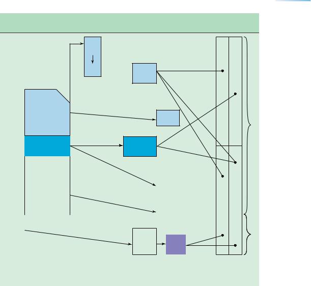
Prechordal plate
Notochordal process
&plate Primitive streak
&node
Site of future cloacal membrane
Allantoic diverticulum
Ectoderm
Mesoderm |
Endoderm |
Figure 7–1. Reconstruction of an embryo at stage 7 (nearly 3 weeks), immediately before the neural groove and folds become visible. (A) Dorsal view and (B) median view. The various features are identified in the keys below the drawing.
EARLY STAGES |
23 |
TABLE 7–1. The Origin of the Central and Peripheral Nervous Systems, and the Associated Neural Cresta
Neural
ectoderm
Neurosomatic junction
Neural |
C |
P |
|
N |
N |
||
plate |
|||
S |
S |
||
|
folds  Neural tube
Retina |
Optic |
|
|
crest |
1 |
|
|
Neural
crest
|
|
Somatic- |
|
|
|
|
Otic |
Nasal |
|
|
|
ectoderm |
|
|
|
crest |
crest |
|
|
|
|
|
|
|
|
|
|
|
|
|
|
|
|
|
|
|
|
|
|
|
|
|
|
|
|
Placodes |
|
|
|
|
|
|
|
|
|
("islands") |
|
|
|
|
Primi- |
|
|
|
|
|
|
||
|
|
|
|
|
|
|
|
|
|
|
|
|
|
|
|
|
|
|
|
|
tive |
|
|
|
|
|
|
2 |
|
|
|
|
|
|
|
|
|||
|
streak |
|
|
|
Caudal |
|
|
||
|
|
|
|
|
|
||||
|
|
|
|
|
|
|
|
||
|
|
|
|
|
|
Neural |
|
|
|
|
|
|
|
|
eminence |
|
|
|
|
|
|
|
|
|
|
cord |
|
|
|
|
|
|
|
|
|
|
|
|
|
|
Stage |
|
|
|
|
|
|
|
|
|
|
|
|
|
|
|
|
|
|
|
6b |
8 |
8b |
9 |
10 |
12 |
13 |
|
|
|
|
|
|
|
|
|
|
|
|
|
|
|
|
|
|
|
|
|
|
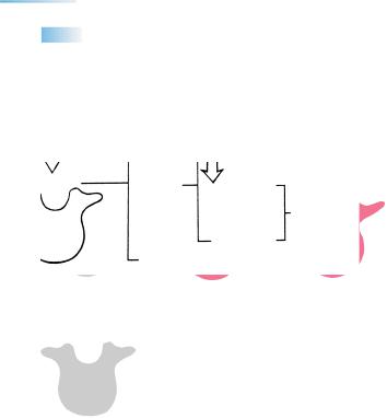
a In primary neurulation (marked 1 at the right) the neural tube is formed by folding of the neural plate and fusion of the neural folds. The neural discs or placodes, which develop in the somatic ectoderm, were thought of by Streeter as islands of neural ectoderm. Secondary neurulation (marked 2 at the right) is the formation of the caudal portion of the neural tube from a cellular mass known as the caudal eminence, and not by fusion of neural folds. The interrupted horizontal line indicates the cerebrospinal junction.
24 |
C h a p t e r 7 : EARLY STAGES |
NEUROTERATOLOGY
It has been shown experimentally in the mouse that excessive cell death in the primitive streak plays a major role in spina bifida aperta caused by nonclosure of the neural folds (Fig. 7-2).
A B C
Myeloschisis
D |
aperta |
|
|
Spina bifida |
(NTD) |
Myelomeningocele
cystica
Meningocele
occulta
Figure 7–2. The completion of a vertebral foramen and its relationship to spina bifida. (A) In the embryo, the cartilaginous neural processes have not yet united. (B) In the newborn, although three ossific centers are present, the spinous process is still cartilaginous, providing a radiological appearance of a normal spina bifida. (C) In the adult, ossification is complete. (D) Spina bifida is a failure of closure of a vertebral foramen from whatever cause. The condition may be concealed (occulta) or evident (aperta) and generally cystic (cystica).
C H A P T E R 8
STAGE 8: THE FIRST
APPEARANCE OF THE
NERVOUS SYSTEM
Approximately 1–1.5 mm in Greatest Length;
Approximately 23 Postfertilizational Days
The embryo is an ovoid (frequently pear-shaped) disc with a longitudinal axis and distinct cephalic and caudal ends. In one-quarter of examples, referred to as
stage 8b, the neural groove can be detected in the neural plate as a very shallow sulcus bounded by faint neural folds. This is the first visible sign of the future nervous system, and most of the folds represent the brain. The neural plate is closely related to two median features: the prechordal plate and the notochordal process.
Stage 8 has been generally listed as 18 days, but is probably more usually about 23 days.
Although few morphological features are visible at the time of initial development of the brain, stage 8 and its
organization are of crucial importance for an understanding of the developing nervous system. It is of interest that the primordium of the brain appears before the heart or any other organs become visible.
Studies of early neurogenesis indicate that differences concerning neural induction among vertebrate classes are present from the beginning of neurulation, and that mammalian neurulation is extremely complex.
Important
The first indication of the neural groove and folds is found during this stage.
The Embryonic Human Brain: An Atlas of Developmental Stages, Third Edition. By O’Rahilly and Muller¨ Copyright C 2006 John Wiley & Sons, Inc.
25
26 |
C h a p t e r 8 : THE FIRST APPEARANCE OF THE NERVOUS SYSTEM |
|
|
|
|
TABLE 8–1. The Neurenteric Canal and Its Site as Important Landmarks |
|
|
Stage |
Rostral to Neurenteric Canal |
Caudal to Neurenteric Canal |
|
|
|
8 |
Notochordal process and notochordal plate induce (in stage 8b) development |
Primitive streak and epiblast |
|
of neural plate from the epiblast in area of future brain as far rostrally as D2 |
|
|
Prechordal plate induces D1 |
|
|
Floor plate may perhaps be present |
|
|
Endoderm incomplete axially |
Endoderm complete |
9 |
Notochordal plate is ventral to brain, including Rh. D |
Primitive streak |
|
Floor plate may perhaps be present |
|
|
Endoderm incomplete axially |
Endoderm complete |
10 |
Rostral part of notochord develops by folding of notochordal plate |
Spinal part of neural plate develops |
|
|
from surface ectoderm |
|
Endoderm gradually continuous axially |
Caudal part of notochord develops |
|
|
directly from mesenchyme |
|
|
|
X
Y
|
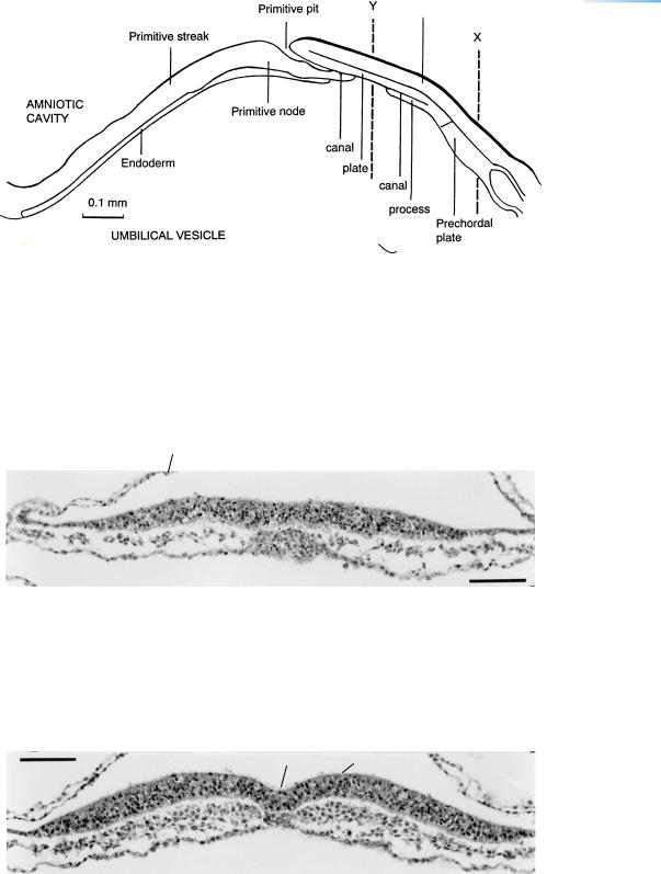
Figure 8–1. Dorsal view of an embryo of stage 8. The rostral |
|
end is above; the caudal end is below and is anchored by the |
|
connecting stalk, at the end of which two chorionic villi have |
|
been sectioned. The amnion has been cut and removed in |
|
order to expose the dorsal surface of the embryo. X and Y are |
|
the levels of the sections shown in Figures 8–3 and 8–4. |
|
The embryo at stage 8 is a slightly vaulted, pear-shaped disc |
|
that displays a longitudinal axis. The axis is indicated (1) by the |
|
primitive groove, which begins at the primitive node and |
|
proceeds caudally, and (2) by the neural groove, which is |
|
situated in the median plane at level Y. This is the region of the |
|
neural plate, which, although sharp boundaries are not |
|
evident, comprises (a) a peripheral rim, the alar plate, and (b) a |
|
more central part capping the notochordal process and |
|
primitive node, namely the basal plate or lamina (Fig. 9–5). |
|
The neural groove, which is shown in Figure 8–4, is the first |
|
visible sign of the future nervous system and, for the most part, |
|
it indicates the site of the brain. |
|
This beautiful drawing of the Heuser embryo is by James F. |
|
Didusch, whose “work is unexcelled in the anatomical |
|
literature” (Crosby and Cody, 1991). In 1917 Max Brodel¨ |
0.14 mm |
referred to Didusch as “my first pupil.” |
THE FIRST APPEARANCE OF THE NERVOUS SYSTEM |
27 |
Figure 8–2. Graphic reconstruction prepared from transverse sections of an embryo of stage 8. The rostral end is to the right. From rostral to caudal, the following are indicated: prechordal plate, neural plate (dorsally), and endoderm.
The neural plate is in close contact rostrally with the prechordal plate, and ventrocaudally with the notochordal process. The rostralmost portion of the neural plate corresponds to the optic area of the diencephalon, neuromere D1.
The notochordal plate is the roof of the notochordal process at levels where the floor has already disappeared during the process of intercalation of the plate in the endoderm. The notochordal process is a cellular rod that contains the notochordal canal and is in close contact with the neural ectoderm dorsally and the mesenchyme laterally. Its canal begins as a pit in the primitive node and then proceeds more or less vertically, as the neurenteric canal, before continuing rostrally. The primitive streak is situated caudal to the primitive node, and it gives rise to numerous mesenchymal cells.
Amnion
Figure 8–3. Transverse section at the level of the prechordal plate corresponding to line X in Figure 8–2. This figure and Figure 8–4 were selected from a histologically superior embryo. The prechordal plate is a multilayered accumulation of cells in close contact with the floor of the neural groove of D1. It is involved in neural induction, which is probably maximal at stage 8. The plate also furnishes mesenchyme for the orbital muscles and probably for the tentorium cerebelli. The location of the prechordal plate in the human would seem to correspond to the axial mesoderm between the first pair of “somitomeres” in the mouse, in which species seven such segmental units have been detected rostral to the first somite. Bar = 0.05 mm.
The prechordal plate is an important source of prenotochordal tissue. It is maintained that the prechordal plate is ventral to a single retinal field at the rostral end of the neural plate and that normally it suppresses the median part of that field, thereby causing bilateral retinal primordia to develop (Fig. 10–13).
Neural groove & fold
Figure 8–4. Transverse section at the level of the notochordal plate corresponding to line Y in Figure 8–2. The shallow neural groove, present in one-quarter of embryos of stage 8, is bounded on each side by a slightly elevated neural fold. Numerous mitotic figures are present in the neural plate and are adjacent to the amniotic cavity—that is, near the future ventricular surface of the later-appearing neural tube.
The multilayered mesoderm interposed between ectoderm and endoderm is separated by the basement membranes of the two epithelia. The median part of the neural epithelium is the future floor plate. It is firmly attached to the notochordal plate and is as thick as the lateral portion of the neural plate. Bar = 0.05 mm.