

Книги по МРТ КТ на английском языке / The Embryonic Human Brain An Atlas of Developmental Stages. Third Edition. 2006. By Ronan O'Rahilly
.pdf88 |
C h a p t e r 1 4 : THE FUTURE CEREBRAL HEMISPHERES |
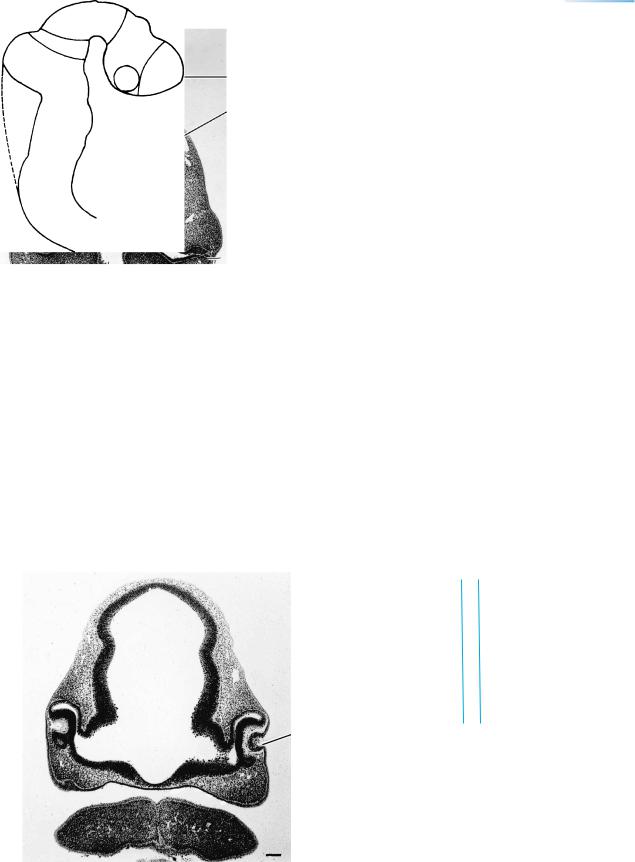
Figure 14–3. Developing fasciculi and nerve exits. The outline of the right optic primordium is indicated by an interrupted line. The preoptico- hypothalamo-tegmental tract (black band indicated by a dagger) and the ventral longitudinal fasciculus are identifiable and are important in the transportation of monoamines from the locus caeruleus to the forebrain. The surface overlying the area of the future amygdaloid nuclei possesses tall horizontal cells of the Cajal–Retzius type. This is the first region where a primordial plexiform layer begins to develop. This is not surprising, because the site is adjacent to the diencephalon, and the only possible access for corticipetal fibers into the embryonic cerebral cortex is through the di-telencephalic sulcus (Mar´ın-Padilla, 1988a). The medial longitudinal fasciculus (thick black band) begins in the interstitial nucleus of the prerubral area (Fig. 14–5) and joins the ventral longitudinal fasciculus of the rhombencephalon. The lateral longitudinal fasciculus is indicated by a wide, stippled band. Dorsally, along the sulcus limitans, the common afferent tract (unshaded) contains sensory fibers from the cranial nerves. Present also are the beginnings of the habenulo-interpeduncular and medial tectobulbar tracts, and the mesencephalic tract of the trigeminal nerve. The exits of cranial nerves 3, 6, and 12 are ventral, whereas those of the others are along the sulcus limitans, where all afferent fibers enter.
1 |
2 |
3 |
A
B
C
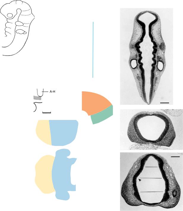
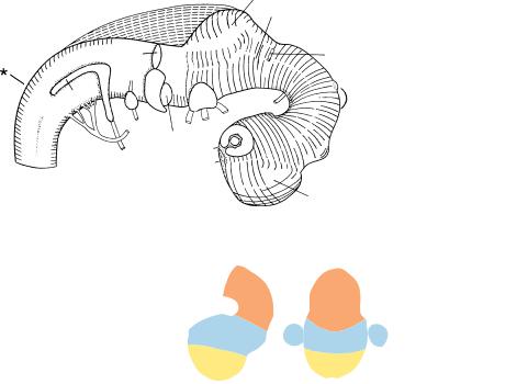
Figure 14–4. The telencephalon, showing the development of the future cerebral hemispheres in (1) right lateral,
(2) median, and (3) head-on views: (A) no indication of cerebral hemispheres is seen; (B) hemispheric evagination has begun; (C) the future hemispheres are evident (C belongs to stage 15). In column 2, the telencephalon is shown in yellow, and the entrance to the left optic stalk in black. In column 3, the telencephalic portion of the third ventricle is delineated between the lateral ventricles.
Interstitial
nucleus
M1
Di.
A-H
THE FUTURE CEREBRAL HEMISPHERES |
89 |
The levels of the sections in Figures 14–5 and 14–6 are shown in the key. The bars represent 0.1 mm.
Sulcus limitans
Primary meninx
Figure 14–5. Section through the midbrain and hypothalamus. The interstitial nucleus is discernible at the ventral transitional area between diencephalon and mesencephalon. The adenohypophysial pouch is at all times closely related to the diencephalic floor. The mesenchyme adjacent to the brain is becoming looser: this is the primary meninx. From Bartelmez and Dekaban (1962).
Fig. 14 –
5 6
Syn.
Di.
Lens pit
Figure 14–6. Section through the optic stalks and optic cups. The optic ventricle still communicates with the interval between the retinal layers (where so-called detachment of the retina can occur). From Bartelmez and Dekaban (1962).
90 |
C h a p t e r 1 4 : THE FUTURE CEREBRAL HEMISPHERES |
A
1
2
3
4
5
6
7
B
C
Syn.
Caudal
par.
Rostral
par.
D1
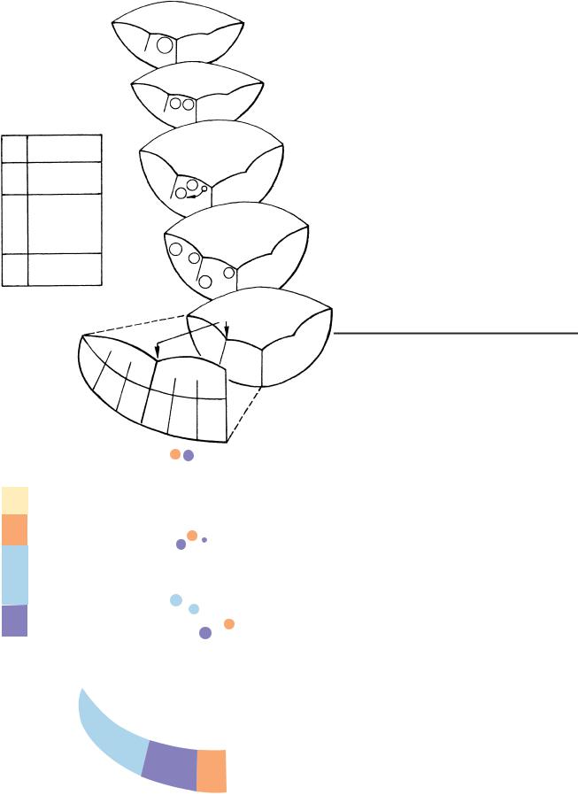
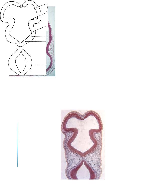
Figure 14–7. The neuromeres at stage 14 as shown in graphic reconstructions. The distinction that can now be made between the rostral and caudal parts of the parencephalon completes the series of 16 neuromeres. The initial appearance of the neuromeres is summarized in Table 10–1. The rostral parencephalon contains the opening of the optic ventricle on each side and is associated with the infundibular region and the ventral thalamus, whereas the caudal parencephalon presents the dorsal thalamus and the mamillary region. (A) Longitudinal section of the brain stem at the level indicated in the key. The mesencephalon is joined to the rhombomeres by the isthmus. Bar: 0.25 mm. (B) Section through the forebrain along the line B. The nasal discs and their neural crest are visible on each side. In the key drawing the asterisks denote the sulcus medius, which separates the dorsal (d) from the ventral (v) thalamus. The hypothalamic cell cord (stippled) indicates longitudinal differentiation in the diencephalon even before the hypothalamic sulcus appears at the next stage. (C) Section through the forebrain along the line C. The four compartments separated by horizontal lines are synencephalon, caudal parencephalon, rostral parencephalon, and D1 (immediately above the chiasmatic plate). The sulcus medius is marked by an arrowhead. Portions of the optic stalk and optic cup are visible on the right-hand side of the photomicrograph. Bar: 0.25 mm.
BRAIN
Ventricle
a |
b c |
Retinal |
|
fissure |
|
|
d |
|
|
e |
|
|
|
BRAIN |
|
a |
Terminal bars |
|
b |
Germinal or |
|
|
ventricular |
|
c |
Mantle |
|
d |
Marginal |
|
e |
ELM |
‘‘Optic
ventricle’’
Layers
EYE
a
b
c
d
e
RETINA
ELM
Proliferative
Mantle
Marginal
ILM
Figure 14–8. Comparison of the wall of the developing forebrain and that of its derivative, the optic cup, at about 41/2 weeks. The cavities (future third ventricle and temporary optic ventricle) are continuous (Fig. 14–6). The corresponding layers are as follows:
a.Adjacent to the cavities is a terminal bar net, known in the retina as the external limiting membrane (ELM).
b.Germinal or proliferative cells, adjacent to the ventricle.
c.Mantle layer.
d.Marginal layer.
e.The basement membrane is the external limiting membrane of the brain. In the eye, however, because of the invagination that forms the optic cup, it is the internal limiting membrane (ILM) of the retina.
Layers
Common
efferent
Somatic
efferent
Somatic
afferent &
Visceral
afferent
Visceral
efferent
G, general S, special
Stage
12,
13
14
15
18
Sulcus
limitans
G
S
A S
G
V G S G
A V
AV
E V S
E E
Figure 14–9. The original position of the somatic efferent nuclei in a ventrolateral cell column and of the visceral efferent nuclei in a ventromedial column was illustrated and clarified previously (O’Rahilly et al., 1984). It is not generally appreciated that the position changes considerably during development (Windle, 1970; Muller¨ and O’Rahilly, 1990c, Table 2).
92 |
C h a p t e r 1 4 : THE FUTURE CEREBRAL HEMISPHERES |
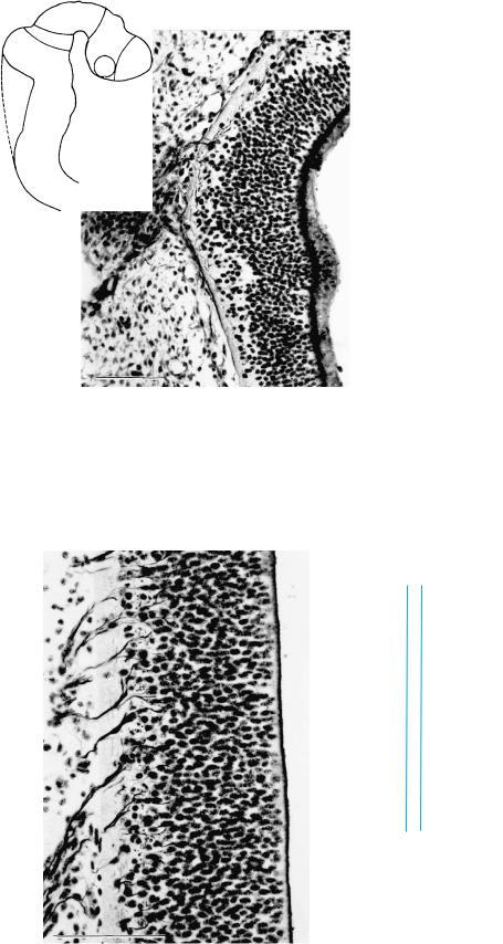
Rh.2
Figure 14–10. Section through the trigeminal ganglion. Nerve fibers from the ganglion enter the rhombencephalic wall and form the common afferent tract. It is maintained that the motor fibers of the pharyngeal nerves leave the even-numbered rhombomeres, in which case (in the mouse) the trigeminal fibers that arise in the motor nuclei of Rh.2 and Rh.3 emerge from rhombomere 2.
Fig. 14 –
11 10
Figure 14–11. Section through hypoglossal rootlets, which emerge from the still laterally placed hypoglossal nucleus and traverse the clear marginal layer of the rhombencephalic wall.
Figures 14–10 and 14–11 are from silver-impregnated sections.

C H A P T E R 15
STAGE 15: LONGITUDINAL ZONING IN THE DIENCEPHALON
Approximately 7–9 mm in Greatest Length;
Approximately 35 Postfertilizational Days
The brain presents five major subdivisions at five weeks (Table 15–1). The neuromeres of the forebrain are still recognizable. Each cerebral hemisphere is
limited externally by the di-telencephalic sulcus and internally by the torus hemisphericus. The medial ventricular eminence of the basal nuclei has appeared in the previous stage and it is diencephalic. The lateral ventricular eminence, which now appears, is telencephalic (Fig. 19–6). The amygdaloid region will be derived mainly from the medial eminence. The wall of the diencephalon presents five longitudinal zones: epithalamus, dorsal thalamus, ventral thalamus, subthalamus, and hypothalamus. The primordium of the epiphysis cerebri is beginning. The sulcus limitans ends rostrally at the midbrain (Ml) and is not continuous with the hypothalamic sulcus. Hence the alar/basal distinction
does not arise in the forebrain. The cerebellum is derived from both the isthmus and rhombomere 1. Most cranial nerves are present. Axodendritic synapses have been detected in the cervical region of the spinal cord, followed by axosomatic synapses early in the fetal period (Okado, 1981). The vertebrae are now first clearly defined. The tapered caudal end of the trunk is not a “tail.”
Important
By this stage all five major subdivisions of the brain are present: myelencephalon, metencephalon, mesencephalon, diencephalon, and telencephalon (the future cerebral hemispheres as well as the telencephalon medium).
The Embryonic Human Brain: An Atlas of Developmental Stages, Third Edition. By O’Rahilly and Muller¨ Copyright C 2006 John Wiley & Sons, Inc.
93
94 |
C h a p t e r 1 5 : LONGITUDINAL ZONING IN THE DIENCEPHALON |
Cerebellum
.
Figure 15–1. Right lateral view of the brain. The asterisk indicates the junction with the spinal cord. The cerebral hemisphere is demarcated from the diencephalon by the di-telencephalic sulcus. The lens vesicle and the optic cup with its retinal (“choroid”) fissure are shown
(Fig. 15–7). Still funnel-shaped, the isthmus blends at its wider end with the cerebellar region and narrows towards M2.
M
Di.
T

LONGITUDINAL ZONING IN THE DIENCEPHALON |
95 |
Figure 15–2. Graphic reconstruction prepared from transverse sections to show a median view of the brain. The asterisk indicates the junction with the spinal cord. The cerebral hemisphere is limited from the diencephalon by an internal crest, the torus hemisphericus (Fig. 15–4), which begins at the velum transversum and ends as a basal thickening in the medial ventricular eminence (Table 15–2). In the development of the basal nuclei (Fig. 19–6), the lateral ventricular eminence, which is telencephalic, has appeared (Fig. 15–2, inset). The medial eminence, however, is entirely diencephalic, although it has been misinterpreted by others as the lateral eminence. The chiasmatic plate is well defined and is limited by preoptic and postoptic recesses. The infundibular recess is appearing adjacent to the adenohypophysial pouch. The caudal part of the diencephalon (formerly D2) is the synencephalon, which will give rise to the prerubrum and the pretectum. At this level, the epiphysis cerebri begins as a thickening, but a recess is not yet present.
A characteristic feature of this stage is the appearance of longitudinal zones in the diencephalon (Fig. 15–4 and Table 18–1). For example, the hypothalamic sulcus delineates the hypothalamus from the thalamus. In addition, the hypothalamus comprises subthalamus and hypothalamus sensu stricto (separated by a faint sulcus); the thalamus consists of epithalamus, dorsal thalamus, and ventral thalamus (Table 18–1). The dorsal and ventral thalami are incompletely separated in more advanced embryos by the beginning formation of the zona limitans intrathalamica, which is marked on the interior relief by the marginal ridge. Blood vessels begin to penetrate the wall of the diencephalon.
The midbrain consists of two neuromeres: M1 and M2. The sulcus limitans ends at the rostral limit of M1. M2 is joined to the hindbrain by the isthmus. The supramamillary commissure is marked by a dagger. In the hindbrain, the cerebellar plate is distinct and extends over two rhombomeres: the isthmus and Rh. 1. The roof of the isthmus forms the superior medullary velum. The locus caeruleus is indicated (Fig. 15–10). The notochord is partly surrounded by sclerotomic material that represents the future basioccipital component of the skull.
The inset drawing below left shows the contiguity of the torus hemisphericus and the medial ventricular eminence. The lateral eminence is visible in the lateral wall of the (left) cerebral hemisphere.
The boundaries between the major divisions of the brain are listed in Table 15–2.
96 |
C h a p t e r 1 5 : LONGITUDINAL ZONING IN THE DIENCEPHALON |
Fig. 15 – 4
Figure 15–3. Median section of the brain. The mesencephalic flexure is now extremely acute.
M
AH
Chiasma
Commissural
plate
Lateral ventricle
Torus hemisphericus
Hypothalamic sulcus
Subthalamus
Hypothalamus
Isthmus
Figure 15–4. Section through the forebrain and isthmus rhombencephali. The hippocampal thickening is distinct on each side of the lamina terminalis.

LONGITUDINAL ZONING IN THE DIENCEPHALON |
97 |
TABLE 15–1. The Sequence of Appearance of Key Features of the Brain, Including the Main Divisions and Subdivisions
|
Stage 9 |
10 |
11 |
12 |
13 |
14 |
15 |
|||
|
|
|
|
|
|
|
|
|
|
|
Flexures |
Mesencephalic |
|
|
|
|
|
|
|
Pontine |
|
|
|
|
|
|
|
|
|
|||
|
|
|
|
|
|
|
|
|
|
|
Neuropores |
|
|
|
Rostral |
Caudal |
|
|
|
|
|
|
|
|
closes |
closes |
|
|
|
|
|
|
|
|
|
|
|
|
|
|
|
||
|
|
|
|
|
|
|
|
|
|
|
|
|
|
Telencephalon |
|
|
|
|
|
|
1 |
|
|
|
|
|
|
|
|
|
||
|
|
|
medium or |
|
|
|
|
|
|
Future |
|
|
|
impar |
|
|
|
|
|
|
cerebral |
|
|
|
|
|
|
|
|
|
|
hemispheres |
|
Prosencephalon |
|
|
|
|
|
|
|
|
|
|
|
|
Diencephalon |
|
|
|
|
|
|
2 |
Neural |
|
|
|
|
|
|
|
|
|
|
folds |
Mesencephalon |
|
|
|
|
|
|
|
3 |
|
|
|
|
|
|
|
|
|
|
||
|
|
|
|
|
Isthmus rhomb. |
|
|
|
|
|
|
|
|
|
|
|
Cerebellar |
|
|
|
4 |
|
Rhombencephalon |
|
|
|
|
primordium |
|
|
|
Metencephalon |
|
|
|
|
|
|
|
|
|||
|
|
|
|
|
|
|
|
|
5 |
|
|
|
|
|
|
|
|
|
|
||
Myelencephalon
TABLE 15–2. Boundaries Between the Main Subdivisions of the Brain
|
Division of Brain |
Limit at Roof |
Limit at Floor |
|
|
|
|
|
|
|
Telencephalon medium |
|
|
|
Velum transversum |
Preoptic recess |
|||
|
|
|||
|
Diencephalon |
|
|
|
|
Between posterior commissure and |
Caudal supramamillary commissure |
||
|
|
|||
|
|
|||
|
|
commissure of superior colliculi |
|
|
|
Mesencephalon |
|
|
|
|
Rostral commissure |
Rostral to nuclei of trochlear nerves |
||
|
|
|||
|
|
of trochlear nerves |
|
|
|
Rhombencephalon |
End of hypoglossal rhombomere (Rh.D) |
|
|
|
|
|
||
|
Spinal cord |
|
||
|
|
|
||
|
|
|
||
|
|
|
|