C H A P T E R 23
STAGE 23: THE BRAIN
AT THE END OF THE
EMBRYONIC PERIOD
Approximately 27–31 mm in Greatest Length;
Approximately 56 Postfertilizational Days
The cortical plate covers almost the whole neopallial surface. The hippocampus has reached the temporal pole; its cells exhibit the largest intercellular distances in the entire brain. The insula appears as an indented area. In the corpus striatum the primordia of the caudate nucleus and the putamen are recognizable, and the globus pallidus externus has moved into a telencephalic position. The anterior commissure begins to develop in the commissural plate, which has gained in thickness. It needs to be stressed that the telencephalon and the diencephalon are not fused. The optic tract reaches the ventral portion of the lateral geniculate body. The dorsal thalamus occupies approximately half of the medial diencephalic surface. In addition to the ventricular layer, which is found in all parts of the brain, the rhombic lip has a germinative role. It produces the external germinal layer of the cerebellum, which has begun to spread over the rostral surface of the flocculus. The rhombic lip also gives off olivo-arcuate and pontine migratory sheets. The cerebellar commissures have appeared. Two cerebellar peduncles, the inferior and the
superior, are well-developed.
Stage 23, 8 postfertilizational weeks, needs to be stressed because this is the close of the embryonic period proper.
The pyramidal decussation appears already during the embryonic period.
The brain at stage 23 is far more advanced morphologically than is generally appreciated, to such an extent that functional considerations are imperative.
The arrangement of the rhombencephalic nuclei and tracts at 8 weeks is very similar to that present in the newborn. It is likely that the rapid growth of the rhombencephalon during the embryonic period proper is associated with correspondingly early functional activity.
Important
The term basal nuclei is best reserved for the basal structures affected pathologically in so-called extrapyramidal motor diseases, i.e., the corpus striatum, the subthalamic nucleus, and the substantia nigra (Fig. 19–6). The inclusion of the claustrum and the amygdaloid complex, however, has little to recommend it.
The Embryonic Human Brain: An Atlas of Developmental Stages, Third Edition. By O’Rahilly and Muller¨ Copyright C 2006 John Wiley & Sons, Inc.
220 |
C h a p t e r 2 3 : THE BRAIN AT THE END OF THE EMBRYONIC PERIOD |
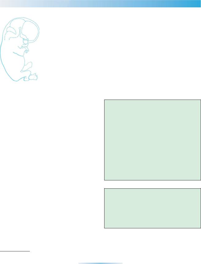
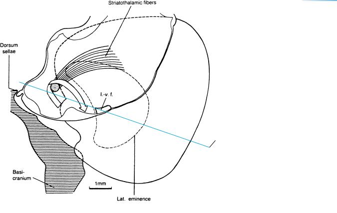
Figure 23–1. A right lateral view of the head accompanied by a reconstruction of the brain of an embryo of stage 23, modified from a drawing by Mary M. Cope (Gilbert, 1957).
This stage was not included by Bartelmez and Dekaban (1962). The insula, previously recognizable by its flat surface (stages 18–20), is now clearly indented. Almost the entire diencephalon is covered by the cerebral hemispheres, so that the epiphysis cerebri is no longer visible in this embryo, although it is in some others of the same stage. Contrary to the opinion of certain authors in the past, the telencephalon and the diencephalon are not fused: They are separated from each other by a thin sheet of mesenchyme.
THE BRAIN AT THE END OF THE EMBRYONIC PERIOD |
221 |
A
b
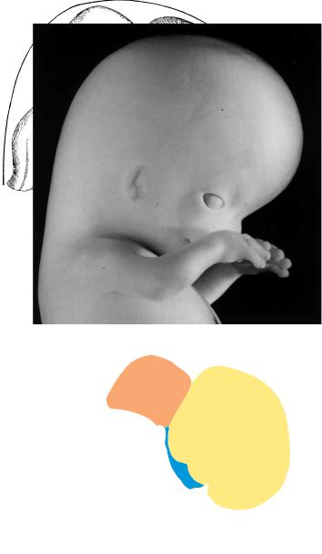
Figure 23–2. External features of the brain.
(A) The interrupted circle around the drawing serves to emphasize that the brain is more compact than in the previous stage. This is related in part to the progression of such C-shaped features as the hippocampus, the corpora striata, and the entire cerebral hemispheres.
The inset shows the topographical changes of the hemispheric poles from stage 18 (continuous line) to stage 23 (dotted line). (B) The three flexures are arranged approximately in the form of the letter M. (C) In this embryo, in contrast to that shown in A, the roof of the diencephalon is still visible externally.
222 |
C h a p t e r 2 3 : THE BRAIN AT THE END OF THE EMBRYONIC PERIOD |
Figs 23 – 19
Figs 23 – 24
16, 17
a
Post. X
b
Habenular X
Figure 23–3. Graphic reconstruction prepared from transverse sections to show a median view of the brain in which the internal relief is based on an unpublished Perspektomat reconstruction. The asterisk indicates the junction with the spinal cord. The outline of the left hemisphere is continued as an interrupted line. Only small portions of the diencephalic surface remain uncovered by the hemispheres. The di-telencephalic sulcus is filled with mesenchyme (Figs. 23–19 and 23–24) and the only bridge is that present since the beginning, which is now termed the hemispheric stalk. The stalk contains fibers between the thalamus and the hemisphere, and these travel in the lateral prosencephalic fasciculus, as seen already in the previous stage (Fig. 22–6). Corticothalamic fibers arise in proximity to the future central sulcus, probably in the area that corresponds to the future precentral and/or postcentral gyrus. The commissural plate is very thick and, in some embryos, is approached by fibers of the anterior commissure. Lateral strands leading to the site of the commissure can be observed before crossing fibers are present. The choroid fissure is now longer. It and the hippocampus are shown as if the prosencephalon were transparent. The height of the diencephalon from the chiasmatic plate to the epiphysis is appreciable. The suprapineal recess has developed. The marginal ridge, which separates the dorsal from the ventral thalamus, ends at the level of the interventricular foramen. In this embryo the fibers of the posterior commissure are separated from those of the commissure of the superior colliculi, which latter has two portions. If not earlier, then at least at stage 23 it would seem to be legitimate to use the terms basal nuclei and corpus striatum, and their various components (Fig. 19–6)
The internal ridge in the mesencephalon does not seem to indicate the border between neuromeres M1 and M2. The isthmic recess has become a shallow groove. Dopaminergic neurons are no longer produced in the ventrocaudal proliferative area of the mesencephalon, as they were from 6 12 to 7 12 weeks (Freeman et al., 1991).
THE BRAIN AT THE END OF THE EMBRYONIC PERIOD |
223 |
←−−−−−−−−−−−−−−−−−−−−−−−−−−−−−−−−−−−−−−−−−−−−−−−−−−−−−−−−−−−−−−−−−−−−−−−−−−−−−−−−−−−−−−−
Figure 23–3. (Continued ) A commissure of considerable size situated adjacent to the isthmic recess seems to be the decussation of the superior cerebellar peduncles (Cooper, 1946a). The cerebellum is not visible in its whole extent because portions of it extend ventrally and laterally, and are hidden by the rhombencephalic tegmentum. The cerebellar commissures contain, from rostral to caudal: (1) decussating fibers of the mesencephalic trigeminal root, (2) decussating vestibulocerebellar fibers, and (3) fibers from the area that includes the dentate nucleus. These ontogenetic data have been partly described by other authors, although only commissure No. 3 seems to correspond with the adult condition. The region with the (cerebellar) commissures is covered by an external germinal layer and so should be regarded already as the primordium of the vermis. The decussation of the trochlear nerves is rostral to the cerebellar commissures.
At the cerebrospinal junction two bundles are found: the fasciculus cuneatus (larger) and the fasciculus gracilis (smaller). They form the decussation of the medial lemnisci. More caudally, the pyramidal decussation is shown. The pyramidal tract and its decussation become definitive bundles only at this stage.
A
Olf. fibers
Insula
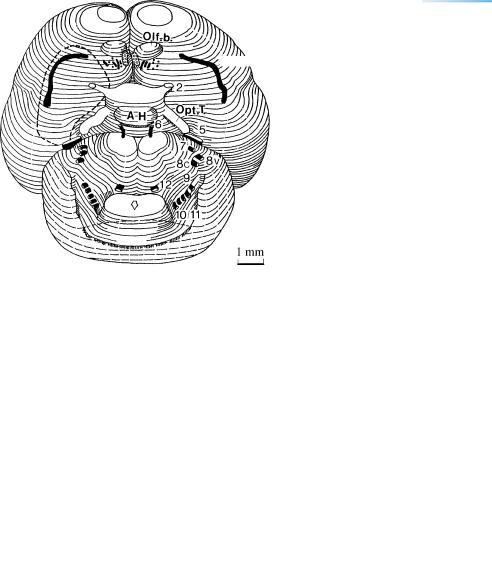
Figure 23–4. The cranial nerves. (A) A basal view of the brain at stage 23 (Perspektomat reconstruction) showing most of the cranial nerves. The mesencephalon and nerves 3 and 4 are hidden. The combined cerebral hemispheres are now wider than the cerebellum.
224 |
C h a p t e r 2 3 : THE BRAIN AT THE END OF THE EMBRYONIC PERIOD |
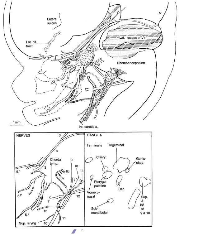
B
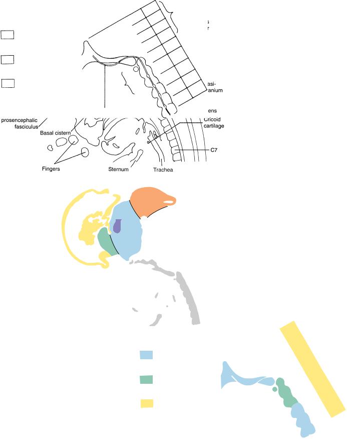
Lat. olf. fibers
Figure 23–4. (Continued ) (B) Reconstruction of the cranial nerves in relationship to some of the “membrane” bones and to the surface of the head. The arrangement of the nerves is now very complex and resembles the adult pattern. The laryngeal nerves are a good example of this (Muller¨ et al., 1981, 1985). Interestingly, small branches that are easily overlooked in the adult can readily be followed in embryos and fetuses. The recurrent branch of the trochlear nerve is a case in point. The skeletal surroundings of the nerves, however, are still in an unfinished state. The facial nerve, for example, is surrounded only by mesenchyme instead of bone, and neither the cribriform plate nor the foramen ovale has formed. All the cranial autonomic ganglia are present. The main divisions of the trigeminal nerve (5) are marked by superscripts 1, 2, and 3. The sheaths of cranial nerves 3, 4, and 5 are partly meningeal and partly peripheral in type (Kehrli et al., 1995).