

Книги по МРТ КТ на английском языке / The Embryonic Human Brain An Atlas of Developmental Stages. Third Edition. 2006. By Ronan O'Rahilly
.pdf138 |
C h a p t e r 1 8 : THE FUTURE CORPUS STRIATUM, THE INFERIOR CEREBELLAR PEDUNCLES |
D
A
B
Lat. em.
C
Med.
em.
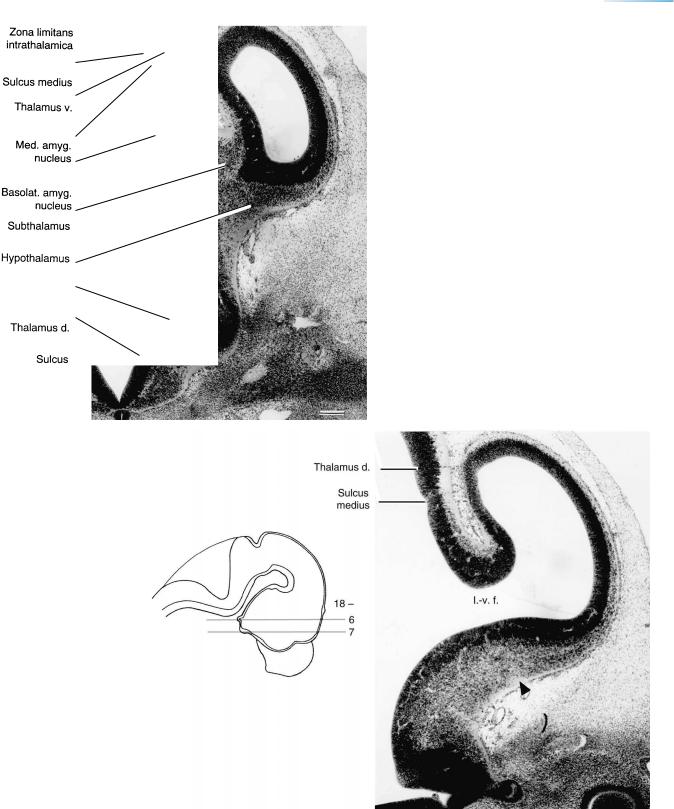
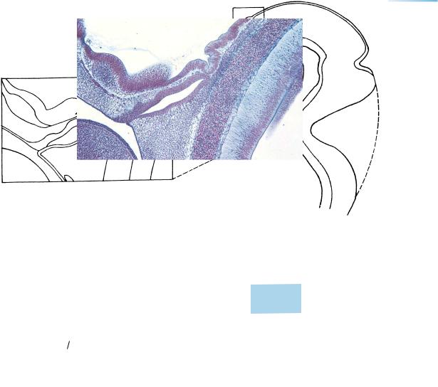
Figure 18–5. The future amygdaloid complex indicates the developing temporal pole. Three blocks through the ventricular eminences are presented, as shown in the key (D). The di-telencephalic sulcus is marked by a long black arrow. The small arrow points to the hypothalamic sulcus. (A) The lateral ventricular eminence, which is visible adjacent to the roof of the interventricular foramen. (B) Four amygdaloid nuclei, which are arising almost entirely from the medial ventricular eminence. The nuclei present at this stage are (1) cortical, (2) medial, (3) anterior, and (4) basolateral. The medial ventricular eminences are clearly visible. Humphrey’s (1968) claim that the human amygdaloid complex is derived exclusively from the telencephalon is incorrect (Muller¨ and O’Rahilly, 1989b, 2006). The lateral and particularly the medial eminence at stages 18–20 contain large cavities that were observed by earlier investigators. (C) The basal part of the cerebral hemispheres, showing the ventral thalami. The optic stalk enters at the rostral part of the chiasmatic plate. (D) The right cerebral hemisphere and the ventricular eminences are projected onto a median section. The medial eminence indicates the site of the future temporal pole.
THE FUTURE CORPUS STRIATUM, THE INFERIOR CEREBELLAR PEDUNCLES |
139 |
||
|
Figure 18–6. The di-telencephalic transition showing |
||
|
the zona limitans intrathalamica, ventral thalamus, |
|
|
|
subthalamus, and hypothalamus. Two amygdaloid nuclei |
||
|
(the medial, No. 2, and the basolateral, No. 4) are |
|
|
|
identifiable. |
|
|
|
The five subdivisions of the diencephalon (Table 18–1) |
||
|
are based on the comportment of their matrix zones, as |
||
|
elucidated by Kahle (1956), adopted by Richter (1965), |
||
|
and confirmed by Muller¨ |
and O’Rahilly (1988c). The |
|
|
subdivision of dorsal and ventral thalami used by |
|
|
2 |
Bartelmez and Dekaban (1962) and Kuhlenbeck (1978) is |
||
|
similar, although based on different grounds. The dorsal |
||
|
and ventral thalami are separated by the sulcus medius, |
||
4 |
which is adjacent to the marginal ridge that overlies the |
||
|
zona limitans intrathalamica. This last is the precursor of |
||
|
the lamina medullaris externa. Hochstetter (1919) |
|
|
marked the marginal ridge by “Leiste” and generally by an asterisk. His S.M. (Sulcus Monroi, hypothalamic sulcus) has been incorrectly taken by many writers to be the sulcus medius.
In Figures 18–6 to 18–13 the bars represent 0.2 mm.
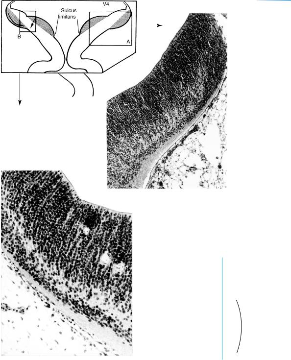
Figure 18–7. A section from the same embryo to show the interventricular foramen, the medial ventricular eminence, and the amygdaloid area. The lateral prosencephalic (forebrain) bundle is indicated by an arrowhead. The optic stalk and a portion of the eye are visible below. The ventricular eminences show a distinct subventricular layer. In addition to their separation by the sulcus medius, the ventral thalamus is distinguishable by its possession of a distinct intermediate layer, whereas the dorsal thalamus has only ventricular and marginal layers. Intramural capillaries penetrate the ventricular layers, but the vessels in the subarachnoid space are not yet distinguishable histologically as arteries and veins.
140 |
C h a p t e r 1 8 : THE FUTURE CORPUS STRIATUM, THE INFERIOR CEREBELLAR PEDUNCLES |
Plexus
fold
Paraphysis
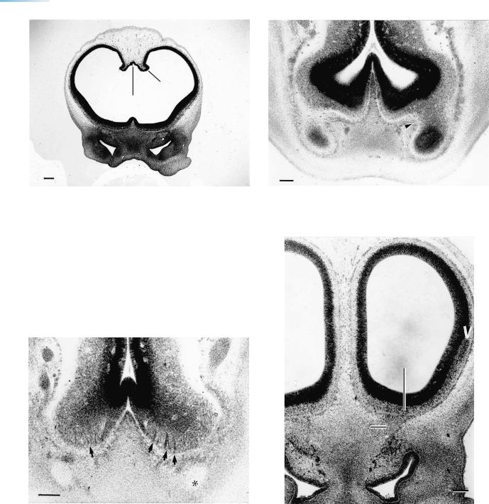
Figure 18–8. The rostral area showing the lateral ventricular eminences, the paraphysis in the roof between the hemispheres, and beginning vacuoles in the subdural space. The roof of the telencephalon medium and the lamina epithelialis of the lateral ventricle form a choroid fold that is lined by a relatively thick epithelial layer of four to five nuclear rows. The nasal sacs are evident, as well as related mesenchymal condensations, especially that of the nasal septum.
The neocortical differentiation of an embryo of stage 18 was illustrated by Mar´ın-Padilla (1983, 1988a).
Figure 18–9. The olfactory bulbs, separated from the septal area by the sulcus circularis (arrowhead). The longitudinal fissure, filled with mesenchyme, can be seen between the hemispheres below.
Olf. bulb
Olf. n.
Figure 18–10. The prosencephalic septum and the olfactory tubercles. The arrowheads point to cellular islands. The light area marked by an asterisk is the olfactory nerve.
Figure 18–11. The cerebral hemispheres and the olfactory nerves. The arrowhead points to the intermediate layer along the lateral ventricular eminence, the primordium of the future caudate nucleus. The dark cells between the olfactory bulbs and the nasal sacs (visible also in Figs. 18–12 and 18–13) are neural crest and ganglionic cells. The olfactory bulb and nerve are indicated.
THE FUTURE CORPUS STRIATUM, THE INFERIOR CEREBELLAR PEDUNCLES |
141 |
Figure 18–12. The caudal part of the nasal sacs. The oronasal membrane (arrowhead) has almost regressed on the other side.
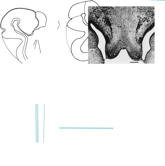
Figure 18–13. The vomeronasal ganglion (v-n), the nerve fibers of which begin in the wall of the vomeronasal organ, which appears bilaterally as an epithelial pocket in the nasal septum. Further details: Muller¨ and O’Rahilly (2004c). The organ continues to enlarge during prenatal life, and measurements have been published (Smith et al., 1997). Its involution postnatally has been queried.
18 – 9
10
18 – |
11 |
8
12
13
142 |
C h a p t e r 1 8 : THE FUTURE CORPUS STRIATUM, THE INFERIOR CEREBELLAR PEDUNCLES |
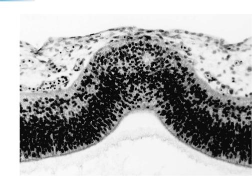
Figure 18–14. The epiphysis cerebri.
A transverse section to show that the epiphysis now appears as a dorsal evagination of the synencephalon. One so-called follicle is visible. The adjacent part of the diencephalic roof is a portion of the habenular region. The epiphysis is now at Stadium III of Turkewitsch (1933). See Figure 19–16(III). ×214.
Chiasma
THE FUTURE CORPUS STRIATUM, THE INFERIOR CEREBELLAR PEDUNCLES |
143 |
Fig. 18 – 14
3rd ventricle |
N |
|
|
|
|
|
|
|
|
|
|
||
|
|
|
. |
|
|
|
|
|
|
|
|
|
|
|
|
|
- |
|
|
|
|
|
|
|
|
|
|
|
|
|
H |
|
|
|
|
|
|
|
|
|
|
|
|
|
. |
|
|
|
|
|
|
|
|
|
|
|
|
|
|
. |
|
|
|
|
|
|
a |
e |
|
|
|
|
Basilar |
a |
|
|
|
|
u |
l |
|
|
|
|
|
. |
|
|
|
|
d |
|
|
|
|||
.- |
|
|
|
|
|
|
|
|
|||||
A |
H |
|
|
|
m |
e |
|
|
|
|
|
|
|
|
|
|
|
|
|
|
|
|
|
|
|||
|
|
|
|
|
m |
|
|
|
|
|
|
|
|
|
|
|
|
|
|
|
|
|
|
|
|
|
|
|
|
|
|
|
u |
|
|
|
|
|
|
|
|
|
|
|
|
|
t |
|
|
|
|
|
|
|
|
|
|
|
|
|
p |
|
|
|
|
|
|
|
|
Not. |
|
|
|
e |
|
|
|
|
|
4 |
th |
||
|
|
|
S |
|
|
|
|
|
|||||
|
|
|
|
|
|
|
|
|
|||||
|
|
|
|
|
|
|
|
vent. |
|||||
|
|
|
|
|
|
|
|
|
|||||
Figure 18–15. The hypophysis cerebri.
A sagittal section. The adenohypophysis and the neurohypophysis are in close apposition; mesenchyme has not yet infiltrated between them. In contrast, vascular mesenchyme has penetrated between the adenohypophysis and the hypothalamic floor. The folded wall of the neurohypophysis seems to be characteristic at this time. Various extensions of the lumen of the adenohypophysial pouch have been described (Conklin, 1968).
144 |
C h a p t e r 1 8 : THE FUTURE CORPUS STRIATUM, THE INFERIOR CEREBELLAR PEDUNCLES |
|
|
|
Figure 18–16. The cerebellar plate at stage |
|
|
18. (A) Medial view of the right half of the brain, |
|
|
indicating the right part of the cerebellar plate |
|
|
shown in B. (B) Medial view, as seen from the |
|
cerebellar |
ventricular cavity, to show the internal |
|
|
cerebellar swelling. The cerebellar primordium |
|
|
is formed not only by rhombomere 1 but also by |
|
|
a part of the isthmus. (C) Left lateral view of the |
|
|
brain, indicating the portion shown |
|
|
in D. (D) Left lateral view to show the external |
|
|
cerebellar swelling. Cranial nerve 8 is in close |
|
|
proximity and vestibular fibers from it already |
|
|
reach the flocculus. (E) A block of tissue shown |
|
- |
in F and G has been removed from D. (F) The |
|
|
posterolateral fissure delimits the rhombic lip |
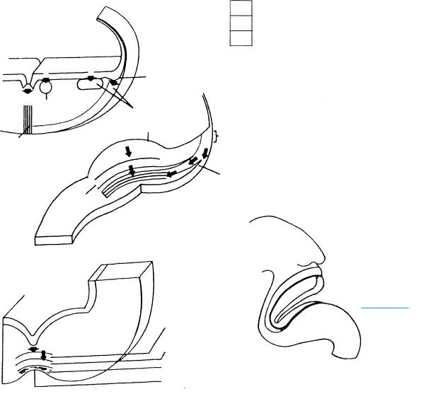
and the primordium of the flocculus from the remainder of the alar lamina. The fibers to and from the cerebellum are arranged in two strata, internal and external; the latter is the marginal layer. The fibers to the primordium of the flocculus are mostly vestibulocerebellar (shown by small black arrows at the level of the external cerebellar swelling). (G) To clarify the two sources of the cells that build up the cerebellum:
(1) radially migrating cells produced by the ventricular layer, and (2) tangentially migrating cells from the rhombic lip. The two streams intermigle. The open (white) arrow marks the direction of the afferent fibers to the cerebellum.
THE FUTURE CORPUS STRIATUM, THE INFERIOR CEREBELLAR PEDUNCLES |
145 |
||
|
|
|
|
A
B
18 – 17
Figure 18–17. Transverse sections through the cerebellum. (A) A laterally situated band of cells that represents the region of the future flocculus. The internal cerebellar swelling (int.) faces the fourth ventricle. (B) The transitional region between the internal (above) and external (below) cerebellar swellings (ext.). Two streams of cellular migration can be recognized and are indicated by arrows in the upper inset drawing:
(1) radially oriented cells produced in the ventricular layer form the main cellular mass, and (2) tangentially oriented cells produced by the rhombic lip form a peripheral band. A layer of nerve fibers is present internal to that band, and another external to it.
Tracts present at stage 18 include the stria medullaris thalami, mamillothalamic tract, medial and lateral tectobulbar, dentatorubral, and the tractus solitarius. The inferior cerebellar peduncles have been reconstructed (O’Rahilly et al., 1988). They contain vestibulocerebellar and trigeminocerebellar fibers, and they reach the region of the dentate nuclei (Fig. 18–3). Collateral vestibular fibers run to the flocculus. The tracts mentioned will be illustrated with the next stage.
146 |
C h a p t e r 1 8 : THE FUTURE CORPUS STRIATUM, THE INFERIOR CEREBELLAR PEDUNCLES |
||
|
|
|
|
|
|
|
|
Figure 18–18. The sites of production and the migration of neurons in (A) the rhombencephalon, (B) the cerebellum, and (C) the mesencephalon. (A) Block from the hindbrain showing migration from the floor plate to the raphe (1), from the ventricular layer for the motor and sensory (e.g., vestibular) nuclei (2), and from the rhombic lip, (e.g., for the cochlear nuclei (3)). The sensory areas are lateral to the sulcus limitans. (B) Cerebellar migration from both the ventricular layer and the rhombic lip for the primordium of the dentate nucleus (stage 21). Cells from the ventricular layer give rise also to the piriform (Purkinje) cells, and those from the rhombic lip form the external germinal (or external granular) layer (stage 23). (C) Migration in the mesencephalic tegmentum from the ventricular layer, producing first the interpeduncular nucleus and later the ventral tegmental area. (D) The plane of the sections.

THE FUTURE CORPUS STRIATUM, THE INFERIOR CEREBELLAR PEDUNCLES
Sup. salivatory nucleus
|
3 |
Red |
|
8v |
|
A |
nucleus |
E |
|
|
|
|
7i |
|
|||
|
|
7m |
|||
|
|
|
|
Interpeduncular nucleus
|
Vestibular nuclear |
|
zone |
|
Intermediate |
|
layer |
B |
F |
4 |
Dorsal & |
|
ventral |
||
|
||
|
cochlear nuclei |
|
|
|
G |
|
|
|
|
12 |
|
|
|
M.I.f. |
9s |
|
C |
Mes. |
|
9m |
|
|
|
|||
|
tr. 5 |
|
Nucleus |
|
|
|
|
||
|
|
Visceral |
ambiguus |
|
5s |
|
efferent |
|
|
|
|
|
||
|
|
|
Tractus |
|
5s |
5m |
L.I.f. |
solitarius |
|
H |
||||
|
|
|||
D |
|
|
|
|
|
|
|
12 |
|
|
|
|
10s |
|
|
|
5m |
10m |
|
|
|
|
||
5s |
|
|
Nucleus |
|
|
|
|
ambiguus |
147
6
7
6
7
Figure 18–19. The nuclei of the cranial nerves. The visceral efferent, ventromedial cell column and the nuclei derived from it are in mauve. The somatic efferent nuclei and the reticular formation are in orange. The afferent nuclei are in blue. Comments on the original site of the nuclei and their migration were included in the legend of Figure 14–9. (A) The oculomotor nucleus lies lateral to the midventral proliferative area and the interpeduncular nucleus. The material of the interpeduncular nucleus has been identifiable since stage 15. (B) The trochlear nucleus in the isthmus has a more medial position than the oculomotor nucleus. (C) and (D) The trigeminal area is continuous with the cerebellar plate. An afferent trigeminal nucleus has appeared. (E) The dorsal nucleus of the trapezoid body or superior olivary nucleus (dagger) is lateral to the reticular formation. The medial cell column for the facial nerve is still voluminous. Only a few fibers of the genu cross the rostral part of the abducent nucleus. (F) Fibers of the cochlear nerve with the basal and dorsal cochlear nuclei. Also recognizable is the vestibular zone with fibers to the medial longitudinal fasciculus. The nuclei of the abducent (6) and (in G and H) hypoglossal (12) nerves are in a similar position, but are not continuous. (G) The glossopharyngeal nerve with its motor and sensory components, and the cell mass derived from the rhombic lip. (H) The nucleus ambiguus of the vagus. Abbreviations: m and s (after the numeral of a cranial nerve) motor and sensory components, respectively. The levels of A to H are indicated in the key drawing on page 148.