

Книги по МРТ КТ на английском языке / The Embryonic Human Brain An Atlas of Developmental Stages. Third Edition. 2006. By Ronan O'Rahilly
.pdf68 C h a p t e r 1 2 : CLOSURE OF THE CAUDAL NEUROPORE AND THE BEGINNING OF SECONDARY NEURULATION
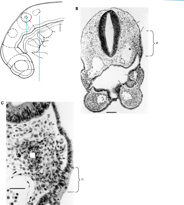
Figure 12–10. Development of the ganglia of cranial nerves. Neural crest gives rise to the main, proximal portions of the ganglia of nerves 5, 7, 9, and 10, whereas epipharyngeal discs (incorporated in the ectodermal ring) contribute to the distal portions. Shown here are examples from an embryo of 22/23 pairs of somites. (A) Transverse section showing the otocyst and the facial ganglion, both of which are very close to the wall of the rhombencephalon. (B) Transverse section through rhombomere 7 and the pharynx, showing the superior vagal ganglion (10s), which arises from neural crest, and the inferior ganglion (10i), which is arising from the epipharyngeal disc (asterisk) of pharyngeal arch 3. Bar: 0.15 mm. (C) A more rostral section, through a part of the otic vesicle (Ot.), showing the geniculate (or facial) ganglion, which arises from both neural crest and the epipharyngeal disc (asterisk) of pharyngeal arch 2. Bar: 0.2 mm.
Abbreviations: Ao., dorsal aorta; 5, 7/8, 9/10, cranial ganglia; 10s, superior vagal ganglion.
CLOSURE OF THE CAUDAL NEUROPORE AND THE BEGINNING OF SECONDARY NEURULATION |
69 |
CB
10s
10i
Ao.
2
1
Ot.
7
Figure 12–10. (Continued )
70 C h a p t e r 1 2 : CLOSURE OF THE CAUDAL NEUROPORE AND THE BEGINNING OF SECONDARY NEURULATION
TA
CC
RV
LV
LA
RA
SV
PLACENTA
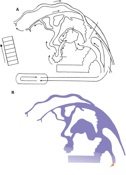
Figure 12–11. The early blood vessels of the head.
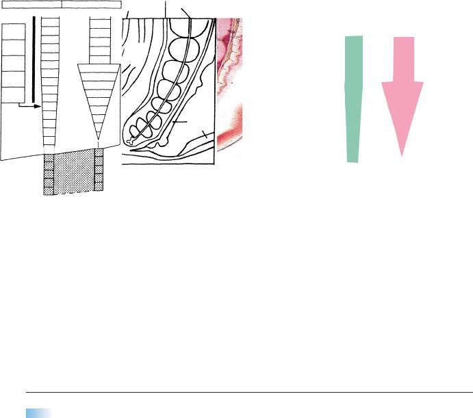
(A) The vascular system at stage 12 in an embryo with 28 paired somites. The surface of the brain and the cranial ganglia have a good blood supply. Three aortic arches (the second is marked 2) are present. The capital plexuses (a, b, c) and the capital vein are now well-developed, and the cardinal venous system is established: precardinal, postcardinal, and common cardinal. Bar: 0.15 mm. Based on Streeter in O’Rahilly and Muller¨ (1987a) and on Padget (1957).
Abbreviations: a, b, c, rostral, middle, and caudal capital venous plexuses; Ao., dorsal aorta; Ot., otic vesicle; 5, trigeminal ganglion. (B) The blood flow indicated by arrows. Oxygenated blood from the placenta reaches the body by way of the umbilical vein. The rectangle at the left shows the order of the various chambers and their abbreviations: sinus venosus, right atrium, left atrium, left ventricle, right ventricle, conus cordis, and truncus arteriosus.
|
Common |
|
cardinal |
LA |
vein |
|
SV |
|
RV |
|
Omphalo- |
|
mesenteric plexus |
|
Umbilical vein |
Aorta |
−−−−−−−−−−−−−−−−−−−−−−−−−−−−−−−−−−−−−−−−−−−−−−−−−−−−−−−−−−−−−−−−−−−−−−−−−−−−−−−−−−−−−−−→
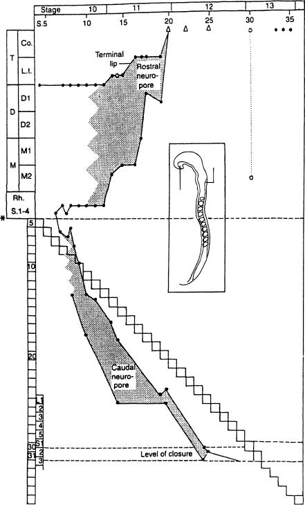
Figure 12–12. The closure of the neuropores in the human, based on data from 19 embryos. The future neuropores are recognizable as soon as fusion of the neural folds begins, when approximately five to seven pairs of somites are visible (Heuser and Corner, 1957).
In the rostral region the dorsal lip formed by the roof progresses rapidly and has a much longer track than the terminal lip, formed by the floor. This can be seen from the steep rise of the line representing the roof. The line of the floor, by contrast, remains horizontal during stage 10 and then rises during stage 11, indicating the bidirectional closure of the rostral neuropore. Although closure seemingly may occur in several places independently at stage 10 at least in some instances, no regular pattern of multiple sites, such as has been described in the mouse, has yet been shown in the human embryo (O’Rahilly and Muller,¨ 2002).
Two instances of dysraphia (open circles) are included for comparison. In the first example an embryo of 14 somitic pairs showed complete failure of fusion of the neural folds (Dekaban, 1963), although the terminal lip had progressed normally. In the second case, believed to be future anencephaly (Muller¨ and O’Rahilly, 1984), an embryo of stage 13 showed a well-developed terminal lip but a marked deficiency in the progression of the dorsal lip (Fig. 13–9). Both these examples show that development of one lip can proceed more or less independently of the other. The triangles represent the situs neuroporicus; the asterisk, the cerebrospinal junction.
The caudal region differs considerably from the rostral. Both the floor and the roof become increasingly elongated as further pairs of somites (the stepwise line) become visible. They remain approximately parallel, however. The neuropore, which remains small throughout, becomes displaced more and more caudally until it disappears at a level corresponding approximately to future sacral vertebra 2. This is taken to be the level of the junction between primary and secondary neurulation. Fetal ascensus of the spinal cord would be expected to result in a higher level postnatally.
Dorsal
lip
Terminal
lip
V e r t e b r a
e
Neuropore closed Secondary
neurulation
Figure 12–12. (Continued ) Once the caudal neuropore is closed, the whole caudal area of the embryo is covered by surface ectoderm. The continuing formation of the spinal part of the neural tube takes place without involvement of ectoderm and is termed secondary neurulation. The caudal part of the tube is present first as a concentration of tissue in the caudal eminence. Secondary neurulation is the continuing formation of the sacrocaudal part of the spinal cord without direct involvement of the surface ectoderm, i.e., without the intermediate phase of a neural plate. It begins once the caudal neuropore has closed during stage 12. The caudal eminence is a mass of pluripotent mesenchymal tissue covered by ectoderm. It is recognizable already at stages 9 and 10, and gradually replaces the primitive streak. It provides structures that are comparable to those formed more rostrally by the three germ layers. The derivatives include the caudal portion of the digestive tube, blood vessels, notochord, somites, and spinal cord. At stage 12 the caudal eminence gives rise to a compact cellular mass known as the neural cord, which forms the nervous system in the sacrocaudal part of the body. The cavity (central canal) of the spinal cord, present already more rostrally, extends into the neural cord. The caudal eminence gives rise to at least somitic pair 32 (corresponding to future sacral centra 3 and 4) and those following. The mesenchyme for somitic pairs 30–34 is the material for sacral vertebrae 1–5. Secondary neurulation is important in regard to timing and localization of spina bifida aperta.

72 C h a p t e r 1 2 : CLOSURE OF THE CAUDAL NEUROPORE AND THE BEGINNING OF SECONDARY NEURULATION
Hindgut
Axial mesenchyme
Neural cord
Hindgut
Notochord
Spinal cord
Cloacal
memb.
H
Cloacal
membrane
Caudal
eminence
Somitic plate
H
Somite
Figure 12–13. Secondary neurulation.
(A)A reconstruction of the caudal end of an embryo of stage 12 with 29 pairs of somites. The site of the now closed caudal neuropore is indicated by an arrow. Somite 25 is shown as a dotted oval outline, and the position of the more caudal somites, including future No. 30, is shown. The closure of the neuropore is opposite future somite 31. The cross sections, from an embryo with 24 pairs of somites, show some of the structures derived from the mesenchyme of the caudal eminence, e.g., the neural cord, spinal cord, and notochord of the caudal region.
(B)An embryo of stage 13 with 33 pairs of somites. The somites have not been included. The cross section, from another embryo also with 33 pairs of somites, shows the somitic plates that unite ventral to the hindgut.
CLOSURE OF THE CAUDAL NEUROPORE AND THE BEGINNING OF SECONDARY NEURULATION |
73 |
|||||
|
|
|
|
|
Closure |
|
|
|
|
|
|
of caudal |
|
|
|
|
|
|
neuropore |
|
Weeks |
2 |
3 |
|
4 |
5 |
6 |
Stage |
|
|
|
|
|
|
3 |
|
|
8 |
10 |
12 |
17 |
|
|
Primary |
|
Development |
|
|
|
|
|
|
|
S1 – 29 |
|
|
|
|
|
Pry neurulation |
|
|
|
|
|
|
|
Secondary development |
|
|
|
|
|
Caudal |
S29 – 38 |
|
|
|
|
|
part of |
|
|
|
|
|
|
|
|
|
|
|
|
|
notochord |
Secondary neurulation |
|
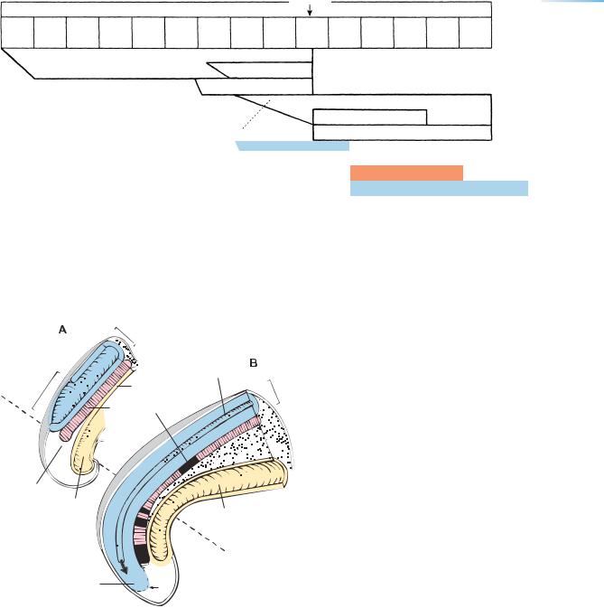
Figure 12–14. Primary and secondary developmental modes in the caudal region related to stages. From Muller¨ and O’Rahilly (2004a, Cells Tissues Organs) by permission of S. Karger AG, Basel.
|
Neural |
|
tube |
|
Central |
Caudal |
canal |
neuropore |
Endoderm |
|
|
|
Notochord |
Caudal eminence
Hindgut
Neural cord


 Mesenchyme


 Intestine
 PRIMARY


 NEURULATIONSECONDARY
Figure 12–15. Primary and secondary neurulation. Median sections showing the left half of the caudal end of the body. (A) At stage 10. (B) At stages 12, 13. Beyond the caudal neuropore, the caudal eminence gives rise to the neural cord (blue), into which the canal penetrates (arrow). Based on reconstructions by the authors.
74 C h a p t e r 1 2 : CLOSURE OF THE CAUDAL NEUROPORE AND THE BEGINNING OF SECONDARY NEURULATION
|
Median sacral a. |
|
Paramesonephric ducts |
Coelom |
Notochord |
|
||
|
Rectum |
|
Neural Genital tube tubercle
Urogenital sinus
Figure 12–16. Median section of spinal cord and vertebral column at stage 21. The neural tube and the vertebral column are coextensive during the embryonic period.
|
SOMITES |
CENTRA |
|
|
L 1 |
Occ. 4 |
|
|
C |
8 |
|
T |
12 |
S 1 |
|
|
|
L |
5 |
|
S5
34 Co.1
38
Co. 6
10
44
Figure 12–17. Scheme to show that the somites (green) are out-of- phase in relationship to the vertebral centra (mauve). The distribution of the first 34 somites is indicated at the left. The maximum number of somites is now believed to be 38 or 39 (Muller¨ and O’Rahilly, 1994; O’Rahilly and Muller,¨ 2003), but was formerly thought to be greater (shaded area).
NEUROTERATOLOGY
Agenesis of the hypoglossal nerve would be expected from an isolated absence of the hypoglossal nucleus at stage 12. Although the lingual musculature would not be affected primarily, lack of innervation would lead to atrophy and fasciculation. The hypoglossal cell cord, which becomes recognizable at stage 12, gives rise to the muscles of the tongue and possibly to some of those of the larynx, although their connective tissue is believed to come from the neural crest.
Albinism is related to a defect in the production of neural crest, including probably the optic neural crest, which latter is noticeable at stage 12.
Sacral agenesis may result from a localized defect within the caudal eminence, although it has also been
attributed to an exaggeration of the normal process of caudal regression that occurs later in the embryonic period. Complete lack of the caudal eminence would result in cloacal deficiency and consequently an incomplete separation of the two lower limb buds, i.e., symmelia (O’Rahilly and Muller,¨ 1989b).
Caudal Aplasia. The caudal tip of the trunk appears particularly tapered at 5 weeks because it contains merely neural tube, but it is in no sense a future vertebrated “tail.” Neuroschisis. The claim that “neural clefts (neuroschisis)” existed in 100 of more than 200 Carnegie embryos from stage 10 to stage 23 (Padget, 1970) has not been
substantiated (O’Rahilly and Muller,¨ 1988).

C H A P T E R 13
STAGE 13: THE CLOSED
NEURAL TUBE AND THE
FIRST APPEARANCE OF
THE CEREBELLUM
Approximately 4–6 mm in Greatest Length;
Approximately 32 Postfertilizational Days
Normally, at 4 to 5 postfertilizational weeks, in embryos of some 5 mm in length, both neuropores are closed, so that the future ventricular sys-
tem no longer communicates with the amniotic cavity. The choroid plexuses will not appear for another fortnight, so that the liquid in the neural tube is “ependymal fluid.” The terminal-vomeronasal crest is arising from the nasal discs, the retinal and lens discs are beginning to develop, and the adenohypophysial pouch is distinct. Three diencephalic neuromeres are present: D1, parencephalon, and synencephalon. The isthmus rhombencephali is visible as a neuromere between M2 and Rh. 1. A marginal layer is distinguishable in the wall of the mesencephalon and rhombencephalon. The primordia of various somatic and visceral efferent nuclei, the common afferent tract, and the ganglia of most of the cranial nerves can be discerned. The first indication of the cerebellum appears in Rh.1 during stage 13.
Precision
A double m is not used in mamillary because the term is derived from the Latin mamilla, which in turn is a diminutive of mamma, which does have a double m.
First Appearance of the
Cerebellum
Stage 13. The cerebellum commences as a thickening in the alar plate of rhombomere 1.
Stage 14. It extends into the alar portion of the isthmus.
Stage 17. The rhombic lip begins to participate.
Precision
Median plane is the correct term, not midsagittal nor midline. The term parasagittal is superfluous because all planes parallel to the median are simply sagittal.
The Embryonic Human Brain: An Atlas of Developmental Stages, Third Edition. By O’Rahilly and Muller¨ Copyright C 2006 John Wiley & Sons, Inc.
75
76 |
C h a p t e r 1 3 : THE CLOSED NEURAL TUBE AND THE FIRST APPEARANCE OF THE CEREBELLUM |
M
Di.
T
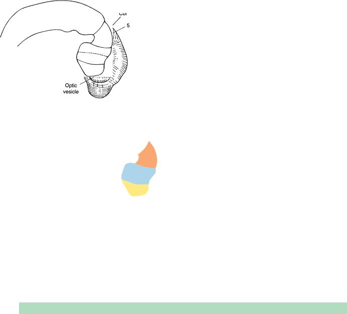
Figure 13–1. Right lateral view of the brain. The number of somitic pairs in this extensively studied embryo is now known to have been 32. The mesencephalon is well-delimited. The cerebellum is at the alar plate of Rh.1. The roof of the fourth ventricle is translucent. The otic vesicle is closed. The wall of the optic vesicle related to the surface ectoderm becomes flattened and constitutes the retinal disc, and the lens disc becomes distinguishable concomitantly.
The arteries to the head at stage 13 were reconstructed by Padget (1948, Fig. 1), who included the internal carotid and the caroticobasilar anastomoses.
TABLE 13–1. Mean Measurements (in mm) in 93 Embryos of Stages 6a–13 (ca 21/2–41/2 Weeks)
Stage |
6a |
6b |
7 |
8a |
8b |
9 |
10 |
11 |
12 |
13 |
|
|
|
|
|
|
|
|
|
|
|
Greatest embryonic length |
0.23 |
0.31 |
0.59 |
0.90 |
1.30 |
1.44 |
2.1 |
3.2 |
3.86 |
4.88 |
Greatest embryonic width |
|
|
0.44 |
0.68 |
0.81 |
0.68 |
|
|
|
|
Prechordal plate |
|
|
0.87 |
1.35 |
1.4 |
0.09 |
0.11 |
|
|
|
Length of primitive streak |
|
0.08 |
0.23 |
0.28 |
0.43 |
0.27 |
|
|
|
|
Length of caudal eminence |
|
|
|
|
|
|
0.57 |
0.31 |
0.34 |
0.67 |
Length of neural groove/tube |
|
|
|
|
0.31 |
0.97 |
1.63 |
3.1 |
6 |
8.5 |
Length of neural cord |
|
|
|
|
|
|
|
|
0.15 |
0.6 |
n |
18 |
13 |
13 |
7 |
4 |
5 |
13 |
12 |
3 |
5 |
|
|
|
|
|
|
|
|
|
|
|
THE CLOSED NEURAL TUBE AND THE FIRST APPEARANCE OF THE CEREBELLUM |
77 |
Telencephalon medium
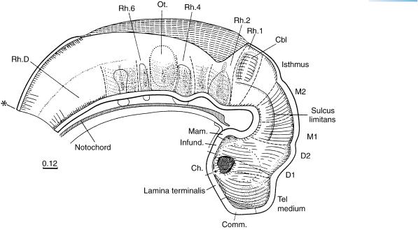
Figure 13–2. Graphic reconstruction from transverse sections to show a median view of the brain. The asterisk indicates the junction with the spinal cord. Both neuropores are now closed, so that, at approximately 4 to 5 postfertilizational weeks, the cavity of the neural tube is a closed (future ventricular) system, normally no longer in communication with the amniotic cavity. The contained “ependymal fluid” is presumably formed by the lining cells and later, when the choroid plexuses have appeared, will become the cerebrospinal fluid. Once the neural tube has closed, its walls become subject to the pressure of the contained fluid, provided fluid formation is greater than absorption.
The telencephalon is of approximately the same length as D2. The commissural plate is the median area at the level of the nasal plates. The terminal-vomeronasal crest is appearing (O’Rahilly, 1965). Cells leave the still convex or flat nasal plates and form cellular buds, which will participate in forming the ganglia of the nervus terminalis and the vomeronasal nerve, as well as in the development of the olfactory nerve.
The roof of D2 bulges slightly at the site of the future synencephalon. The adenohypophysial pouch and the mamillary region are now distinct. At the transition between D2 and M1, the interstitial nucleus is forming ventrally, and its fibers constitute the uncrossed part of the medial longitudinal fasciculus. The oculomotor and trochlear nuclei are present, the former in M2, the latter in the isthmus.
The isthmus rhombencephali, so named by His, lies between M2 and Rh.1, and is clearly separated from the latter. This is a newly formed neuromere. The rhombencephalon is considerably enlarged dorsoventrally in comparison with stage 12, and the floor of the fourth ventricle is bent in the region of Rh.2. Although the wall of the brain remains histologically simple for the most part, being formed by the pseudostratified epithelium of the ventricular layer, a marginal layer has developed in the mesencephalon and rhombencephalon (Figs. 13–6 and 13–7).
The primordia of the somatic and visceral efferent nuclei of cranial nerves 5, 6, 7, 9–11, and 12 (Fig. 13–6) are present. The common afferent tract is being formed by descending fibers of nerves 5, 7, 9, and 10.
Rhombomeres 2 to 7 are characterized by internal grooves. The first indication of the cerebellum (marked Cbl) can be found in the alar lamina of Rh.1 in most embryos of stage 13. Loosely arranged cells begin to constitute its intermediate layer.