

Книги по МРТ КТ на английском языке / The Embryonic Human Brain An Atlas of Developmental Stages. Third Edition. 2006. By Ronan O'Rahilly
.pdf28 |
C h a p t e r 8 : THE FIRST APPEARANCE OF THE NERVOUS SYSTEM |
A
|
|
Chor. |
|
|
villi |
|
Embryonic |
|
|
disc |
|
|
Amniotic |
|
|
cavity |
Body |
|
|
|
Umbilical |
|
stalk |
vesicle |
|
Allantoic |
|
|
diverticulum |
B
Amnion
Neural plate
Artifact
Primitive pit
Primitive Notochordal streak
process
Neurenteric
canal Endoderm
C
Ectoderm |
Primitive |
|
groove & streak |
||
|
Mesoderm
Endoderm
Figure 8–5. The formation of the primitive streak at stage 8a. (A) Median section of an embryonic disc of nearly 1 12 mm in length. (B) A higher powered view of the section shown above. The neurenteric canal connects the amniotic cavity with the umbilical vesicle temporarily. It is an important landmark in stages 8 to 10 (Table 8–1). (C) Transverse section through the primitive groove and primitive streak, which attains its greatest length at this stage.
THE FIRST APPEARANCE OF THE NERVOUS SYSTEM |
29 |
A
B
A
B
C
C
D
E
D
E
Figure 8–6. Serial sections through an excellent example of stage 8. The level of the sections is indicated on the reconstructed median section.
(A) The prechordal plate in transverse section shows as many as eight rows of cells that differ from the endodermal cells in being larger and more nearly spherical, and in containing numerous granules. The neural ectoderm at this level shows a very faint indentation, the beginning of the neural groove. The prechordal and neural plates are separated by a basement membrane. (B) The neural groove in an area rostral to the notochordal process. (C) The primitive pit and its continuation with the notochordal canal of the notochordal process. (D) The primitive node. (E) The primitive groove and streak. At this caudal level the mesenchyme between the surface ectoderm and the endoderm is far greater in quantity than at rostral levels. Blood vessels are not yet present. Bar = 0.03 mm.

30 |
C h a p t e r 8 : THE FIRST APPEARANCE OF THE NERVOUS SYSTEM |
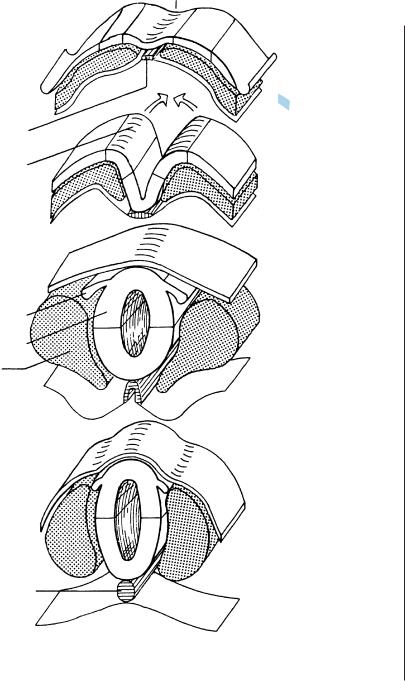
EARLY NEURAL EVENTS
After the neuroepithelium is induced, the neural plate forms and is characterized by cellular movements mediad (convergent extension). The edges of the plate form bilateral neural folds (Fig. 8-4) that undergo median bending accompanied by expansion of the underlying mesenchyme. The folds are separated by the neural groove (Figs. 8-4, 9-3, and 10-11). The lengthened neural plate is broader rostrally, where the three major divisions of the brain are
identifiable (Fig. 9-2). The free edges of the neural folds turn mediad (sharply so in rodents at what are termed dorsolateral bending points) resulting in apposition of the neural folds. Normal bending of the neural folds is believed to depend on contraction of actin microfilaments, emigration of neural crest, and neuroepithelial apoptosis (as studied in rodents and summarized by Copp, 2005). The neural folds then fuse in the median plane (Figs. 10-2A, 10-5, and 11-9) and this is the beginning formation of the neural tube (primary neurulation).

C H A P T E R 9
STAGE 9: THE MAJOR DIVISIONS OF THE BRAIN
Approximately 1.5–2.5 mm in Greatest Length;
Approximately 26 Postfertilizational Days
The embryonic disc is now an elongated body characterized by a long and deep neural groove.
The first somites have appeared and are occipital. The three major divisions of the brain are distinguishable in the folds of the completely open neural groove: prosencephalon (mostly diencephalon), mesencephalon (at the mesencephalic flexure), and rhombencephalon (comprising four subdivisions, A to D). Rhombomere D is related to the (occipital) somites and was not recognized by previous authors. No neural tube has yet formed, and “brain vesicles” are not present. Neural crest is beginning to develop.
The otic discs, which are the first indication of the (internal) ears, are first visible at this stage.
The primitive streak is replaced by the caudal eminence. The neural folds of the spinal cord begin to develop.
Important
The three major divisions of the brain can be distinguished in the completely open neural folds, when no so-called vesicles are present.
The Embryonic Human Brain: An Atlas of Developmental Stages, Third Edition. By O’Rahilly and Muller¨ Copyright C 2006 John Wiley & Sons, Inc.
31

32 |
C h a p t e r 9 : THE MAJOR DIVISIONS OF THE BRAIN |
Figure 9–1. Left lateral and dorsal views of an embryo of stage 9. Striking changes have occurred since the preceding stage.
The embryonic disc is now clearly an elongated embryonic body, set off from the surface of the umbilical vesicle (the so-called yolk sac). Various features are indicated in Figure 9–2 A,B.
In the lateral view, the “waist” of the embryo can be seen to be the site of considerable lordosis. The cephalic and caudal portions of the body are elevated from the surface.
In the dorsal view, the region of the head, which is broadest at the level of the mesencephalon, is clearly joined to the caudal eminence by a “waist,” in which the first (occipital) somites are appearing. Caudally the greatest width is at the level of the primitive node, the site of which is shown in Figure 9–2. (In the present figure the artist has placed it too far caudally.) Cephalic to the node, the embryo has elongated greatly. Beyond the embryo, the umbilical vessels and the allantoic diverticulum have been sectioned. The neural folds bound a long and deep neural groove. Preotic and postotic sulci are very faint (compared with those in rodents).
This is another example of an elegant drawing by James F. Didusch, artist to the Carnegie Collection.

THE MAJOR DIVISIONS OF THE BRAIN |
33 |
A
Rhombencephalon
B
Primitive |
Somites |
|
|
||
|
|
|
streak |
node |
|
C |
Ot. |
|
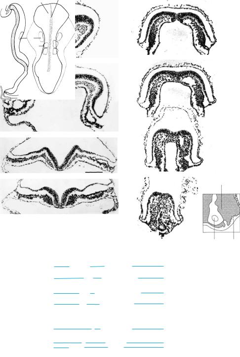
Figure 9–2. (A) dorsal, (B) right lateral, and (C) median views of an embryo of stage 9. The cephalic end is to the right. The first (occipital) somites are stippled. The otic discs are indicated by interrupted lines; C is a graphic reconstruction prepared from transverse sections.
Although no part of the neural tube has yet formed and no so-called brain vesicles are present, the three major divisions of the brain recognized by von Baer are distinguishable. The prosencephalon is almost exclusively diencephalic. The mesencephalon is at the site of a marked bend, the mesencephalic flexure. In C, the rhombencephalon, which extends from the otic to the somitic region, can be seen to comprise four subdivisions: rhombomeres A, B, C, and D, the last (first recognized by Muller¨ and O’Rahilly, 1983) representing the hypoglossal region. These rhombomeres are real entities (as shown by immunological methods in the chick). The asterisk indicates the future cerebrospinal junction. A faint groove is visible between the mesencephalon and Rh.A. It is clearest in a dorsal view (Fig. 9–2A), where it separates the wider midbrain from the narrower hindbrain. It corresponds to the preotic sulcus in rodents.
The first sign of the production of neural crest can be found in a few embryos of stage 9. It occurs at the level of the mesencephalon and the rhombencephalon. In addition, the production of general mesenchyme continues. The prechordal plate participates in the formation of the head and probably provides cardiac mesenchyme. The primitive streak, however, seems to have ceased as a site of proliferation, and its functional role has been replaced by that of the caudal eminence. The primitive node is shown in its correct position (more rostrally than in Fig. 9–1).
A E
F
B
G
C
D H
Neural fold & groove
A
B
C
D
E, F
G
H
Mesenchyme
Allantoic
diverticulum Epiblast
Head fold
Mesencephalon
Rhombomere B
Rhombomere D
Primitive pit & node
Primitive groove
Caudal eminence
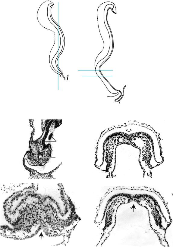
Figure 9–3. Sections through an embryo of stage 9, a stage that is rarely seen.
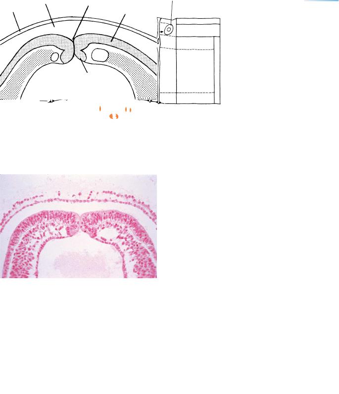
Key drawings, a median section and a dorsal view, are the authors’ reconstructions. The levels of the sections reproduced are indicated from A to H. The degree of lordosis of human embryos of this stage seems to be less pronounced than in those of some other primates and of rodents. (A) The head fold, including the most rostral part of the neural plate, a little caudal to the prechordal plate and rostral to the cardiac primordium. The foregut is a closed tube at this level. The amnion and the amniotic cavity are well shown in this and in the next section. (B) The deep neural groove here is at the level of the mesencephalon. The floor plate lies on the broad notochordal plate, which is intercalated in the endoderm. The paired pericardial cavities are clearly visible. (C) Rhombomere B. The neural groove is widely open to the amniotic cavity. The floor plate is in contact with the notochordal plate and has the same thickness as the lateral walls of the future neural tube. The thick ectodem lateral to the sumit of each neural fold is the otic plate. (D) Rhombomere D at the level of the caudal part of somite 1. This embryo possessed two somitic pairs. (E) The primitive pit and the umbilical vesicle communicate with each other through the neurenteric canal.
(F) The primitive node and the beginning of the primitive groove. Venous channels on each side are the umbilical veins and that on the left side of the embryo (right side of the photograph) is wider, an asymmetry also seen in other embryos of this stage. (G) The primitive grove is deeper here. Mesenchyme invaginating from the primitive streak is visible. (H) Caudal region of the primitive streak. Invaginating cells and a pseudostratified epiblast are visible. Mesenchyme forms also from the surface of the body stalk near the allantoic diverticulum as emphasized in the key drawing.
A
Umbilical vesicle
C, D
B
Allantoic diverticulum
A B
Neural ectoderm
Umbilical
vesicle
C
D
Figure 9–4. Regression of the primitive streak in stage 9. (A) A coronal section. The epiblast has mostly been replaced by cuboidal surface ectoderm. The dark tissue rostral to the umbilical vesicle (and mostly at the right side of the photograph) is neural ectoderm. (B) A transverse section through the rostral part of the primitive streak. The median, invaginating flask-shaped and spherical cells are still numerous, and the epiblast is still thick laterally. The open arrow indicates the primitive groove. (C) A transverse section through the neurenteric canal (black arrow) in an other embryo in which the cuboidal surface ectoderm has almost replaced the former epiblast. (D) The neurenteric canal (black arrow), epiblast, and mesenchyme of the primitive streak in the embryo shown in B. Modified from Muller¨ and O’Rahilly (2004a, Cells Tissues Organs) by permission of S. Karger AG, Basel.
36
Notochordal
Floor
Roof
Somites
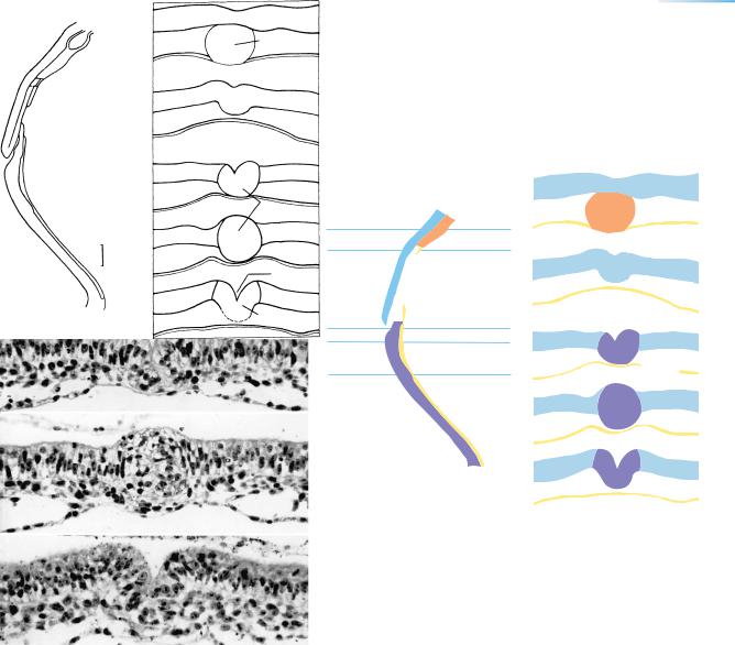
C h a p t e r 9 : THE MAJOR DIVISIONS OF THE BRAIN
Neural |
|
groove |
Stage |
|
|
|
8 |
9
10
11
Figure 9–5. Transverse sections at stages 8 to 11 to show in a purely didactic manner the formation of the neural tube and the notochord. The neural primordium is closely related ventrally to the precursor of the notochord (stages 8–10). Later, when the notochord becomes surrounded by the developing centra of the vertebrae, the latter become and remain an important ventral relation of the spinal cord. Blue, the future alar plate (dorsal lamina); Orange-brown, the future basal plate (ventral lamina) of the neural tube. The floor, basal, alar, and roof plates all express different genes.
Inductional processes between the future notochord and the neural tube would likely be greatest during stages 8 and 9, when the future notochord is present as a plate and hence presents a relatively wide surface. The contact is particularly close at stages 8–10 because, as has been shown in the macaque, a basement membrane is lacking between the two tissues at that time.
|
|
NEURAL TUBE AND NEURENTERIC CANAL |
||
|
|
|
|
Immature |
|
|
|
|
neuron |
S |
G2 |
M |
G1 |
S |
|
|
|||
|
|
|
|
|
BM |
|
|
|
Marginal zone |
|
|
|
|
Intermediate |
|
|
|
|
zone |
Synthesis
of DNA
Ventricular zone
TBN |
Mitotic zone |
|
Cavity of neural tube |
||
|
37
Figure 9–6. A portion of the wall of the neural tube (small drawing at left) showing interkinetic nuclear migration. When DNA is replicated (S phase), the nuclei migrate towards the lumen, where a terminal bar net (TBN) is situated. A mitotic figure is shown at the ventricular surface during the mitotic (M) phase. After division, the nuclei migrate away from the lumen (G1 phase). The ventricular (matrix), intermediate (mantle), and marginal zones are indicated.
|
Amniotic |
Neurenteric |
|
|
cavity |
|
|
|
canal |
Neural |
|
|
|
||
Amnion |
|
|
|
|
|
plate |
|
|
|
|
BV
Notochordal
plate
Umbilical
vesicle
Figure 9–7. The neurenteric canal at stage 9.