

Книги по МРТ КТ на английском языке / The Embryonic Human Brain An Atlas of Developmental Stages. Third Edition. 2006. By Ronan O'Rahilly
.pdf78 |
C h a p t e r 1 3 : THE CLOSED NEURAL TUBE AND THE FIRST APPEARANCE OF THE CEREBELLUM |
|
|
|
|
TABLE 13–2. Embryonic Development of the Cerebellum |
|
|
Events |
Figures |
|
|
|
|
Cerebellum appears as a slight thickening in alar plate of Rh. 1. |
|
|
The roof of the fourth ventricle is attached at the caudal tip of Rh. 1 |
13–1 |
|
The alar plate of Rh. 1 is up to 3 times longer in rostrocaudal direction than the basal plate of Rh. 1. |
|
|
The cerebellar primordium is the widest part of the brain. |
14–2 |
|
The roof of the fourth ventricle is now attached at the caudal tip of the isthmus. |
|
|
The alar plate of the isthmus becomes part of the cerebellar primordium. Its roof represents the |
15–3 |
|
|
future superior medullary velum. |
|
The width of the cerebellum is approximately the same as that of the two cerebral hemispheres |
16–2 |
|
|
together up to ca stage 21. |
|
The posterolateral fissure defines the future flocculus, which is the earliest part of the future vermis. |
17–3 |
|
Extraand intraventricular cerebellum can be distinguished. |
17–4 |
|
The rhombic lip of Rh. 1 shows strong mitotic activity. |
|
|
The inferior cerebellar peduncle begins to form from vestibuloand trigeminocerebellar fibers. |
17–10 |
|
The dentate nucleus can be distinguished. |
18–2 |
|
Dentatorubral fibers are present and initiate the superior cerebellar peduncle. |
18–16 |
|
The external germinal layer begins. |
23–19 |
|
Tegmental decussation of the superior cerebellar peduncles. |
23–3 |
|
|
|
|
M2
M2
Syn.
|
4th |
Isthmus |
ventricle |
|
|
Rh.2 |
|
Primary |
meninx |
|
|
|
|
AH |
Notochord |
|
|
||
Tongue
Ao.
Heart
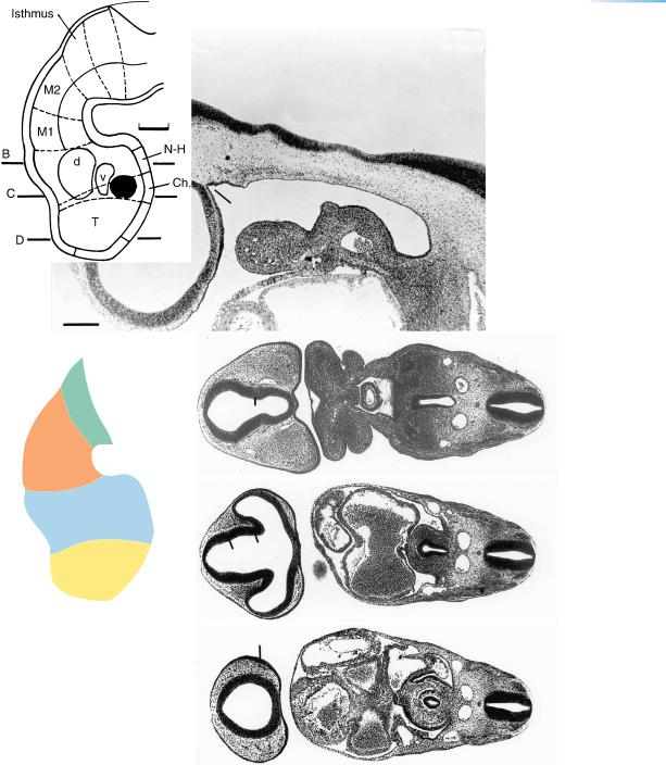
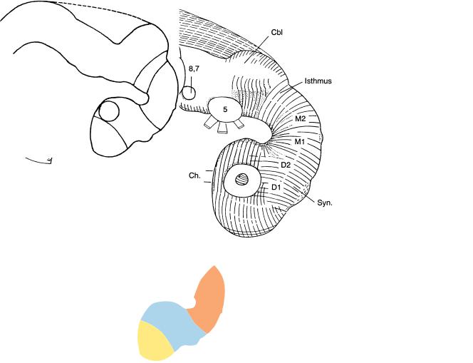
Figure 13–3. Median section of the brain at stage 13. The notochord still extends as far rostrally as the caudal wall of the adenohypophysial pouch. The isthmus rhombencephali is evident. The rhombencephalon, the mesencephalon, and part of D2 (Syn.) are epinotochordal in position and possess a floor plate. The rostral parts of D2 and D1 are prenotochordal.
A B
C D
F
Uveocapillaris
E
1
3
Inverted layer of optic cup
Lens
disc
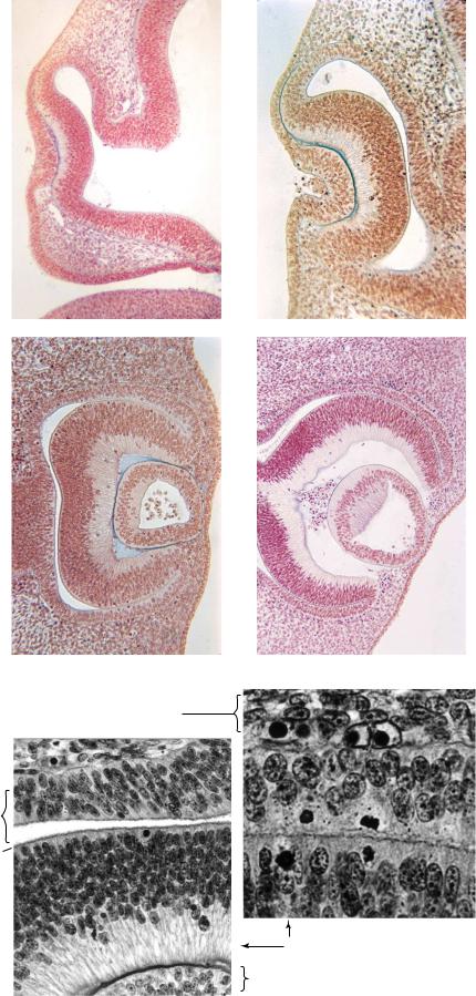
Figure 13–4. The development of the eye from 41/2 to 6 weeks. (A) Stage 13. Early indentation of the retinal and lens discs.
(B) Stage 14. The optic cup and lens pit are evident. (C) Stage 15. The lens pit has become closed off from the surface, forming the lens vesicle. (D) Stage 17. Retinal pigment can be seen in the external layer of the optic cup. The inverted layer of the cup will develop in a manner comparable to that of the wall of the brain (Fig. 14–8). The cavity of the lens vesicle is beginning to become obliterated. Hyaloid blood vessels are visible behind the lens. Early development of the retina. (E) At stage 14, showing layer 1 (the future pigmented layer), layer 3 (the terminal bars of the external limiting membrane), and the thick inverted layer. (F) At stage 15, showing the onset of pigmentation (minute black dots) in layer 1 (as well as two mitotic figures). Only the most external portion of the inverted layer is included in the field selected. Further development can be seen in Figure 23–13. The inverted layer develops in a manner comparable to that of the wall of the brain, as shown in Figure 14–8.
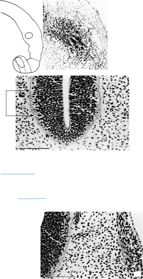
Figure 13–5. Silver-impregnated section showing ganglion 7/8v containing two long afferent fibers.
Rh.4
12
Figure 13–6. The bilateral hypoglossal nuclei are formed by unipolar neurons that at this stage are situated peripherally, being more laterally placed than the visceral efferent nuclei of nerves 5,7, and 9–11. The extent of the hypoglossal nucleus on the left is indicated by arrowheads.
Figs 13 – 6, 7
13 – 5
S.3
Figure 13–7. A hypoglossal root from the neural tube (at left) to somite 3 (at right). The nerve fibers are accompanied by neural crest cells. The hypoglossal roots are not rigidly segmental, some somites receiving two roots. The cells of the primordial hypoglossal nucleus are relatively lateral in position, but that changes during development, as described with Figure 14–9.
The hypoglossal artery participates in the carotico-basilar anastomosis, which is prominent until stage 15 (Fig. 21–22).
THE CLOSED NEURAL TUBE AND THE FIRST APPEARANCE OF THE CEREBELLUM |
81 |
A
2
M2 |
4 |
|
6
M1
A-H
Syn.
B
d
C
D2
v
d
DNas.
Figure 13–8. The neuromeres at stage 13, as shown in a graphic reconstruction. D2 now consists of the synencephalon caudally and the parencephalon rostrally, and the isthmus rhombencephali constitutes a distinct neuromere. (A) Sagittal section near the median plane to show the rhombomeres (Rh. 2, 4, and 6 having ventral evaginations that are numbered). The mamillary region is distinguishable, and the notochord can be seen to end close to the incipient adenohypophysial pouch. (B)–(D) The dorsal and ventral eminences that represent the future dorsal and ventral thalami (d and v). The more caudal portions of the sections include the spinal cord (with the sulcus limitans), the dorsal aortae, as well as the truncus arteriosus and pharyngeal arches 1 and 2 (in B), the pharynx (in B and C), and the atria (in C). The bar for A–D represents 0.5 mm.

82 |
C h a p t e r 1 3 : THE CLOSED NEURAL TUBE AND THE FIRST APPEARANCE OF THE CEREBELLUM |
NEUROTERATOLOGY: CEREBRAL DYSRAPHIA (ANENCEPHALY)
The most frequent neural tube defects (NTD) are anencephaly and myelomeningocele, which are multifactorial, and the genetic and environmental influences have been reviewed (Detrait et al., 2005).
Cerebral Dysraphia. Anencephaly develops in three phases. The first, cerebral dysraphia (encephaloschisis), is believed to arise as a failure of closure of the neural groove rostrally, an idea supported by many authors. Rupture of the already closed neural tube cannot be entirely excluded in some instances. Cerebral dysraphia arises early, probably as a mesenchymal defect, even as early as stages 8 or 9, i.e., before fusion of the neural folds begins, because elevation of the folds depends on the production of sufficient mesenchyme.
Fusion of the neural folds commences during stage 10, when production of neural crest is only starting. The ganglia of the cranial nerves, derived largely from neural crest, are only minimally affected in anencephaly. A reduction in the size of the ganglia, however, as well as an incomplete chondrocranium, would point to defective distribution of the cephalic mesenchyme, caused probably by a derangement of the mesencephalic neural crest in stages 10 and 11. Moreover, stage 10 is characterized by the first appearance of the optic primordium, and, rarely, anencephaly may be combined with anophthalmia. Usually, however, the eyes are very well developed in anencephaly, indicating that development of the diencephalon at first proceeds normally, although later it degenerates.
It would be extremely difficult to detect a pure cerebral dysraphia at stage 11, because it is only during that stage that the rostral neuropore normally closes. Nevertheless, an example of complete cerebrospinal dysraphia at stage 11, presumably the forerunner of craniorhachischisis totalis, has been described in detail (Dekaban, 1963; Dekaban and Bartelmez, 1964).
Cerebral dysraphia has been described in detail in a twin embryo of stage 13, and reconstructions are available (Muller¨ and O’Rahilly, 1984). The neural tube was open over part of the midbrain and forebrain, although the situs neuroporicus (at the locus of the future commissural plate) was closed. Hence, in future anencephaly, fusion at the “terminal lip” of the rostral neuropore to form the embryonic lamina terminalis (Figs. 11–7A and 13–9) may not be affected. It is possible that a causal relationship exists between neural tube defects and monozygotic twinning.
Communication between the amniotic fluid and the ependymal (future cerebrospinal) fluid is normally eliminated by stage 13 because of neuroporal closure, so that α-fetoprotein no longer diffuses into the amniotic cavity,
except in such conditions as anencephaly and spina bifida cystica.
Phases 2 and 3 of anencephaly are discussed in Chapters 22 and 24 respectively.
MISCELLANEOUS
ANOMALIES
Cebocephaly. Indications of this condition have been found in an embryo attributed to stage 13 (Fig. 13–10). A graphic reconstruction was prepared, from which it could be seen that the notochord did not reach the adenohypophysis, as normally it would. A convergence of the nasal discs indicated a narrow median region of the head. The nasal septum would not develop, and the single nasal cavity would possess a single nostril. Cebocephaly arises earlier than arhinencephaly, in which the brain is almost normal except for absence of the olfactory bulbs. It has been found experimentally in the mouse that in cebocephaly little or no tissue from the medial nasal processes is involved in the development of the nose, lips, and palate. The single nostril in cebocephaly ends blindly, and the eyes are closely set. The brain is holoprosencephalic in type and the forebrain septum is absent or reduced. The corpora striata are not separated from each other.
Arhinencephaly. This term sensu stricto refers to absence of the olfactory bulbs and tracts, irrespective of associations with other developmental malformations, such as holoprosencephaly, which is commonly combined with arhinencephaly. Isolated arhinencephaly would be expected to arise at the time when the nasal discs should begin to form: thickenings normally appear at stage 11, and a nasal field is outlined at stage 12 (Bossy, 1980). This timing, however, depends on the unproven assumption that the olfactory bulb is induced by the olfactory nerve. Holoprosencephaly and arhinencephaly may develop independently of synophthalmia, probably at about stage 11, or perhaps even 10. Although they are commonly linked, holoprosencephaly and arhinencephaly can also arise independently of each other. Siebert et al. (1990) studied 103 cases of this condition and found that arhinencephaly is extremely heterogeneous both clinically and pathologically.
Cerebellar agenesis, although rarely total, could arise as early as stage 13, or even in stage 12. The first part of the cerebellum to develop is material for the hemispheres, whereas the primordium of the vermis becomes defined only at stages 18 and 19.
Abducent-facial palsy (Mobius¨ sequence) presents as a nonspecific, commonly bilateral, mask-like facies. The nuclei of cranial nerves 6 and 7 are usually degenerated, but they may be absent. Most of the motor nuclei of the cranial nerves begin to develop at about stage 13. Other cranial nerves may be involved in the sequence,

MISCELLANEOUS ANOMALIES |
83 |
A |
Somites |
13 |
|
|
|
|
|
||
|
|
14 |
|
|
|
|
|
17 |
|
|
|
M |
|
Dorsal |
|
|
|
|
|
|
|
|
|
lip |
|
|
Di.2 |
|
|
|
|
|
|
19 |
|
|
|
Di.1 |
|
|
|
Somites |
|
20 |
|
|
13 17 |
19 |
|
|
|
Terminal lip |
|
|
B |
|
|
|
|
|
Ot. |
|
|
|
|
8, 7 |
|
M |
|
|
|
|
|
|
|
5 |
|
|
|
|
|
|
|
O |
|
|
|
|
p |
|
|
|
Di. |
e |
|
|
|
n |
|
T
Figure 13–9. Comparison of normal and deficient closure. (A) The normal sequence of closure of the rostral neuropore involves (1) a caudorostral fusion of the neural folds at the dorsal lip and (2) a fusion at the terminal lip proceeding towards the neuropore. (B) In an embryo of stage 13 believed to be incipiently anencephalic, the fusion of the neural folds is at a standstill. The open region indicates where fusion should have taken place during stage 11. Fusion at the terminal lip, however, has occurred.
and limb defects are frequent. Some of the variable features (e.g., micrognathia and microglossia) are probably secondary. Moreover, some instances of the sequence are considered either to be caused by involvement of peripheral nerves or as a myopathy. Abnormal torsional
and vertical eye movements could be produced by disturbance of the interstitial nucleus (Crawford et al., 1991). That nucleus appears in stage 13 at the ventral transitional area between diencephalon and mesencephalon (Fig. 14–5).
84 |
C h a p t e r 1 3 : |
A
11
12
THE CLOSED NEURAL TUBE AND THE FIRST APPEARANCE OF THE CEREBELLUM
14
13
17
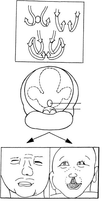
Figure 13–10. Development of cebocephaly. (A) Schematic representation of the change in position of the nasal plates at stages 11–13 and 14–17, and also a combined view of stages 11–17. Modified from Muller¨ and O’Rahilly (2004c, Cells Tissues Organs) by permission of S. Karger AG, Basel. (B) An abnormal embryo of stage 13 in which the nasal plates remained as a single structure at stage 12 instead of migrating bilaterally. This condition would lead to cebocephaly. (C) Two examples of cebocephaly drawn from photographs (in Siebert et al., 1990). None of the 43 holoprosencephalic embryos studied by Yamada et al. (2004) possessed olfactory structures.
B
D2
Opt.
D1
Neuropore
Nasal plate
Phar. arch 1
C

C H A P T E R 14
STAGE 14: THE FUTURE CEREBRAL HEMISPHERES
Approximately 5–7 mm in Greatest Length;
Approximately 33 Postfertilizational Days
The future cerebral hemispheres (arrow) become identifiable during stage 14 and are delimited from the telencephalon medium by the torus hemispher-
icus internally and by the di-telencephalic sulcus externally. Particularly advanced in the prosencephalon are the hypothalamic, amygdaloid, hippocampal, and olfactory regions. The synencephalon is identifiable. The mesencephalic flexure is less marked and is occupied by the
future tentorium cerebelli. The pontine flexure appears. The cerebellum is formed by the alar plate of the isthmus as well as by that of rhombomere 1. Blood vessels now penetrate the wall of the brain. Various bundles (e.g., the medial and lateral longitudinal fasciculi, the common afferent tract) and a number of nuclei of cranial nerves are present. All 16 neuromeres are now identifiable.
The Embryonic Human Brain: An Atlas of Developmental Stages, Third Edition. By O’Rahilly and Muller¨ Copyright C 2006 John Wiley & Sons, Inc.
85
86 |
C h a p t e r 1 4 : THE FUTURE CEREBRAL HEMISPHERES |
M
Di.
T
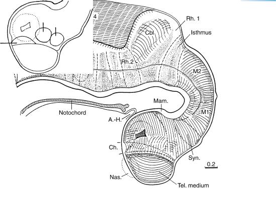
Figure 14–1. Right lateral view of the brain. The asterisk indicates the junction with the spinal cord. The future cerebral hemispheres begin to form during stage 14, but they were not evident in this embryo. The lens pit is present (Fig. 14–6). The synencephalon is recognizable. A minute, dorsal mesencephalic evagination (between M1 and M2) is frequently found: vergangliche¨ epiphysenahnliche¨ Anlage des Mittelhirndaches (Hochstetter, 1919). The isthmus participates in the formation of the cerebellum. The hypoglossal roots and/or rootlets begin to unite usually into two trunks.
The arteries to the head of this embryo were reconstructed by Padget (1948, Fig. 2), who showed that the carotico-basilar anastomoses are beginning to regress.
THE FUTURE CEREBRAL HEMISPHERES |
87 |
Ventral thal. Dorsal thal.
Medial eminence
Figure 14–2. Graphic reconstruction based on transverse sections to show a median view of the brain. The dividing line between the telencephalon medium and the future cerebral hemispheres is the torus hemisphericus, in the basal part of which the medial ventricular eminence (Fig 19–6) develops. Externally, the groove that accompanies the torus is the di-telencephalic sulcus. In the median plane, the borderline between diencephalon and telencephalon extends from the velum transversum to the preoptic recess. The velum transversum is a ridge in the roof of the prosencephalon, situated between D1 and the telencephalon medium. The telencephalon is surrounded by some mesenchyme, even ventrally.
The prosencephalon occupies almost one-quarter of the length of the brain. Four areas are particularly advanced: (1) the hypothalamic region at the level of the chiasmatic plate as well as lateral to it [this is the area of the hypothalamic cell cord, from which the preoptico-hypothalamic tract arises (Fig. 14–3)]; (2) the amygdaloid region of the medial ventricular eminence; (3) the primordium of the hippocampus; and (4) the olfactory region.
Topographically the hypothalamic cell cord corresponds to the “medial zone of the anterior hypothalamus,” which participates postnatally in the regulation of sexual comportment.
The mesencephalic flexure has diminished to about 50◦ , so that only a narrow space (the Mittelhirnpolster of Hochstetter) exists between the diencephalon and the rhombencephalon. The contained mesenchyme will form the tentorium cerebelli, the medial part of which now appears and is continuous with the cellular sheath of the notochord. A few extracerebral oculomotor and abducent fibers are present, and intracerebral trochlear fibers are developing. Blood vessels are much more numerous and, for the first time, penetrate the wall of the mesencephalon and rhombencephalon.
The pontine flexure is beginning as a slight bulging of the ventral wall of the hindbrain. The cerebellum, which is the widest part of the brain, is formed by the alar plate of two neuromeres: the isthmus rostrally, and Rh.1 caudally. An intermediate layer is present in most of the cerebellum, and the future rhombic lip is discernible. The sclerotomes of the four occipital somites form two components near the base of rhombomere D. These will give rise to the basioccipital and exoccipital portions of the skull.
The lower drawing shows the torus hemisphericus between the velum transversum and the preoptic recess. The diencephalon, which is thereby delineated from the telencephalon, possesses three elevations: the medial ventricular eminence, and the ventral and dorsal thalami. The entrance into the optic ventricle and the (dotted) outline of the optic cup are included.