Книги по МРТ КТ на английском языке / The Embryonic Human Brain An Atlas of Developmental Stages. Third Edition. 2006. By Ronan O'Rahilly
.pdf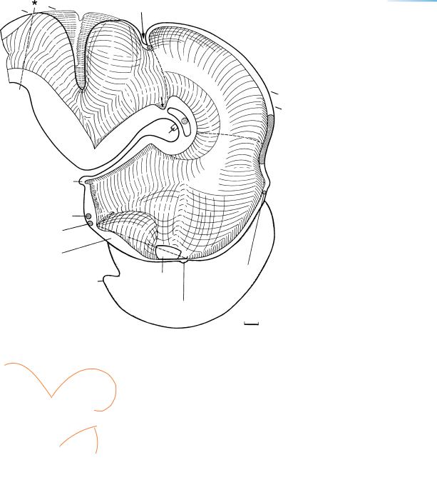
THE FIRST APPEARANCE OF THE CORTICAL PLATE IN THE CEREBRAL HEMISPHERES |
189 |
Isthmic
21 –
18
17
.
a
b
c
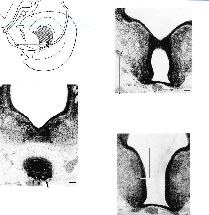
Figure 21–3. Graphic reconstruction from transverse sections to show a median view of the brain. The asterisk indicates the junction with the spinal cord. The pineal recess has formed. Ependymal cells over the posterior commissure form the subcommissural organ. The floor of the rhombencephalon is bent sharply, as described by Hochstetter (1919), and a transverse groove, the sulcus transversus rhombencephali, results. The isthmic recess (short arrow) is a guide to the interpeduncular nucleus. Interrupted lines delimit the rhombencephalon, mesencephalon, diencephalon, and telencephalon.
Striatal arteries in this embryo were shown by Padget (1948, Fig. 21).
As shown in the inset, the main internal grooves are (a) the sulcus limitans, (b) the hypothalamic sulcus, and (c) the marginal ridge.

190 |
C h a p t e r 2 1 : THE FIRST APPEARANCE OF THE CORTICAL PLATE IN THE CEREBRAL HEMISPHERES |
Figure 21–4. (A) The overlap between the left cerebral hemisphere and the left half of the diencephalon. The positions of the subthalamic nuclei (medial and lateral mamillary nuclei, subthalamic nucleus proper, entopeduncular nucleus which is the future globus pallidus internus, and the globus pallidus externus) are shown schematically. The ventral part of the lateral geniculate body has developed (Table 20–1). The hippocampus, area dentata, area epithelialis, and the choroid fissure are indicated on the medial wall of the left hemisphere, which is shown as if transparent. The hippocampus reaches almost as far as the temporal pole.
The site of the future lamina affixa, which at this stage overlies the ventral thalamus, is marked by a dagger. The asterisk at the base of the telencephalon is at the site of a cistern. Parts not visible from the surface are shown by interrupted lines. The horizontal interrupted line delimits the diencephalon from the telencephalon. (B) Levels of Figures 21–11 to 21–17, which, because they are from a different embryo, do not necessarily show a
perfect fit.
Fig. 21 –
11
12
13
14 6, 15 17
16 9, 10
THE FIRST APPEARANCE OF THE CORTICAL PLATE IN THE CEREBRAL HEMISPHERES |
193 |
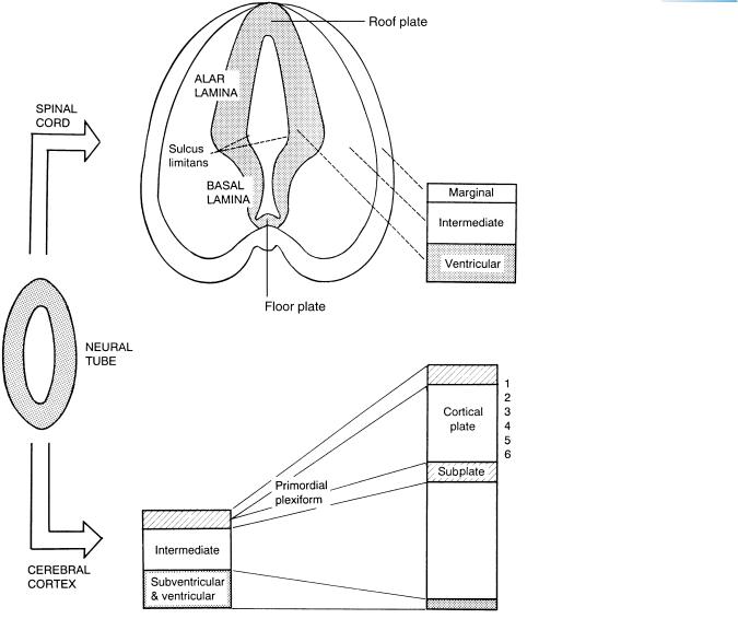
Figure 21–7. Developmental comparison between the spinal cord and the brain wall as derivatives of the neural tube. The cortical plate develops within the primordial plexiform layer (preplate), so that it comes to lie between superficial and deep laminae, namely layer 1 and the subplate of the neocortex. The stages illustrated are 11 for the neural tube, and 17 for the spinal cord. The neocortical blocks are (left) up to stage 20, and (right) from stage 21.


THE FIRST APPEARANCE OF THE CORTICAL PLATE IN THE CEREBRAL HEMISPHERES |
195 |
Subpial layer
Cortical plate
Subplate
Intermediate
layer
Ventricular & subventricular layers
Figure 21–9. The cortical plate is visible within the cell-poor primordial plexiform layer at the periphery of the ventricular eminences. The plate separates the former primordial plexiform layer into the subpial layer, which is future layer 1, and the subplate (Fig. 21–7). The subplate contains thalamocortical fibers that arrived in the primordial plexiform layer already at the previous stage. Penetration of the cortical plate by monoamine fibers occurs at the end of trimester 1 (Verney et al., 1993). It is to be noted that the subpial layer and the subplate appear during the embryonic period and not, as has been claimed (Kostovic´ and Rakic, 1990), in the middle third of prenatal life. Transient fibers in the subplate are illustrated in Figure 23–22. Glial production begins from stem cells in the subventricular layer. From Muller¨ and O’Rahilly (1990b).