

Книги по МРТ КТ на английском языке / The Embryonic Human Brain An Atlas of Developmental Stages. Third Edition. 2006. By Ronan O'Rahilly
.pdf118 |
C h a p t e r 1 7 : THE FUTURE OLFACTORY BULBS AND THE FIRST AMYGDALOID NUCLEI |
A
a
b
c
B
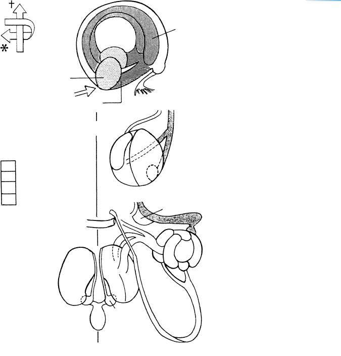
Figure 17–4. Graphic reconstruction prepared from transverse sections to show a median view of the brain. The asterisk indicates the junction with the spinal cord. (A) The sulcus limitans (a), the hypothalamic sulcus (b), and the marginal ridge (c). The sulcus limitans separates the alar and basal laminae of the rhombencephalon and mesencephalon. However, it does not continue into the diencephalon. The hypothalamic sulcus of the diencephalon separates the thalamus sensu lato from the hypothalamus sensu lato (Table 18–1). (B) The medial surface of each cerebral hemisphere has greatly increased and a hemispheric stalk can be identified. This is the junctional area between the telencephalon and the diencephalon.
Shown here are the boundaries between the different parts of the brain, the newly developed recesses (arrows), the commissures, the sulci, and the entrance into the optic ventricle (black dot in the diencephalon). Four small arrows indicate the isthmic, supramamillary, inframamillary, and infundibular recesses. The boundaries are summarized in Table 15–2.
The posterior commissure has suddenly appeared and consists of two subcommissures: the posterior commissure sensu stricto with fibers of the medial longitudinal bundle, and the commissure of the superior colliculi. Other commissures are the supraoptic in the caudal part of the chiasmatic plate, the supramamillary (dagger), and the commissure of the oculomotor nerves. The ventral commissure of the rhombencephalon, which consists of fibers that cross throughout the length of the floor-plate, has not been indicated here. The commissural plate itself does not yet contain crossing fibers.
In the inset, features of the medial aspect of the lateral wall of the left cerebral hemisphere have been superimposed on the median reconstruction. The interventricular foramen is surrounded by the hippocampal area of the telencephalon and by the medial ventricular eminence of the diencephalon, as well as by the ventral thalamus. The medial eminence is invading telencephalic territory and is penetrated by blood vessels. The lateral ventricular eminence (Fig. 19–6) is visible in the left hemispheric wall. The internal cerebellar swelling lies between the sulcus limitans and the rhombic lip. Reconstructions from a different embryo were published by Hines (1922).
Sulcus limitans
A
B
Ep. |
AH |
Di. |
Sclerotomes |
Tel.
Velum transversum
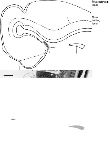
Figure 17–5. Median section. All mesenchymal components of the head have increased greatly. Many skeletal elements can already be reconstructed: the stapes, the styloid process, the cartilage (Meckel’s) of pharyngeal arch 1, the thyroid laminae, and the cricoid cartilage. The primordia of the so-called hypophysial cartilages (trabeculae; polar cartilages) are present in the area of the future hypophysial fossa. The brain sits on a firm basal condensation of mesenchyme, which, in its rostral part, represents the future nasal septum. This restrains the growth of the cerebral hemispheres in a basal direction. Bar: 1 mm.
The mesenchyme is beginning to form the future dural limiting layer, especially in basal areas, and, beginning at the rostral tip of the notochord, it continues to spread between the diencephalon and the rhombencephalon, i.e., at the site of the mesencephalic flexure. The condensed mesenchyme at the flexure is the primordium of the tentorium cerebelli, which is compressed between the basilar artery and the hypothalamus (Fig. 16–5). The dural limiting layer already contains openings (pori durales) for cranial nerves 3, 4, 5, and 12.
The presence of the dural limiting layer between the pia mater and the skeletogenous sheath initiates the formation of the subarachnoid space. The space clearly begins its development before the presence of choroid plexuses. The roof of the fourth ventricle is very thin. Two areas are beginning to become the areae membranaceae rostralis et caudalis. They are formed by flattened ependymal cells, and their position is indicated by two arrows. Such areas (in the adult mouse) are thought to permit the passage of cerebrospinal fluid containing macromolecules, by way of interependymal cellular clefts. The “ependymal fluid” within the ventricles is believed to be produced by the lining cells that will develop later into ependymal and choroid plexus cells.
Already in the previous stage, the vertebral arteries join the basilar, and an anastomosis is present between the basilar and the internal carotid arteries (Fig. 16–13D). An anterior choroid artery is now developing and was reconstructed by Padget (1948, Fig. 5). The main cerebral arteries are now well established.
The caudal part of the spinal cord, which forms by secondary neurulation, reaches the caudal tip of the body. The definitive number of somites (usually 39 pairs: O’Rahilly and Muller,¨ 2003) seems to be present at this and the next stage. The normal regression of the caudalmost part of the spinal cord may begin at approximately this time.
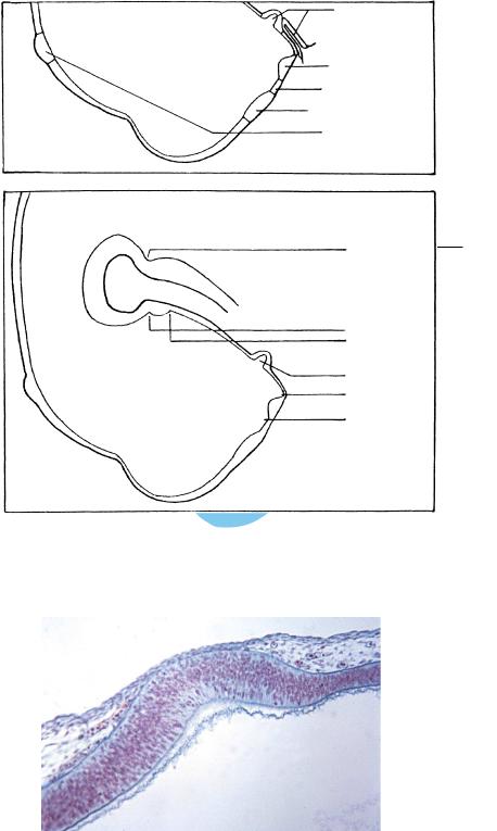
The key shows the position of the sulcus limitans and the alar and basal laminae (cf Fig. 21–8). The neurohypophysis (characterized by folded walls) and the infundibular recess are present in all embryos of this stage.
Cartilage appears in occipital sclerotomes 1–4, and the hypoglossal foramen has formed in sclerotome 4. The sclerotomic mass is shown in the key.
120
A
B
C h a p t e r 1 7 : THE FUTURE OLFACTORY BULBS AND THE FIRST AMYGDALOID NUCLEI
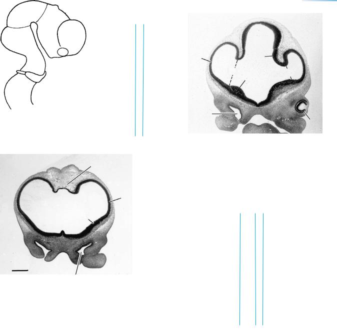
Figure 17–6. Six features and six recesses. (A) The six features that are developing here become more distinct later (Fig. 24–32) but maintain their sequence even in the adult.
(B) Six named, ventricular recesses, one in the midbrain and five in the forebrain, can be identified.
Figure 17–7. The epiphysis is detectable already in stage 16 as an evagination of the synencephalic roof of the diencephalon. In stage 17 mitotic figures of the ventricular layer become numerous and an intermediate layer forms, which is the precursor of the future
Vorderlappen.

THE FUTURE OLFACTORY BULBS AND THE FIRST AMYGDALOID NUCLEI |
121 |
||
|
|
|
|
TABLE 17–1. Development of the Epiphysis Cerebri |
|
|
|
|
Stadium of |
Carnegie Stage |
Figures in |
Features |
Turkewitsch (1933) |
O’Rahilly,1983 |
this Book |
|
|
|
|
Site of future epiphysis may be distinguishable |
|
15 |
19–16(I) |
Epiphysis detectable in diencephalic roof |
I |
16 |
|
Cellular migration in an external direction begins |
II |
17 |
19–16(II) |
Continuing cellular migration forms a Vorderlappen with “follicles” |
III |
18 |
19–16(III) |
The Vorderlappen shows a characteristic step and wedge appearance. |
IV |
19 |
19–16(IV) |
The epiphysis is limited caudally by the posterior commissure. One |
|
|
19–17 |
ventricular recess. |
|
|
|
The habenular commissure appears, and the epiphysis is wedged |
V |
23 |
19–16(V) |
between the habenular and posterior commissures |
|
|
|
Two ventricular recesses |
VI |
23 |
23–3 |
Hinterlappen, “ducts,” etc. |
VI |
Fetal period |
19–16(VI) |
|
|
|
|
TABLE 17–2. Development and Components of the Hypophysis Cerebri
The adenohypophysial (A–H) and the neurohypophysial (N–H) primordia (upper left-hand corner) are in contact from their first appearance. Hence the adenohypophysial pouch and the neurohypophysial evagination are also in contact and result in the adult pituitary gland (rectangle). The terminology in the adult is that of Rioch, Wislocki, and O’Leary, Res. Publ. Ass. Nerv. Ment. Dis. 20:3. 1940. The variously defined terms “anterior” and “posterior” lobes are best avoided.
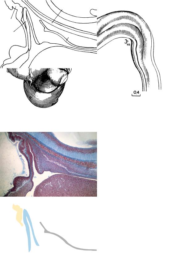
Figure 17–8. Medial view of the brain. In the forebrain, the hypothalamic region can be seen above; below it, from left to right, the elevations of the dorsal thalamus, ventral thalamus, and medial ventricular eminence are visible (cf. Fig. 17–4). Drawn by James F. Didusch from a reconstruction.
NH
AH
Ch.
Pharynx
Basilar a.
Brain
Notochord
Tongue
Figure 17–9. The hypophysis in median section. The elongated adenohypophysial pouch is in contact with the folded wall of the infundibulum.
THE FUTURE OLFACTORY BULBS AND THE FIRST AMYGDALOID NUCLEI |
123 |
Med. forebrain bundle
funiculus
Figure 17–10. The cranial nerves and tracts. All cranial nerves have been present since the previous stage. The last of the regular cranial nerves to appear are the abducent and the cochlear, at stage 16. The olfactory fibers, however, become clear in the present stage. Two bundles of nerve fibers, a lateral and a medial, can be distinguished, and at the site of their entrance into the hemispheres, the olfactory bulb will develop in the next stage. The hypoglossal nerve emerges from the occipital sclerotomic mass as either one (as seen here) or two trunks. The ventral ramus of the first cervical nerve joins the hypoglossal. Cervical nerves 1 and 2 unite to form the inferior root of the ansa cervicalis, and the loop is completed by the superior root, which descends from the hypoglossal nerve. Ganglia are indicated by small circles.
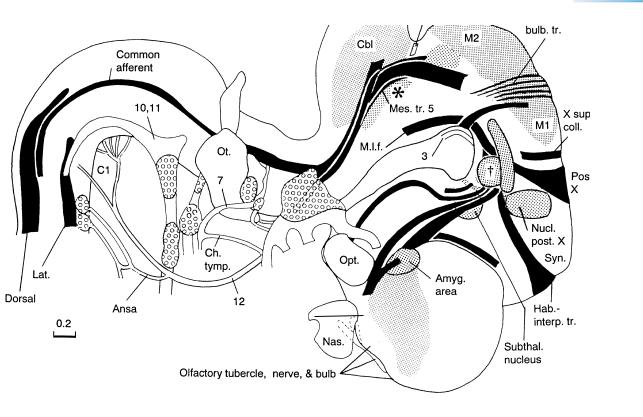
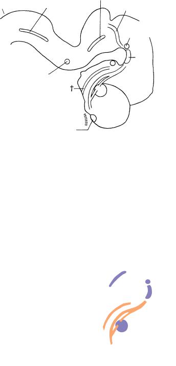
Many tracts have appeared during the previous week (from stage 14 onwards). The order of their appearance as determined from graphic reconstructions is: lateral longitudinal fasciculus, common afferent tract, medial longitudinal fasciculus, dorsal funiculus, preopticohypothalamotegmental tract, mesencephalic tract of the trigeminal nerve, medial prosencephalic fasciculus, medial tectobulbar tract, and mamillotegmental and habenulo-interpeduncular tracts. The medial prosencephalic fasciculus (medial forebrain bundle) contains mainly ascending fibers that terminate in the amygdaloid and preoptic regions. The habenulo-interpeduncular tract (fasciculus retroflexus) delineates the synencephalon rostrally. Some of the nuclei and areas of loose intermediate layer are indicated by stippling. The term “nucleus” refers here to zones of lower cellular density and slightly larger cellular size. The amygdaloid nuclei form the most histologically advanced area of the cerebral hemispheres. The amygdaloid region is related to the medial ventricular eminence (Fig. 19–6). The subthalamic nucleus, red nucleus, interstitial nucleus, nucleus of the posterior commissure, and nucleus of the mesencephalic tract of the trigeminal nerve, and the locus caeruleus (asterisk) are recognizable. The cells of the locus caeruleus occupy the isthmus and rhombomere 1. Its tall cells, twice the size of ordinary neurons, lie at the innermost aspect of the intermediate layer, ventral to the sulcus limitans. The interstitial nucleus is indicated by a dagger. The gustatory fibers begin to separate from the common afferent tract as the tractus solitarius.
The tectal area of the mesencephalon, shown in stippling, has developed an intermediate layer.
In the cerebellum, cells of the rhombic lip participate in the formation of the dentate and isthmic nuclei, and later (stage 23) in the development of the external germinal layer.

124 |
C h a p t e r 1 7 : THE FUTURE OLFACTORY BULBS AND THE FIRST AMYGDALOID NUCLEI |
Figure 17–11. Photomicrograph showing the periphery of the amygdaloid area (the cortex of the future temporal pole), which contains multiple tall cells with long fibers; rostral is to the left-hand side of the page. The neurons for the amygdaloid complex belong to the earliest postmitotic neurons. An initial burst of cell formation for the septal nuclei occurs at this time. Bar: 0.1 mm.
THE FUTURE OLFACTORY BULBS AND THE FIRST AMYGDALOID NUCLEI |
125 |
Figure 17–12. Relationship of the olfactory bulb to other parts of the developing limbic
system at approximately 61 –7 weeks.
/2
(A) Three-dimensional right lateral view of the amygdaloid complex, hippocampal primordium, septal area, and olfactory bulb of right and left sides. The amygdaloid complex is at the future temporal pole of the cerebral hemisphere. (B) Schematic representation of the right amygdaloid complex, seen from the direction of the arrow in (A), with four main nuclei. Two chief fiber connections exist: (1) between olfactory bulb via olfactory tubercle to the medial nucleus and (2) between olfactory bulb and the basolateral and anterior nuclei. The arrows at the right indicate a lateral to medial ( ) and a dorsal to ventral (†) “rotation” during the fetal period. (C) A posterosuperior view of the adult amygdaloid nuclei showing that the definitive arrangement differs from the embryonic (After Neuwenhuys et al., cited in Benninghoff’s Anatomie).
From Muller¨ and O’Rahilly (2006) by permission of the Journal of Anatomy.
126 |
C h a p t e r 1 7 : THE FUTURE OLFACTORY BULBS AND THE FIRST AMYGDALOID NUCLEI |
|
|
|
|
TABLE 17–3. Monoaminergic Centers |
|
|
Stage |
Cells and Nuclei |
Fascicles |
|
|
|
15 |
TH-IR cella |
Medial forebrain bundle (Fig. 15–5) from |
|
Locus caeruleus (A6, Fig. 15–2, 15–10) |
locus caeruleus rostrally |
|
Ventral tegmental area (A9, 10, Figs. 17–13, 20–11) |
Preoptico-hypothalamic tract (Fig. 15–5) |
16 |
Mesencephalic tegmentum (Figs. 17–13, 20–12) |
|
17 |
3–4 amygdaloid nuclei (Fig. 17–12) |
Medial forebrain bundle (Figs. 17–10, 17–13) |
|
Noradrenergic cells in locus caeruleus |
TH-IR-reaction of fibers of medial forebrain |
|
TH-IR neurons in A8, A9, and A10 and along |
bundle from basal mesencephalon to |
|
vomeronasal organ |
amygdaloid regionb |
|
Elongated band of TH-IR cells from nasal sac to |
|
|
septuma,b |
|
|
Olfactory bulb (Figs. 17–10, 17–13) |
|
18 |
4 amygdaloid nuclei (Fig. 18–5) |
|
19 |
4 amygdaloid nuclei (Figs. 19–8, 19–9) |
|
20 |
Interpeduncular nucleus (Figs. 17–13, 20–9) |
|
|
Nuclei solitarius and ambiguus (Fig. 20–19) |
|
Before or at 23 |
TH-IR penetrate telencephalic wall and [lateral] |
|
|
ventricular eminence |
|
|
Fibers in subplate, but not in cortical platea,b |
|
|
|
|
a Verney et al. (1996).
b Zecevic and Verney (1995).
|
A6 |
|
|
A2 |
|
A4 |
||
|
Interpeduncular nucleus
A10
A5
A
A15
Figure 17–13. Monoaminergic centers that are present in stage 17 and later. Those in stage 17 or even earlier (marked in purple) are locus caeruleus (A6), ventral tegmental area (A 10), and amygdaloid body (A). The tracts (orange) related to these centers and also present in stage 17 are the preoptico-hypothalamic tract (dagger) and the medial forebrain bundle. Centers that arise later include the nucleus solitarius (A2), the area along the superior cerebellar peduncle (A4), the area near the facial nucleus (A5), and the external layer of the olfactory bulb (A15).
A, |
Central nucleus of amygdaloid |
A8, |
Retrorubral cells |
|
|
body |
|
|
|
A2, |
Nucleus solitarius |
A9, |
Substantia nigra |
|
A4, |
Cells along superior cerebellar |
A10, |
Ventral |
|
|
peduncle |
|
tegmental area |
|
A5, |
Area near facial nucleus |
A15, |
External layer of |
|
|
|
|
olfactory bulb |
|
A6, Locus caeruleus |
|
|
||
|
|
|
|
|
THE FUTURE OLFACTORY BULBS AND THE FIRST AMYGDALOID NUCLEI |
127 |
Fig. 17 – 15 14
Figure 17–14. The rostralmost section shows only the paired cerebral hemispheres and the telencephalon medium between them. A slight swelling represents the lateral ventricular eminence. The longitudinal cerebral fissure is filled with loose mesenchyme. Here, and elsewhere in the mesenchyme adjacent to the brain, thin-walled blood vessels are numerous, and the anterior choroid artery has appeared. The continuity between the nostril and the nasal sac is visible at the left side. The two nasal sacs are separated by dense mesenchyme that represents the future nasal septum.
Figure 17–15. The second section shows the interventricular foramina, which begin to be apparent at about this time. They become relatively narrower as the medial surface of each cerebral hemisphere develops and as the medial ventricular eminence grows.
Figs. 17– 18 17 16
. Key for Figures 17–16 to 17–18 on page 128.